

Tested Applications
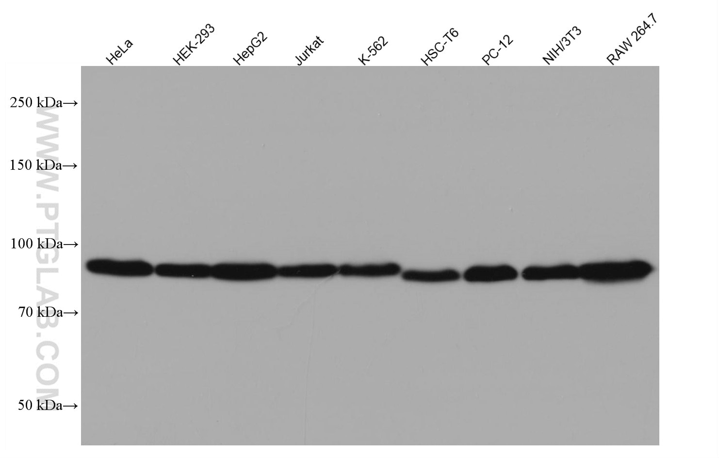
| Positive WB detected in | HeLa cells, HEK-293 cells, HepG2 cells, Jurkat cells, K-562 cells, HSC-T6 cells, PC-12 cells, NIH/3T3 cells, RAW 264.7 cells |
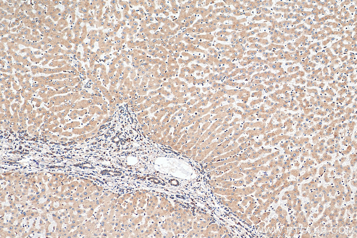
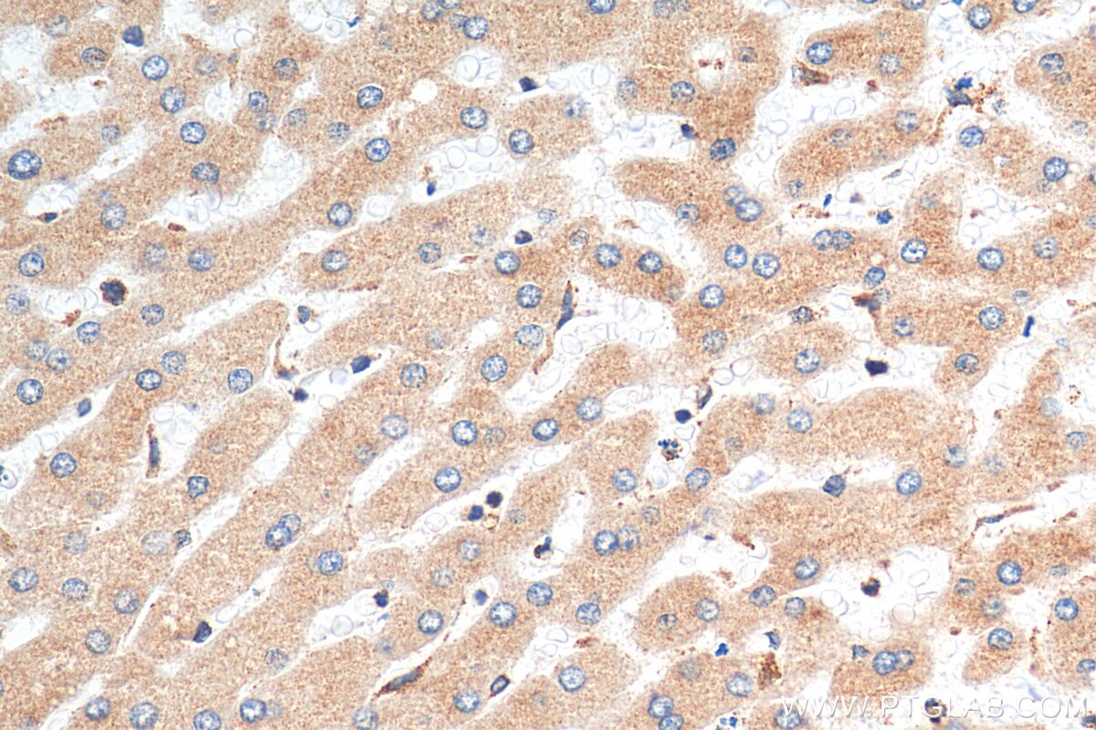
| Positive IHC detected in | human hepatocirrhosis tissue Note: suggested antigen retrieval with TE buffer pH 9.0; (*) Alternatively, antigen retrieval may be performed with citrate buffer pH 6.0 |
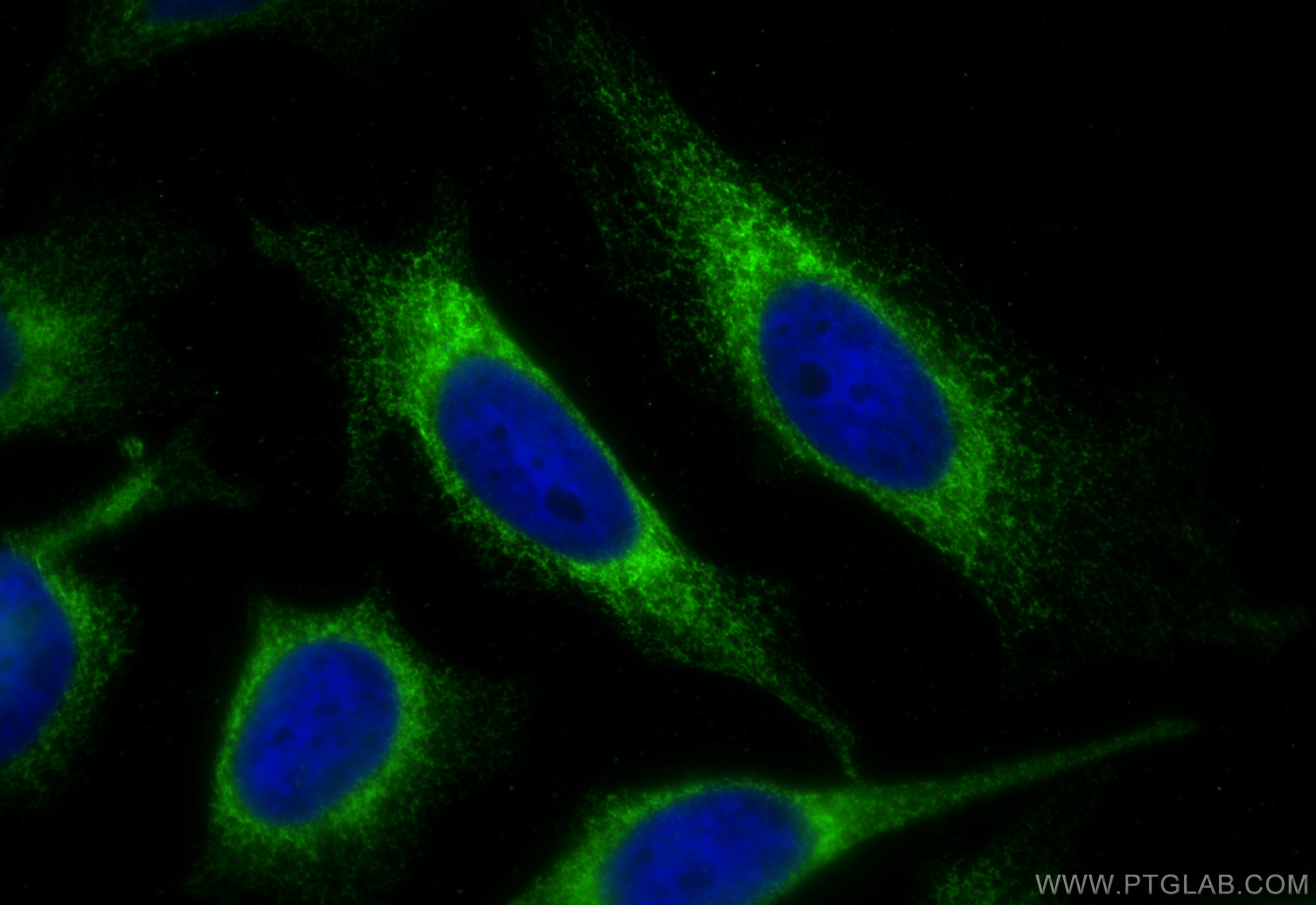
| Positive IF/ICC detected in | HeLa cells |
Recommended dilution
| Application | Dilution |
|---|---|
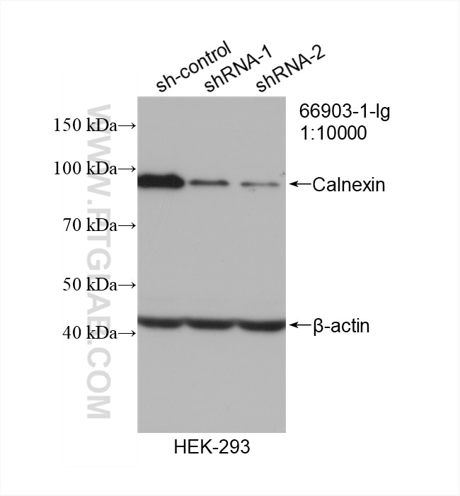
| Western Blot (WB) | WB : 1:5000-1:20000 |
| Immunohistochemistry (IHC) | IHC : 1:1000-1:4000 |
| Immunofluorescence (IF)/ICC | IF/ICC : 1:200-1:800 |
| It is recommended that this reagent should be titrated in each testing system to obtain optimal results. | |
| Sample-dependent, Check data in validation data gallery. | |
Product Information
66903-1-Ig targets Calnexin in WB, IHC, IF/ICC, IP, ELISA applications and shows reactivity with human, mouse samples.
| Tested Reactivity | human, mouse |
| Cited Reactivity | human, mouse, rat, pig, rabbit, hamster |
| Host / Isotype | Mouse / IgG1 |
| Class | Monoclonal |
| Type | Antibody |
| Immunogen |
CatNo: Ag25535 Product name: Recombinant human Calnexin protein Source: e coli.-derived, PET28a Tag: 6*His Domain: 1-273 aa of BC003552 Sequence: MEGKWLLCMLLVLGTAIVEAHDGHDDDVIDIEDDLDDVIEEVEDSKPDTTAPPSSPKVTYKAPVPTGEVYFADSFDRGTLSGWILSKAKKDDTDDEIAKYDGKWEVEEMKESKLPGDKGLVLMSRAKHHAISAKLNKPFLFDTKPLIVQYEVNFQNGIECGGAYVKLLSKTPELNLDQFHDKTPYTIMFGPDKCGEDYKLHFIFRHKNPKTGIYEEKHAKRPDADLKTYFTDKKTHLYTLILNPDNSFEILVDQSVVNSGNLLNDMTPPVNPS Predict reactive species |
| Full Name | calnexin |
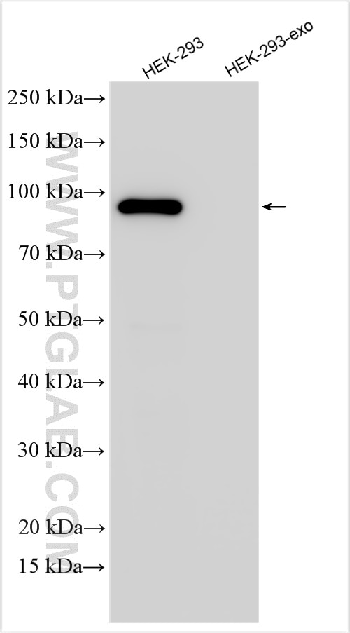
| Calculated Molecular Weight | 90 kDa |
| Observed Molecular Weight | 90 kDa |
| GenBank Accession Number | BC003552 |
| Gene Symbol | Calnexin |
| Gene ID (NCBI) | 821 |
| RRID | AB_2882231 |
| Conjugate | Unconjugated |
| Form | Liquid |
| Purification Method | Protein G purification |
| UNIPROT ID | P27824 |
| Storage Buffer | PBS with 0.02% sodium azide and 50% glycerol, pH 7.3. |
| Storage Conditions | Store at -20°C. Stable for one year after shipment. Aliquoting is unnecessary for -20oC storage. 20ul sizes contain 0.1% BSA. |
Background Information
Calnexin is a molecular chaperone that resides in the endoplasmic reticulum (ER) and participates in the folding and assembly of newly synthesized proteins. Calnexin is highly abundant in ER and is frequently used as an ER marker.
Protocols
| Product Specific Protocols | |
|---|---|
| IF protocol for Calnexin antibody 66903-1-Ig | Download protocol |
| IHC protocol for Calnexin antibody 66903-1-Ig | Download protocol |
| WB protocol for Calnexin antibody 66903-1-Ig | Download protocol |
| Standard Protocols | |
|---|---|
| Click here to view our Standard Protocols |
Publications
What published studies show
The Calnexin Monoclonal antibody (catalog number 66903-1-Ig) has 156 total citations. These appear across high-impact journals including Science, Cell Metabolism, Cell Research, Nature Communications, Autophagy, Molecular Cell, EMBO Journal, and Cell Reports. Research fields span extracellular vesicle/exosome biology, endoplasmic reticulum stress, autophagy and mitophagy, ER-mitochondria contact sites, cancer, neurodegeneration, and infectious disease. Applications are predominantly Western blot and immunofluorescence, with some immunoprecipitation and IHC, across human, mouse, rat, and other species.
| Species | Application | Title |
|---|---|---|
Science Stereo-cell: Spatial enhanced-resolution single-cell sequencing with high-density DNA nanoball-patterned arrays | ||
Cell Metab Adipocyte-derived glutathione promotes obesity-related breast cancer by regulating the SCARB2-ARF1-mTORC1 complex | ||
Cell Res Targeting ATAD3A-PINK1-mitophagy axis overcomes chemoimmunotherapy resistance by redirecting PD-L1 to mitochondria | ||
J Extracell Vesicles The 8-oxoguanine DNA glycosylase-synaptotagmin 7 pathway increases extracellular vesicle release and promotes tumour metastasis during oxidative stress | ||
J Extracell Vesicles Engineered Nestin(+) TSPC-Derived Exosomes Promote Tendon Repair Via Metabolic Reprogramming. | ||
Autophagy RETREG1-mediated reticulophagy is activated by an ATF4-CEBPG/C/EBPγ heterodimer and confers protection against lipotoxicity |
Reviews
What customers say
Both reviewers rated this antibody 5 stars for Western Blot at a 1:10,000 dilution. One user tested it on HeLa cells and reported clean bands that were successfully excised and analyzed by mass spectrometry. The other confirmed strong, clean band detection in HEK293T lysates. Overall, users highlight the antibody's specificity, clean background, and reliable performance in WB applications.
The reviews below have been submitted by verified Proteintech customers who received an incentive for providing their feedback.
Samantha (Verified Customer) (03-18-2024) | Tested on HeLA cells and produced clean bands that were cut out and analysed with Mass Spectrometry
|
Amy (Verified Customer) (11-08-2023) | Clean antibody with strong band for WB in HEK293T lysates.
|







