
Tested Applications
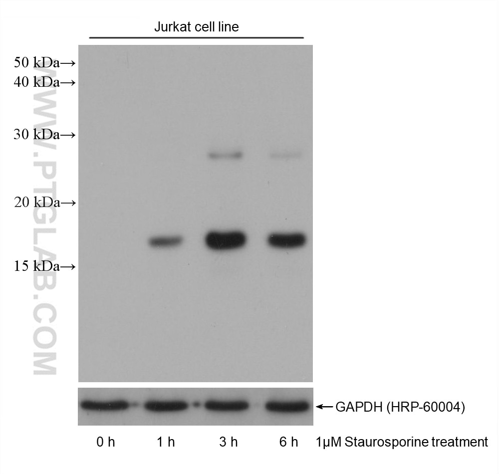
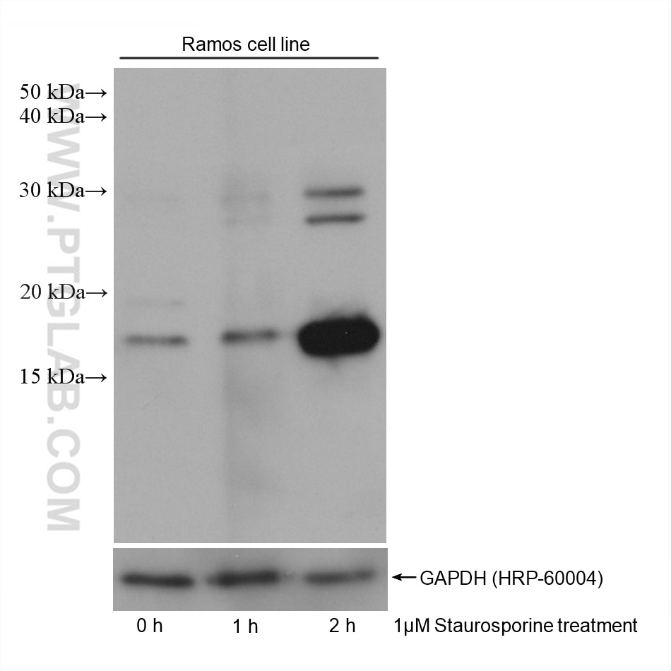
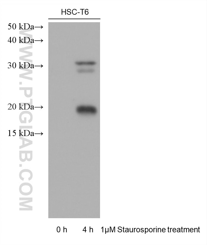
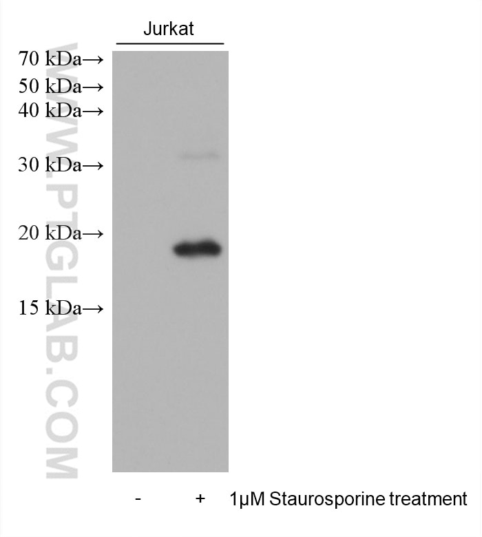
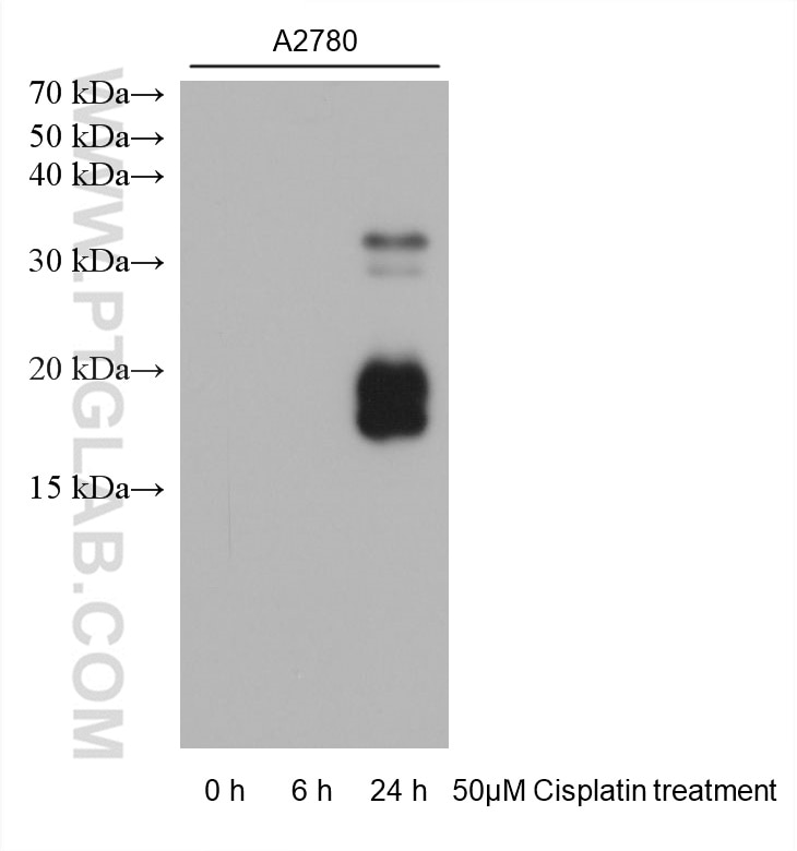
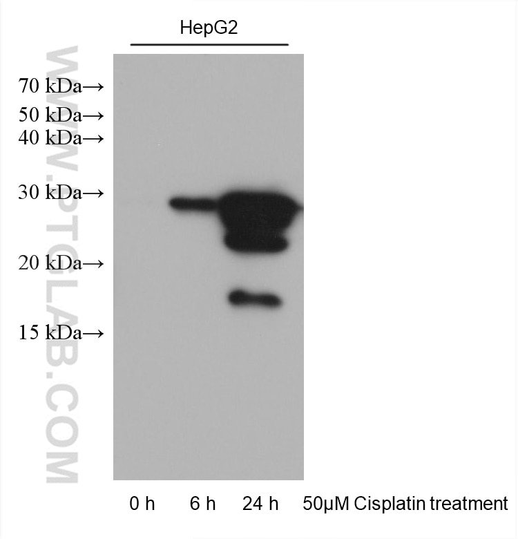
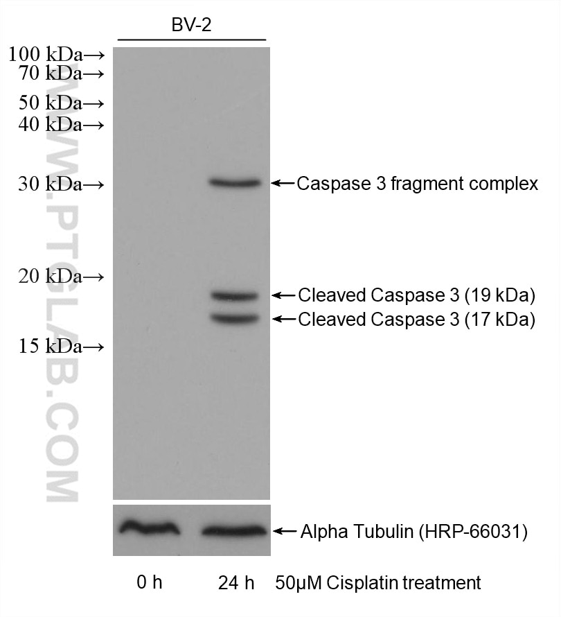
| Positive WB detected in | Jurkat cells, A2780 cells, BV-2 cells, HepG2 cells, HSC-T6 cells, Ramos cells |
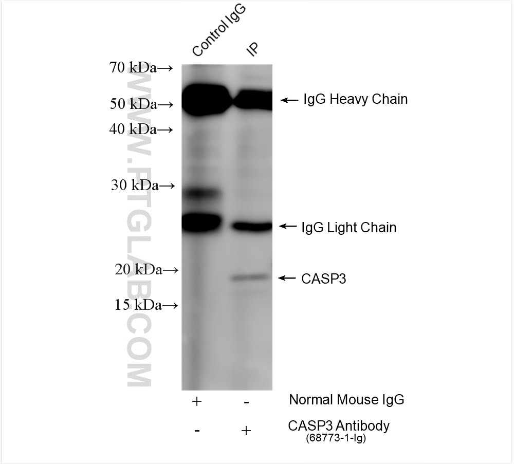
| Positive IP detected in | Staurosporine treated Jurkat cells |
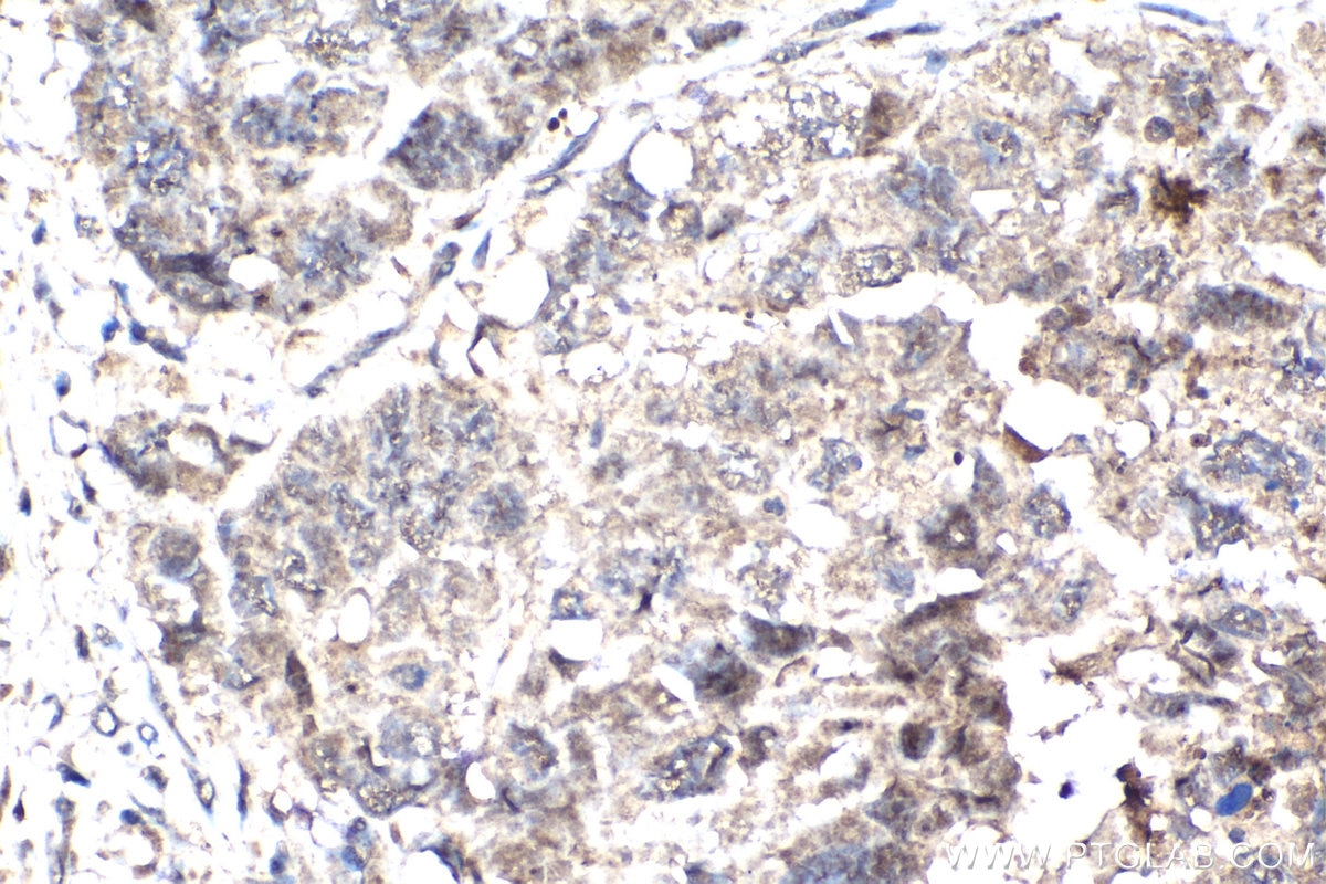
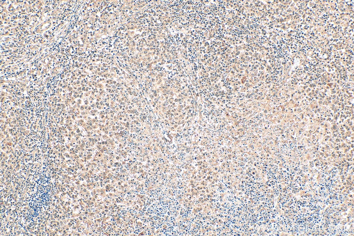
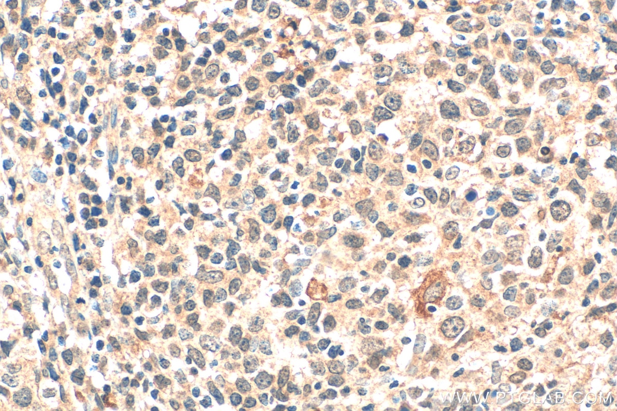
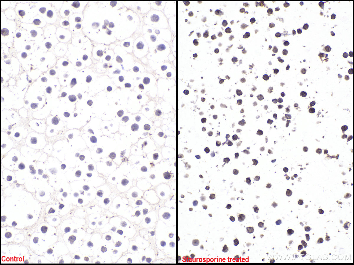
| Positive IHC detected in | human lymphoma tissue, Jurkat cells Note: suggested antigen retrieval with TE buffer pH 9.0; (*) Alternatively, antigen retrieval may be performed with citrate buffer pH 6.0 |
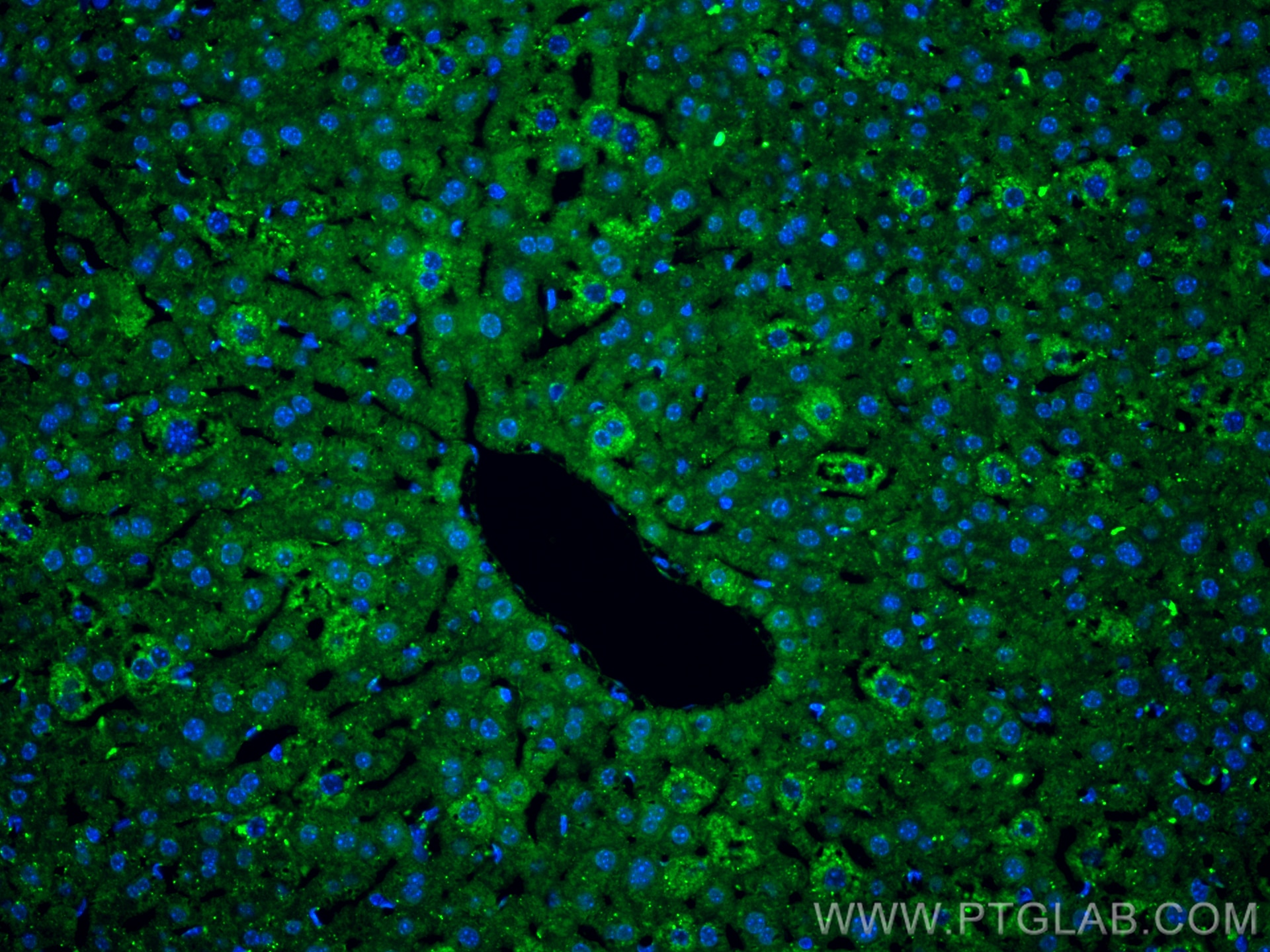
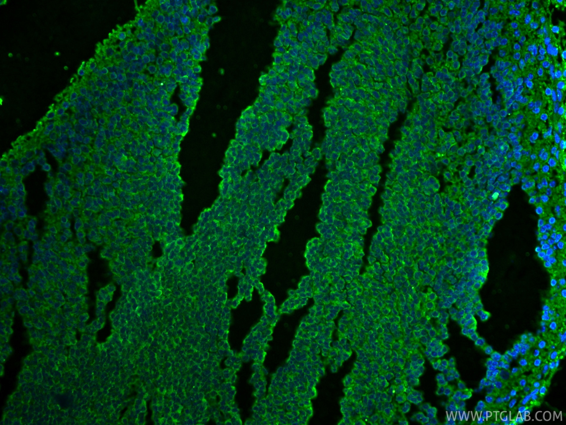
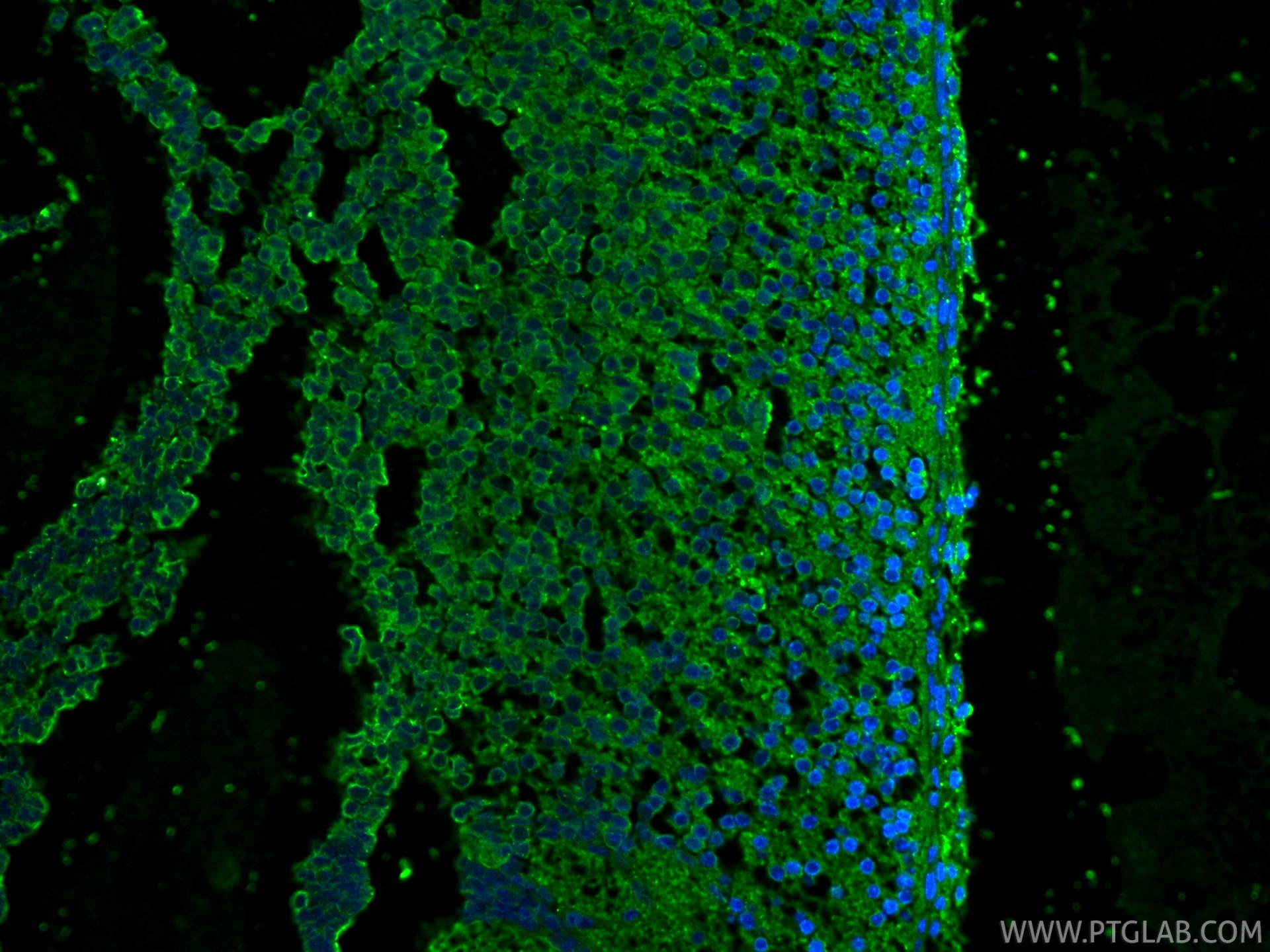
| Positive IF-P detected in | mouse liver tissue, mouse embryo tissue |
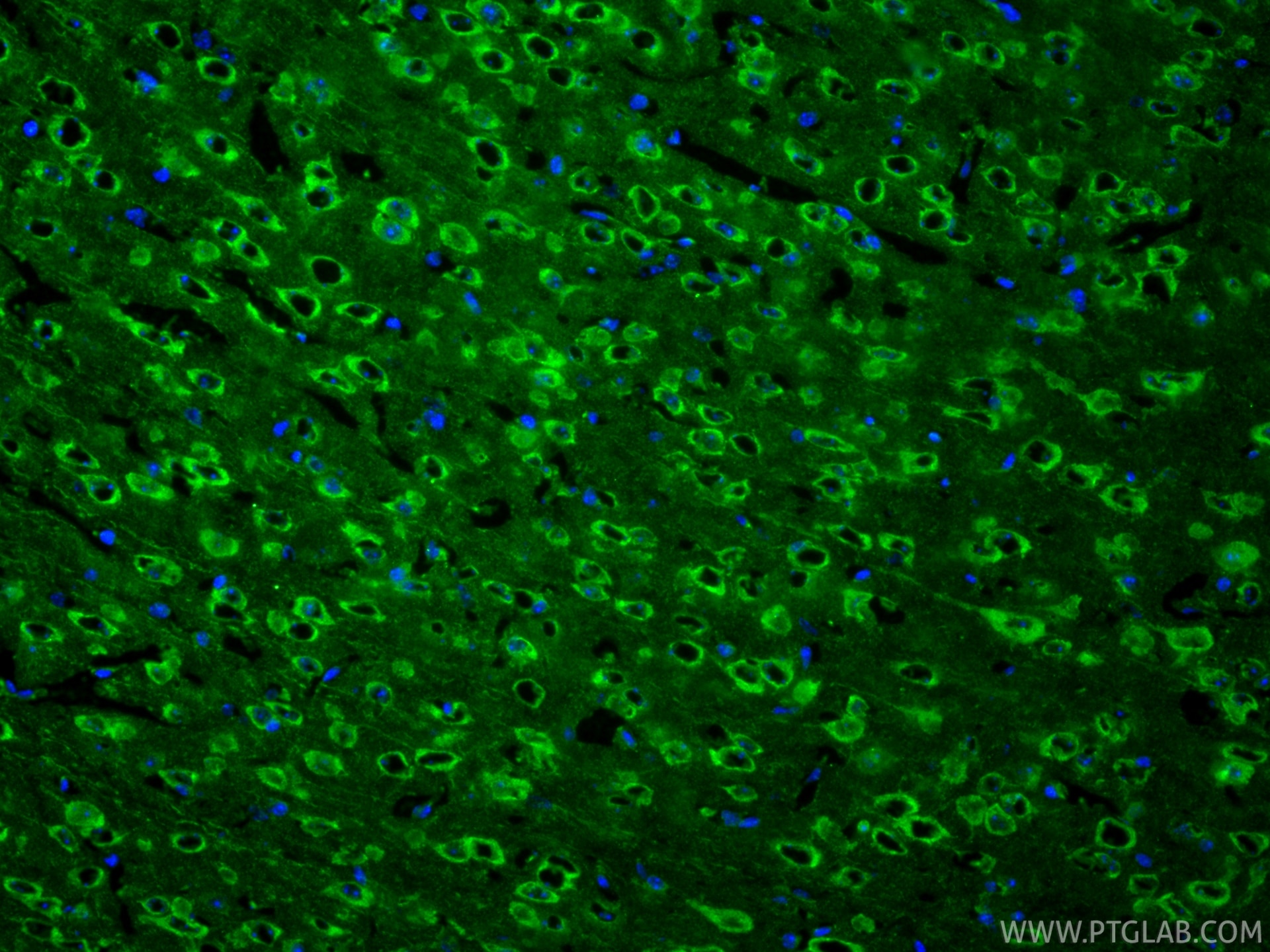
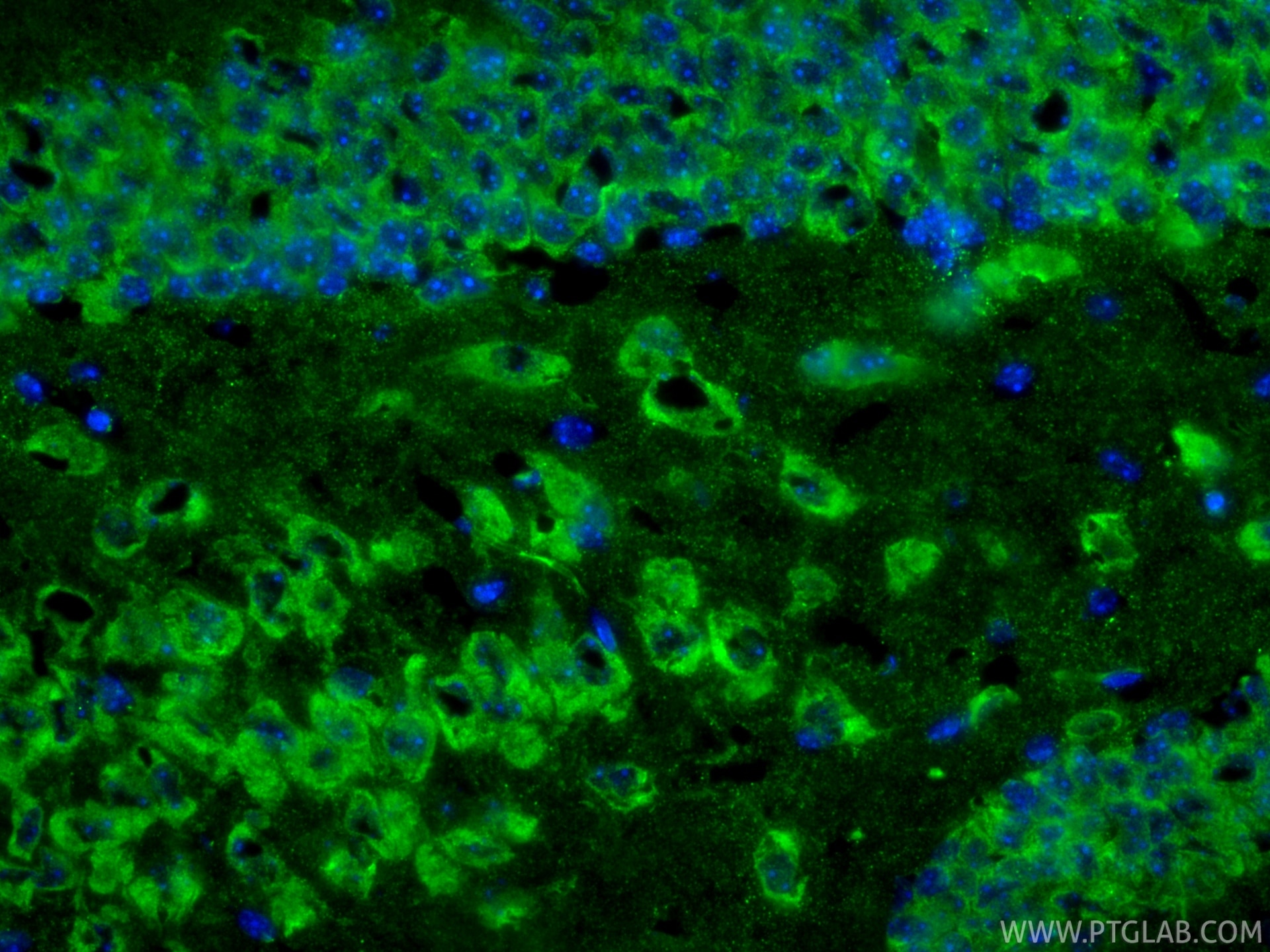
| Positive IF-Fro detected in | mouse brain tissue |
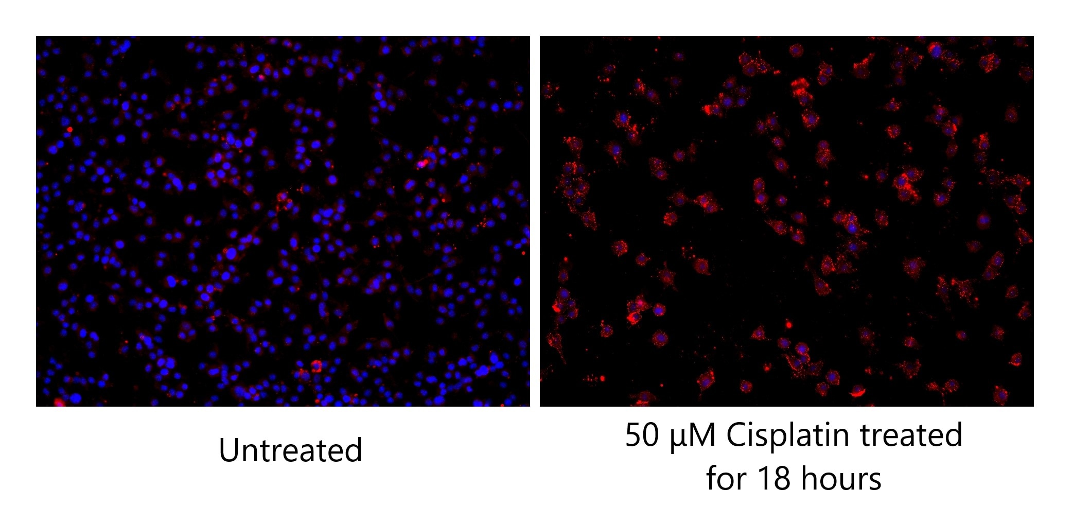
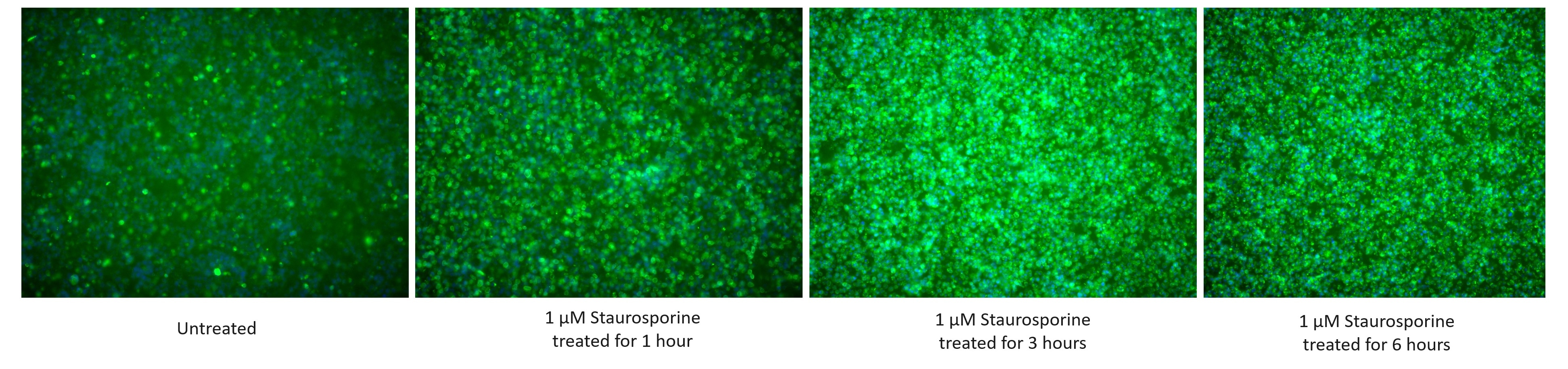
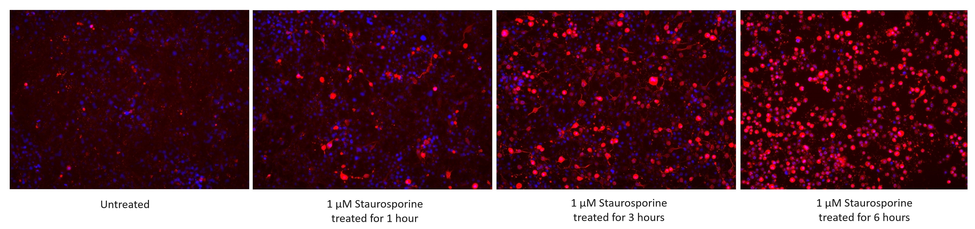
| Positive IF/ICC detected in | 1 μM Staurosporine (1/3/6 hour) treated Jurkat cells, 50 μM Cisplatin (18 hours) treated BV-2 cells, 1 μM Staurosporine (1/3/6 hour) treated HSC-T6 cells |
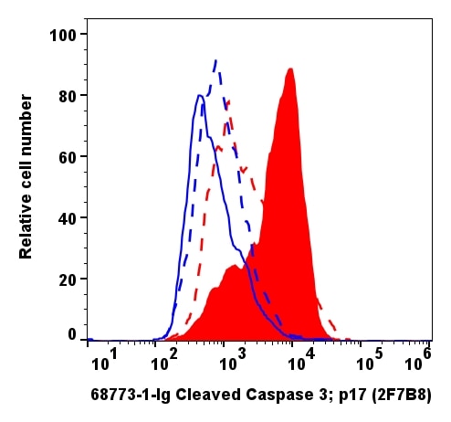
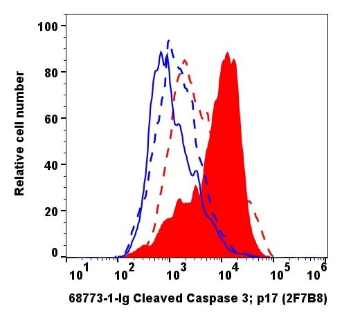
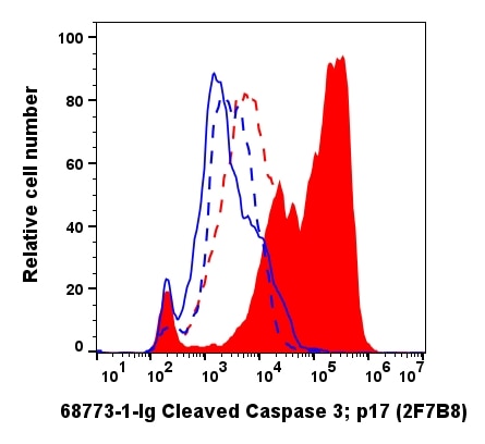
| Positive FC (Intra) detected in | 1 μM Staurosporine (3 hours) treated Jurkat cells, 50 μM Cisplatin(18h)treated BV-2 cells |
Recommended dilution
| Application | Dilution |
|---|---|
| Western Blot (WB) | WB : 1:5000-1:50000 |
| Immunoprecipitation (IP) | IP : 0.5-4.0 ug for 1.0-3.0 mg of total protein lysate |
| Immunohistochemistry (IHC) | IHC : 1:1000-1:4000 |
| Immunofluorescence (IF)-P | IF-P : 1:200-1:800 |
| Immunofluorescence (IF)-FRO | IF-FRO : 1:400-1:1600 |
| Immunofluorescence (IF)/ICC | IF/ICC : 1:500-1:2000 |
| Flow Cytometry (FC) (INTRA) | FC (INTRA) : 0.20 ug per 10^6 cells in a 100 µl suspension |
| It is recommended that this reagent should be titrated in each testing system to obtain optimal results. | |
| Sample-dependent, Check data in validation data gallery. | |
Product Information
68773-1-Ig targets Cleaved Caspase 3/P17/P19 in WB, IHC, IF/ICC, IF-P, IF-Fro, FC (Intra), IP, ELISA applications and shows reactivity with human, mouse, rat samples.
| Tested Reactivity | human, mouse, rat |
| Cited Reactivity | human, mouse, rat |
| Host / Isotype | Mouse / IgG1 |
| Class | Monoclonal |
| Type | Antibody |
| Immunogen |
Peptide Predict reactive species |
| Full Name | caspase 3, apoptosis-related cysteine peptidase |
| Calculated Molecular Weight | 32 kDa |
| Observed Molecular Weight | 17 kDa, 19 kDa |
| GenBank Accession Number | NM_032991 |
| Gene Symbol | Caspase 3 |
| Gene ID (NCBI) | 836 |
| RRID | AB_3665444 |
| Conjugate | Unconjugated |
| Form | Liquid |
| Purification Method | Protein G purification |
| UNIPROT ID | P42574 |
| Storage Buffer | PBS with 0.02% sodium azide and 50% glycerol, pH 7.3. |
| Storage Conditions | Store at -20°C. Stable for one year after shipment. Aliquoting is unnecessary for -20oC storage. 20ul sizes contain 0.1% BSA. |
Background Information
Caspases, a family of endoproteases, are critical players in cell regulatory networks controlling inflammation and cell death. Initiator caspases (caspase-2, -8, -9, -10, -11, and -12) cleave and activate downstream effector caspases (caspase-3, -6, and -7), which in turn execute apoptosis by cleaving targeted cellular proteins. Caspase 3 plays a key role in the activation of sterol regulatory element binding proteins (SREBPs) between the basic helix-loop-helix leucine zipper domain and the membrane attachment domain. Caspase 3 can form heterocomplex with other proteins. This antibody is specific for cleaved caspase 3, and does not recognize full length caspase-3. The cleaved P17 and P19 fragment might form complex and shows at around 30-35 kDa by western blot (PMID: 25501826).
Protocols
| Product Specific Protocols | |
|---|---|
| IF protocol for Cleaved Caspase 3/P17/P19 antibody 68773-1-Ig | Download protocol |
| IHC protocol for Cleaved Caspase 3/P17/P19 antibody 68773-1-Ig | Download protocol |
| IP protocol for Cleaved Caspase 3/P17/P19 antibody 68773-1-Ig | Download protocol |
| WB protocol for Cleaved Caspase 3/P17/P19 antibody 68773-1-Ig | Download protocol |
| Standard Protocols | |
|---|---|
| Click here to view our Standard Protocols |
Publications
What published studies show
| Species | Application | Title |
|---|---|---|
J Extracell Vesicles Small Extracellular Vesicles Engineered Using Click Chemistry to Express Chimeric Antigen Receptors Show Enhanced Efficacy in Acute Liver Failure | ||
Nat Commun Tubulin autoregulation tunes microtubule dynamics to support multicellular architecture and viability. | ||
Nat Commun S100A12 triggers NETosis to aggravate myocardial infarction injury via the Annexin A5-calcium axis | ||
Exp Mol Med Cancer-associated fibroblast-derived extracellular vesicles regulate lipophagy through PLIN2 to modulate dormancy in salivary gland adenoid cystic carcinoma cells. | ||
ACS Nano Mucoadhesive Nanothalidomide Enema: Simultaneous Radioprotection and Barrier Repair in Radiation Enteritis Therapy. | ||
J Nanobiotechnology A combination of a TLR7/8 agonist and an epigenetic inhibitor suppresses triple-negative breast cancer through triggering anti-tumor immune. |
Reviews
What customers say
The reviews below have been submitted by verified Proteintech customers who received an incentive for providing their feedback.
Joana (Verified Customer) (09-02-2026) | Immunohistochemical fluorescence analysis of cryosectioned mouse brain tissue slide using 68773-1-IG (Caspase 3 antibody) at dilution of 1:1000 (under 10x lens). Mus musculus 6-OHDA injection at 2w presents cas3 (white) colocalized with GFAP (Green), Hoescht (blue)
|
Molly (Verified Customer) (10-03-2024) | Worked well in IF on primary rat embryonic neuron/astrocyte co-culture at a range of dilutions.
|
























