

Tested Applications
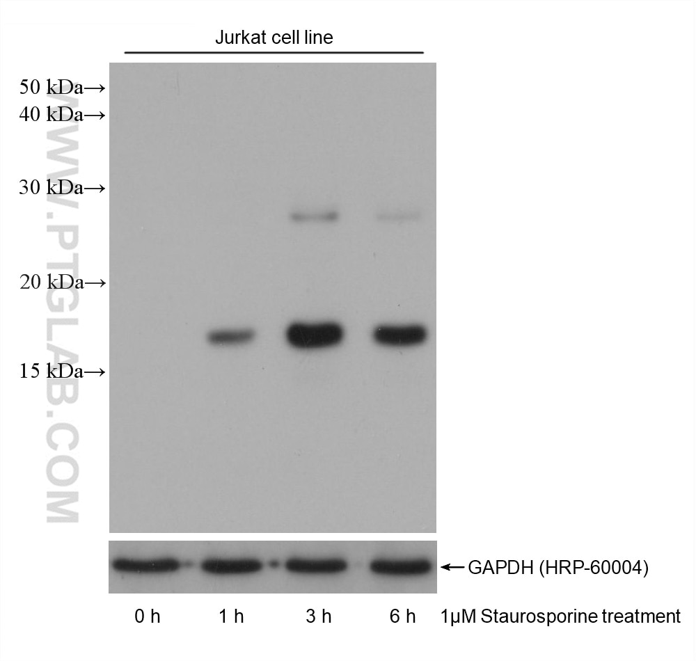
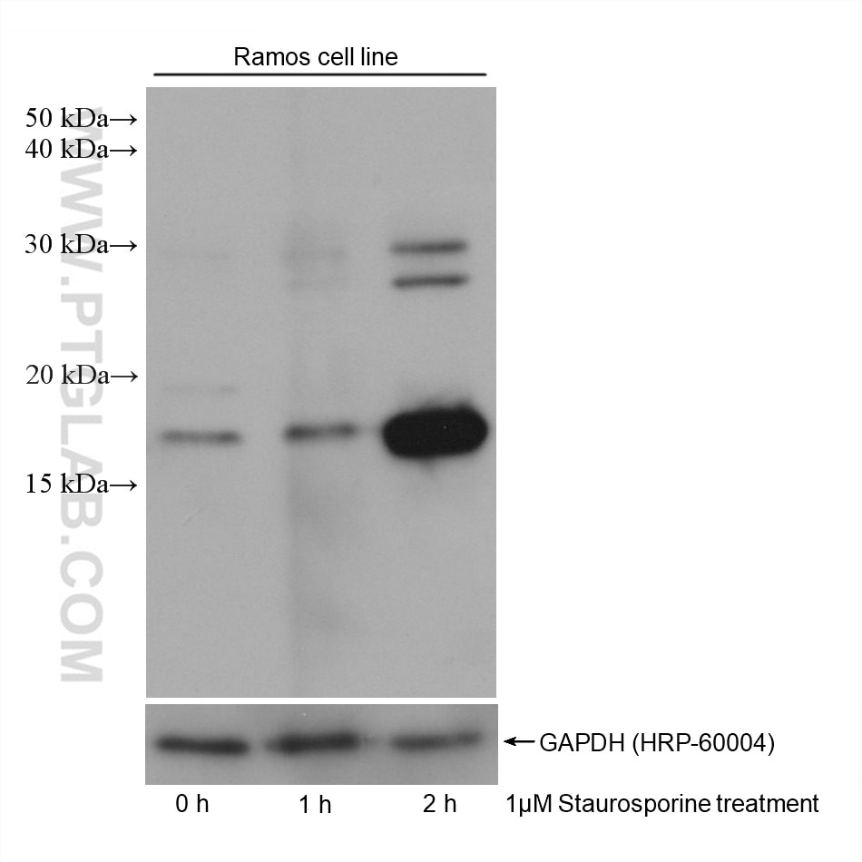
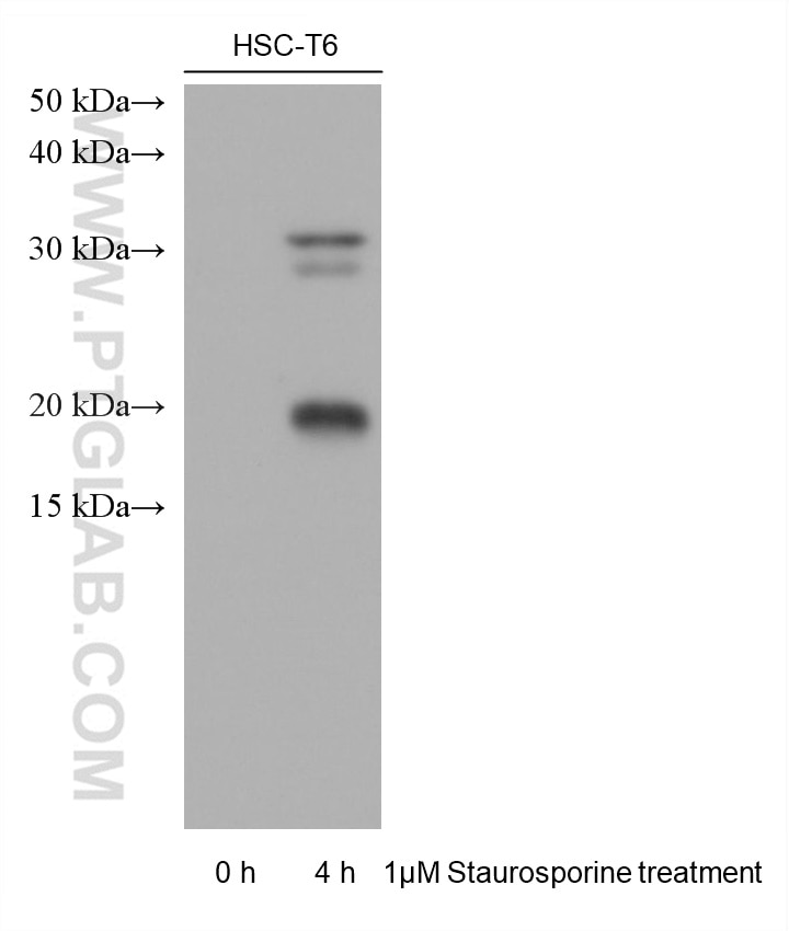
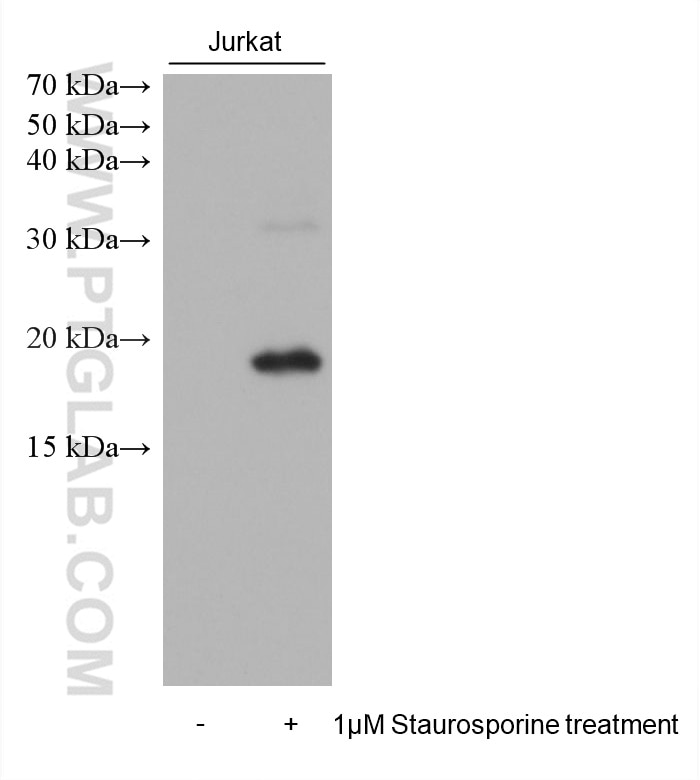
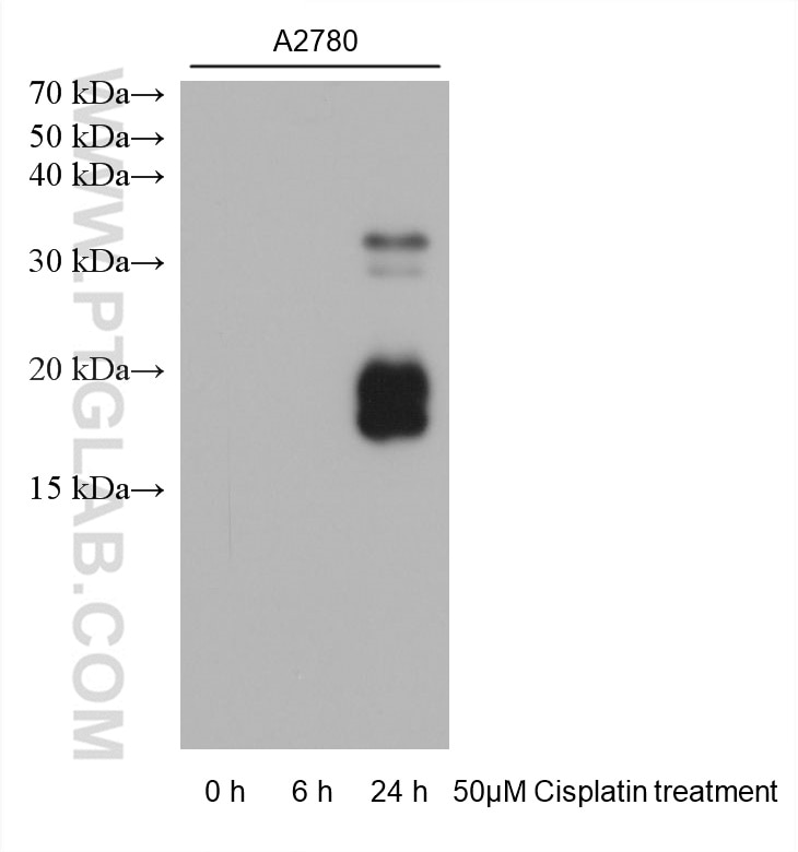
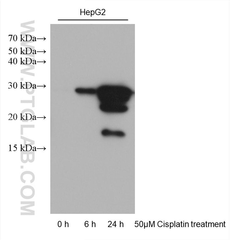
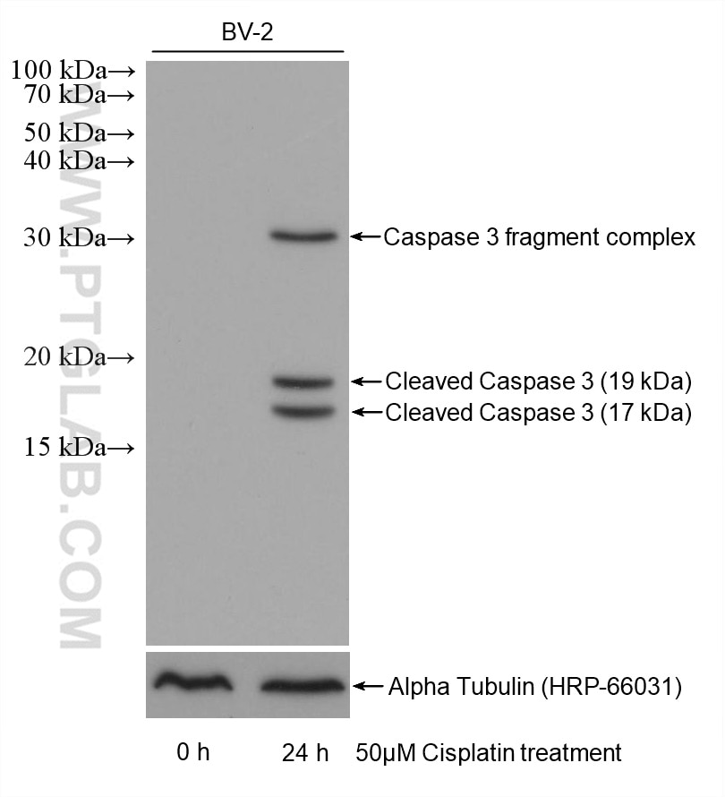
| Positive WB detected in | Jurkat cells, A2780 cells, BV-2 cells, HepG2 cells, HSC-T6 cells, Ramos cells |
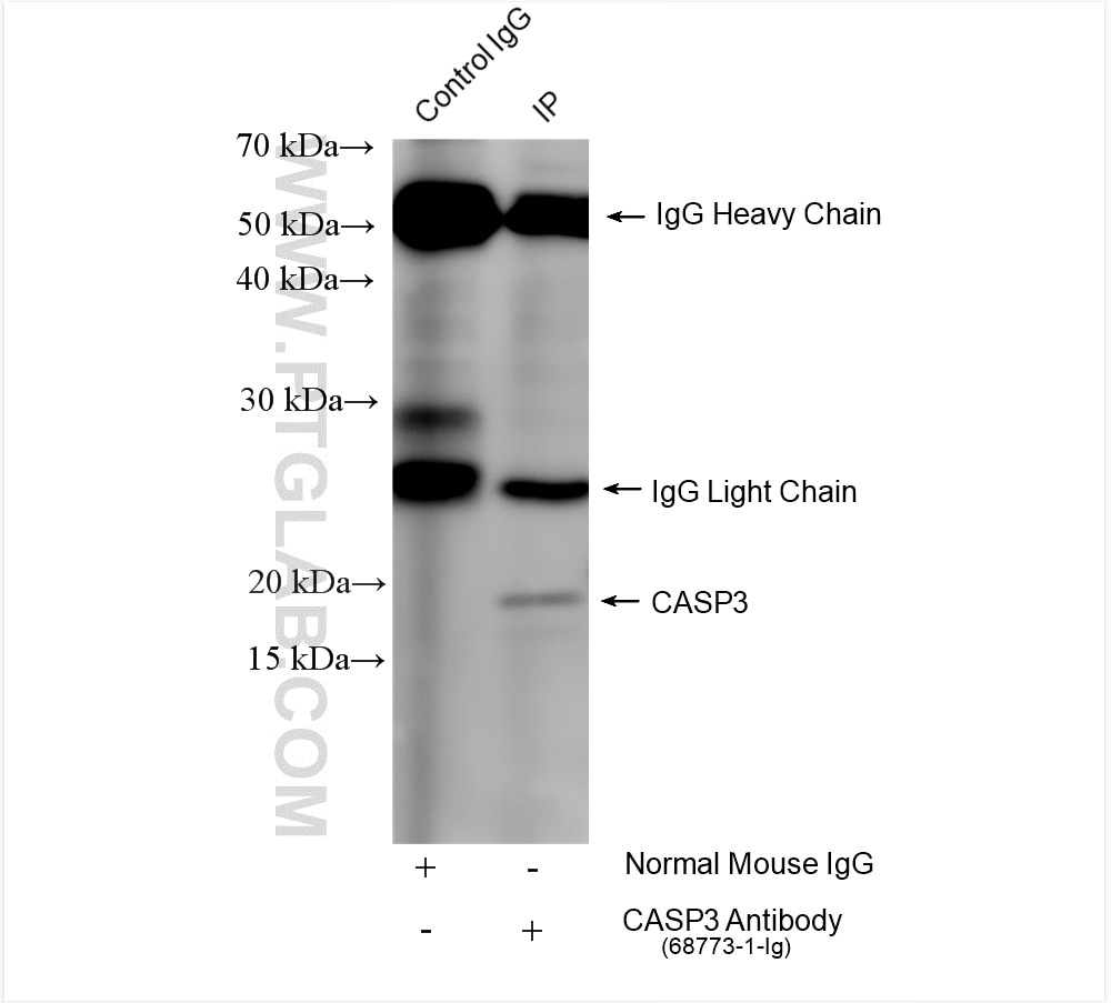
| Positive IP detected in | Staurosporine treated Jurkat cells |
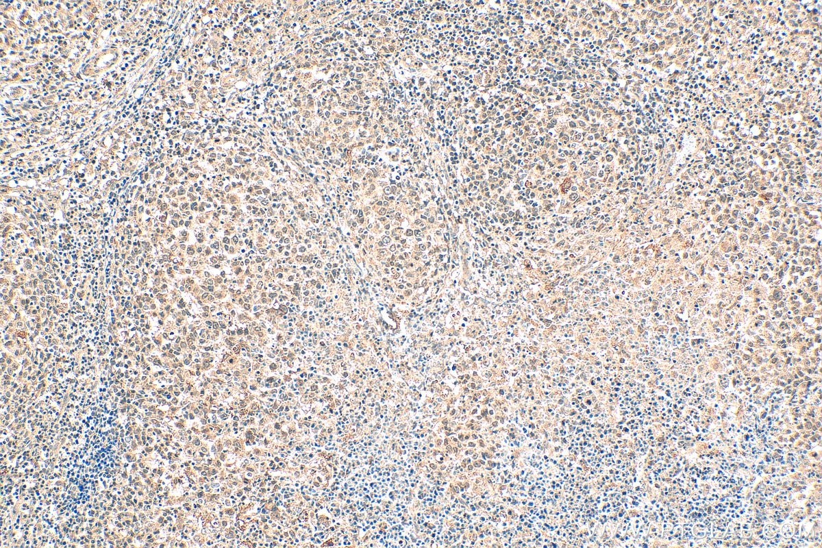
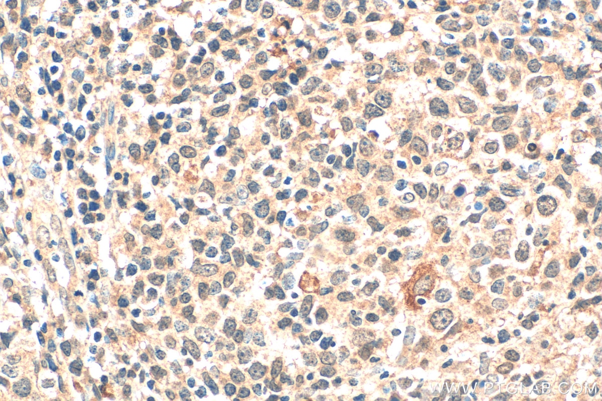
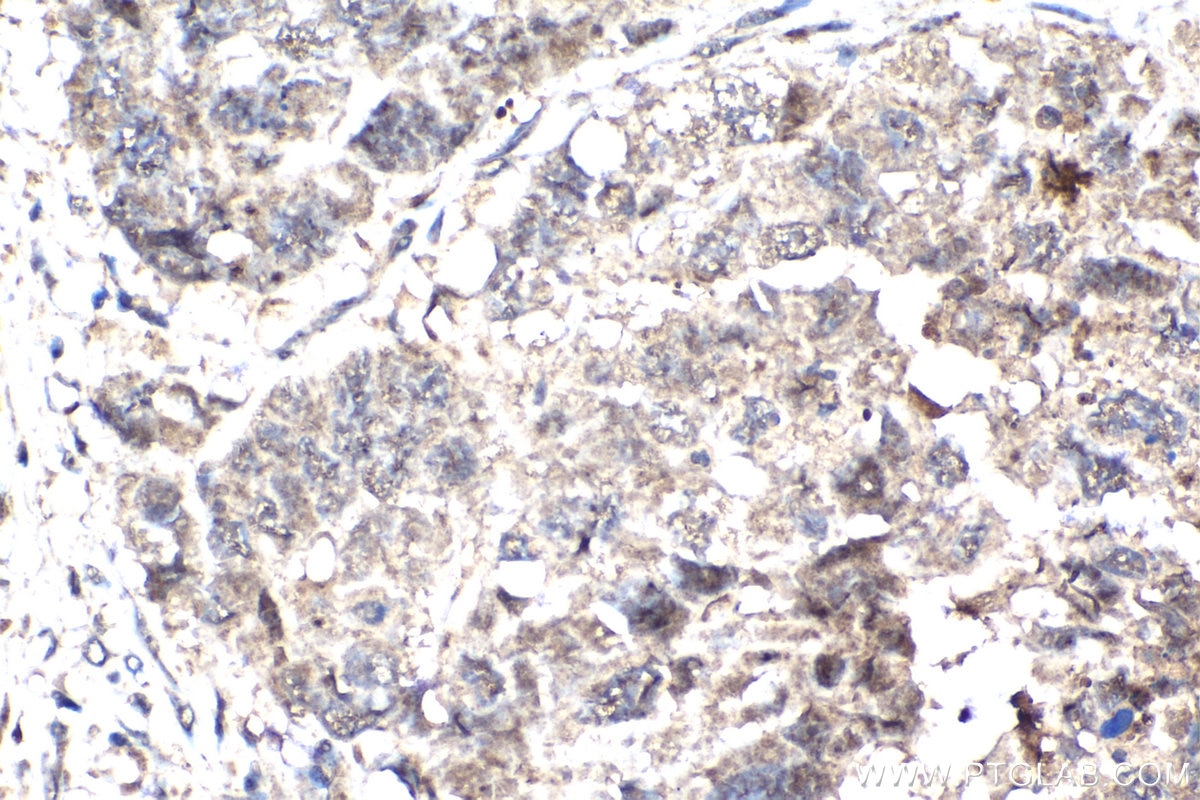
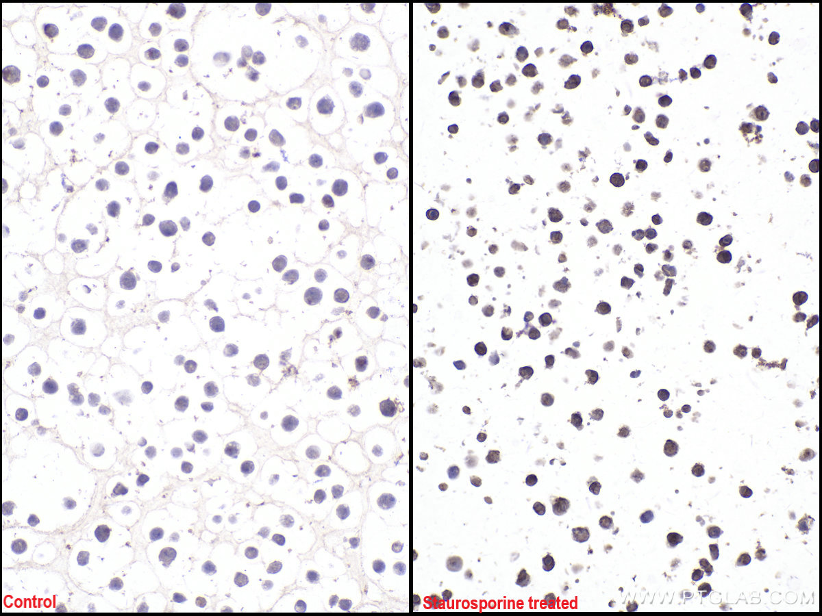
| Positive IHC detected in | human lymphoma tissue, Jurkat cells Note: suggested antigen retrieval with TE buffer pH 9.0; (*) Alternatively, antigen retrieval may be performed with citrate buffer pH 6.0 |
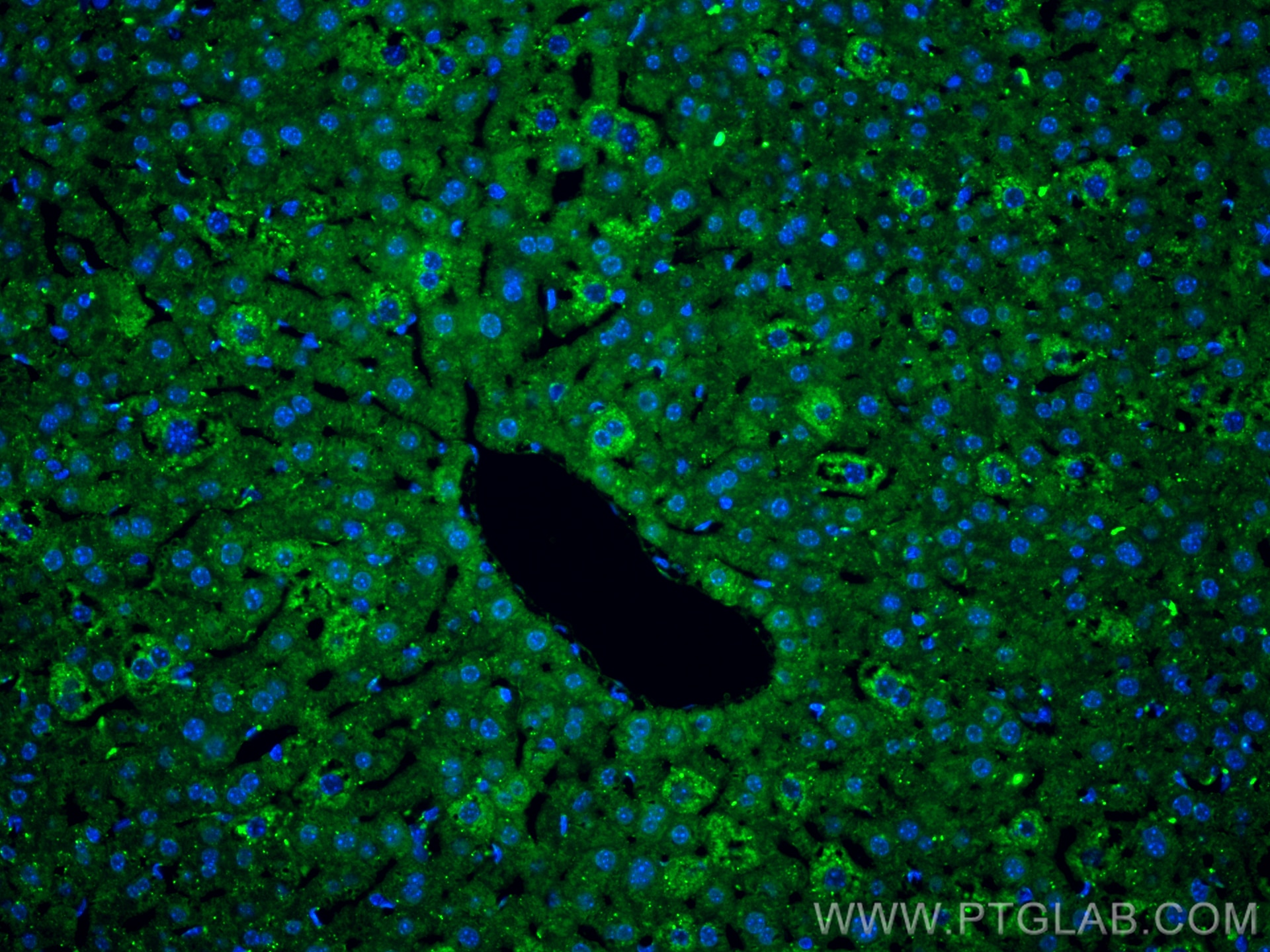
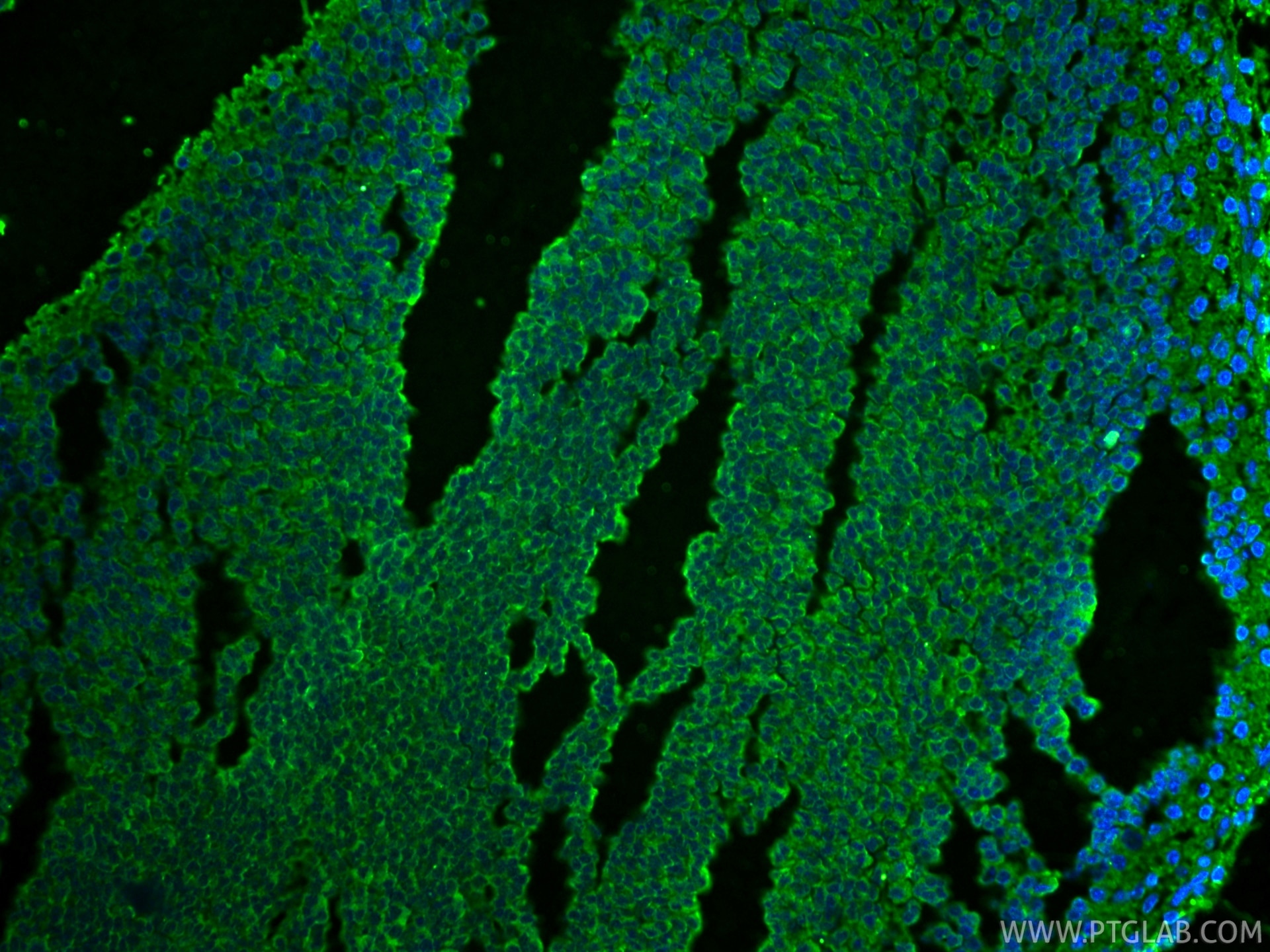
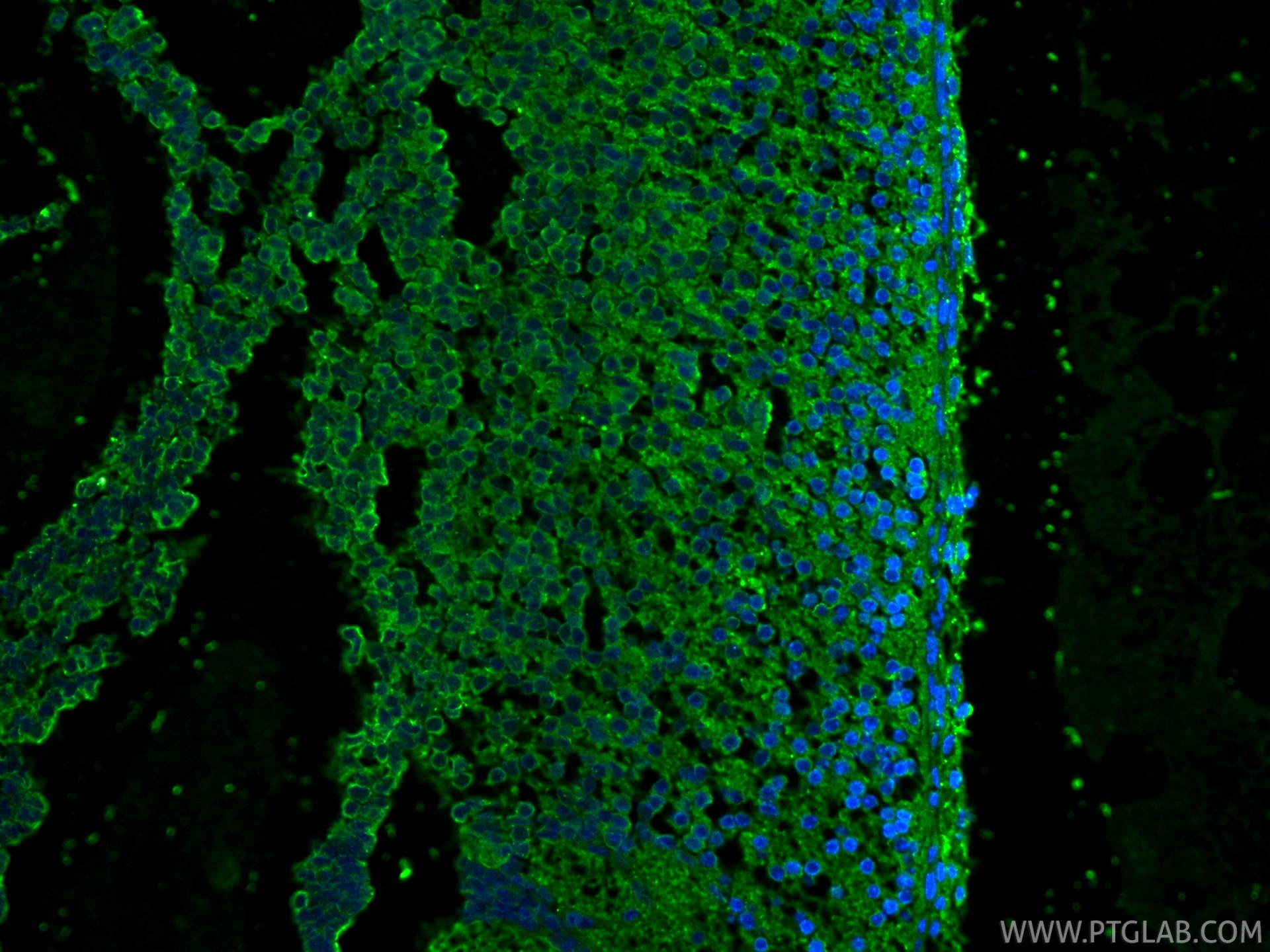
| Positive IF-P detected in | mouse liver tissue, mouse embryo tissue |
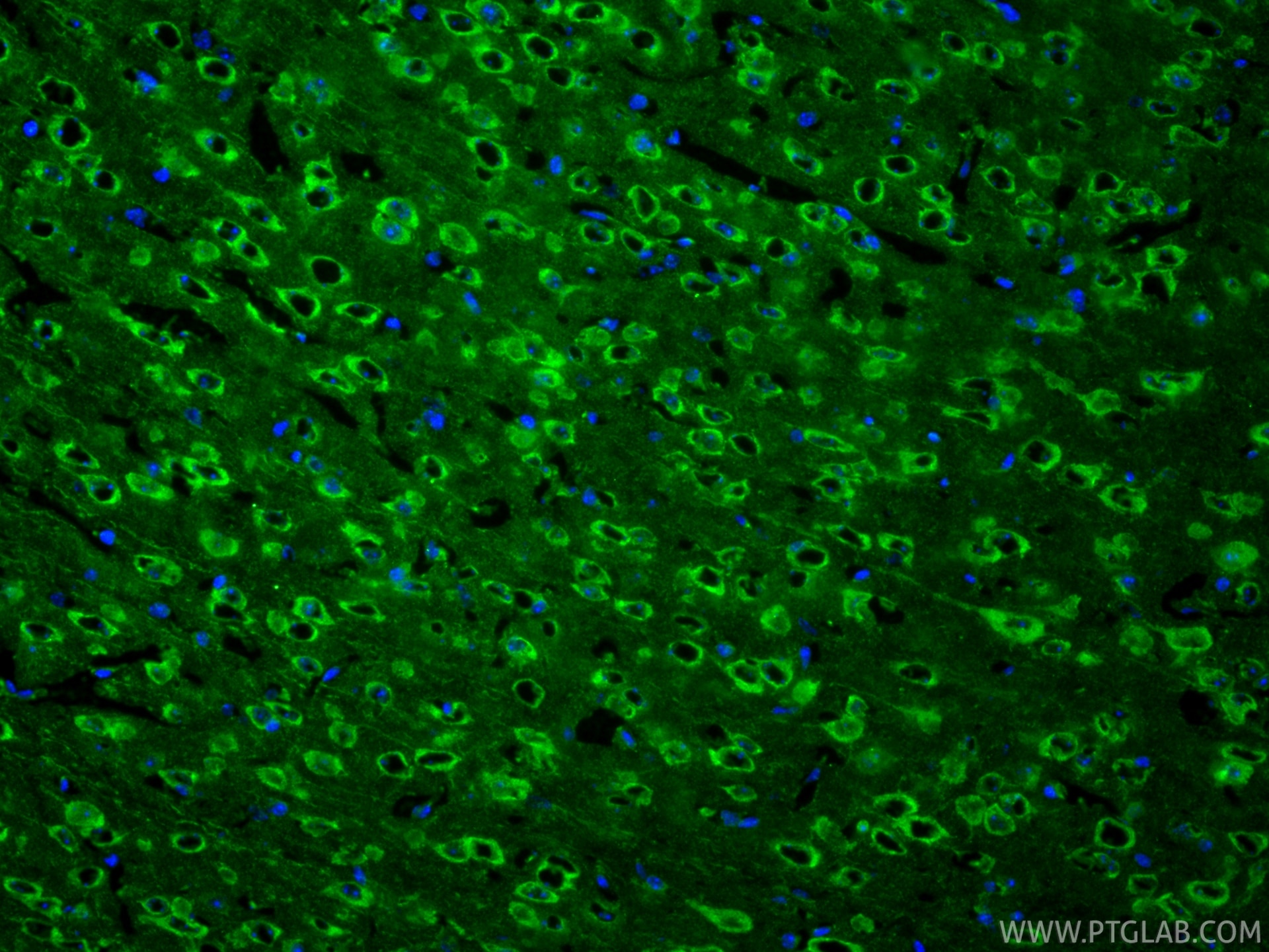
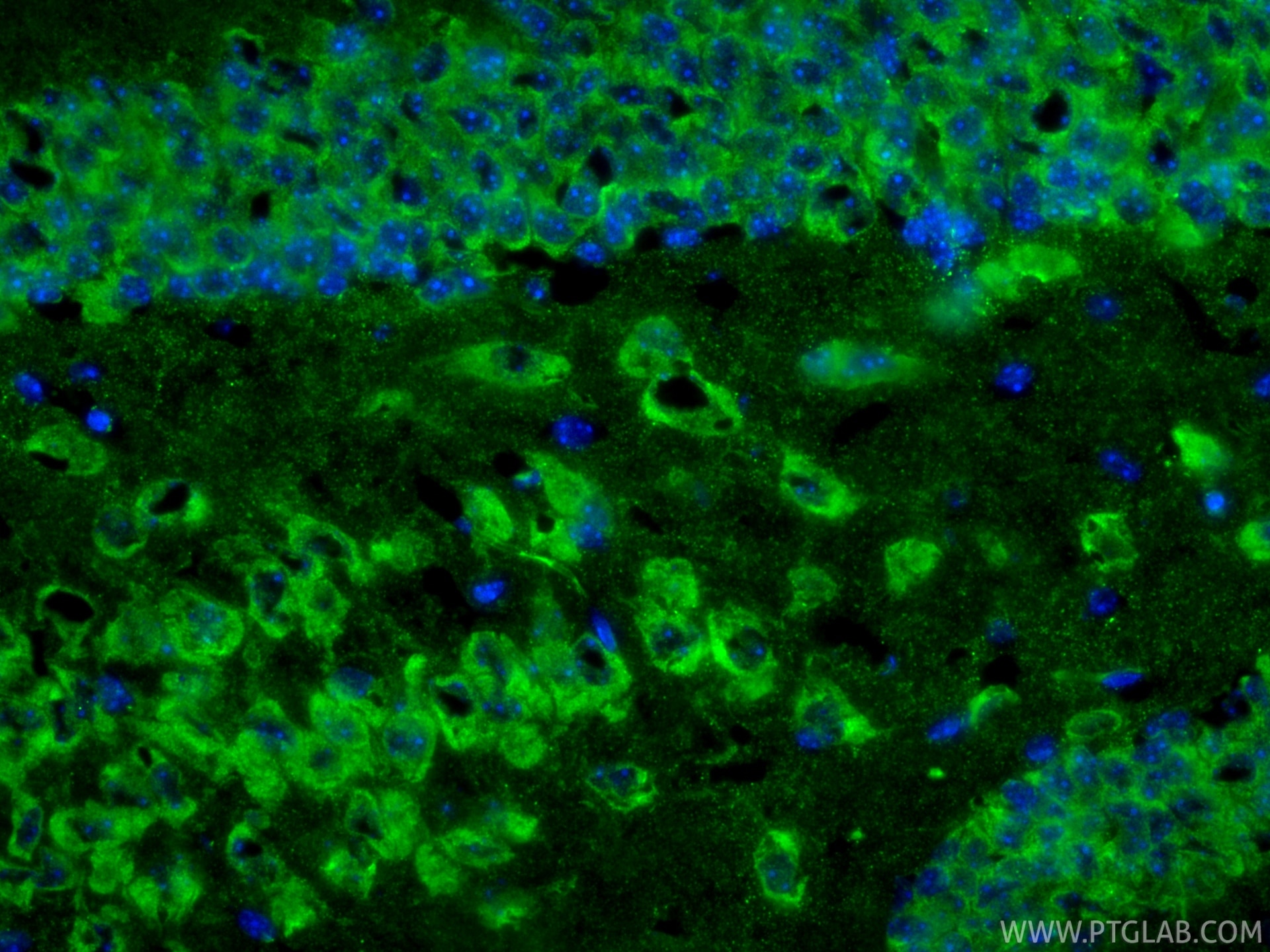
| Positive IF-Fro detected in | mouse brain tissue |
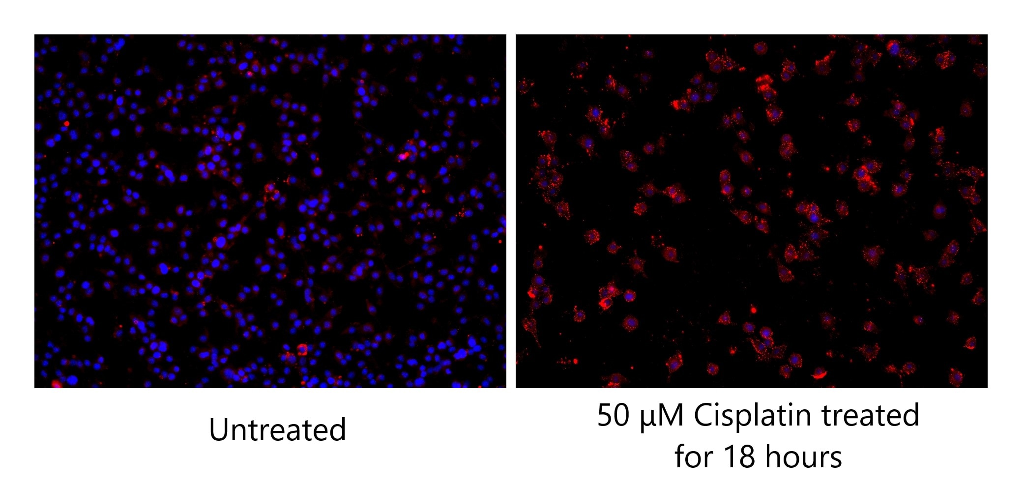
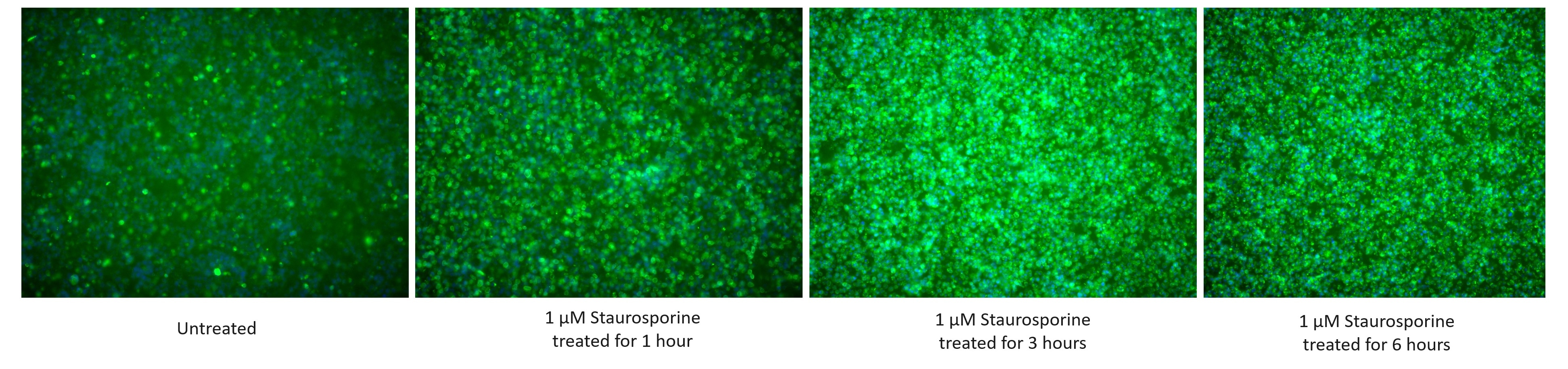
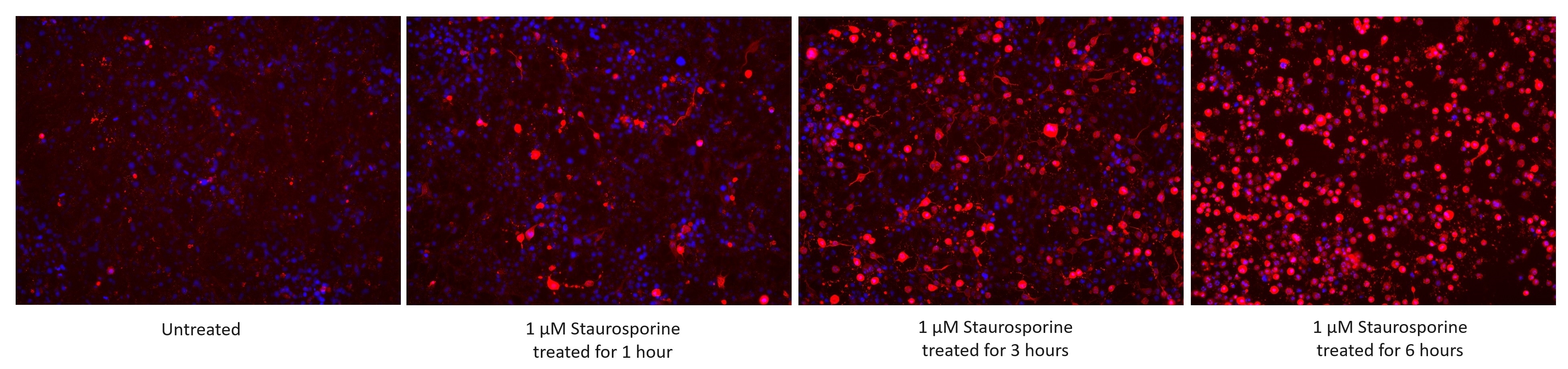
| Positive IF/ICC detected in | 1 μM Staurosporine (1/3/6 hour) treated Jurkat cells, 50 μM Cisplatin (18 hours) treated BV-2 cells, 1 μM Staurosporine (1/3/6 hour) treated HSC-T6 cells |
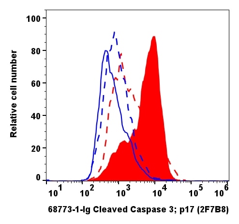
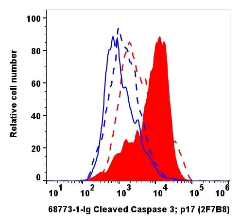
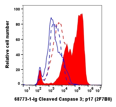
| Positive FC (Intra) detected in | 1 μM Staurosporine (3 hours) treated Jurkat cells, 50 μM Cisplatin(18h)treated BV-2 cells |
Recommended dilution
| Application | Dilution |
|---|---|
| Western Blot (WB) | WB : 1:5000-1:50000 |
| Immunoprecipitation (IP) | IP : 0.5-4.0 ug for 1.0-3.0 mg of total protein lysate |
| Immunohistochemistry (IHC) | IHC : 1:1000-1:4000 |
| Immunofluorescence (IF)-P | IF-P : 1:200-1:800 |
| Immunofluorescence (IF)-FRO | IF-FRO : 1:400-1:1600 |
| Immunofluorescence (IF)/ICC | IF/ICC : 1:500-1:2000 |
| Flow Cytometry (FC) (INTRA) | FC (INTRA) : 0.20 ug per 10^6 cells in a 100 µl suspension |
| It is recommended that this reagent should be titrated in each testing system to obtain optimal results. | |
| Sample-dependent, Check data in validation data gallery. | |
Product Information
68773-1-Ig targets Cleaved Caspase 3/P17/P19 in WB, IHC, IF/ICC, IF-P, IF-Fro, FC (Intra), IP, ELISA applications and shows reactivity with human, mouse, rat samples.
| Tested Reactivity | human, mouse, rat |
| Cited Reactivity | human, mouse, rat |
| Host / Isotype | Mouse / IgG1 |
| Class | Monoclonal |
| Type | Antibody |
| Immunogen |
Peptide Predict reactive species |
| Full Name | caspase 3, apoptosis-related cysteine peptidase |
| Calculated Molecular Weight | 32 kDa |
| Observed Molecular Weight | 17 kDa, 19 kDa |
| GenBank Accession Number | NM_032991 |
| Gene Symbol | Caspase 3 |
| Gene ID (NCBI) | 836 |
| RRID | AB_3665444 |
| Conjugate | Unconjugated |
| Form | Liquid |
| Purification Method | Protein G purification |
| UNIPROT ID | P42574 |
| Storage Buffer | PBS with 0.02% sodium azide and 50% glycerol, pH 7.3. |
| Storage Conditions | Store at -20°C. Stable for one year after shipment. Aliquoting is unnecessary for -20oC storage. 20ul sizes contain 0.1% BSA. |
Background Information
Caspases, a family of endoproteases, are critical players in cell regulatory networks controlling inflammation and cell death. Initiator caspases (caspase-2, -8, -9, -10, -11, and -12) cleave and activate downstream effector caspases (caspase-3, -6, and -7), which in turn execute apoptosis by cleaving targeted cellular proteins. Caspase 3 plays a key role in the activation of sterol regulatory element binding proteins (SREBPs) between the basic helix-loop-helix leucine zipper domain and the membrane attachment domain. Caspase 3 can form heterocomplex with other proteins. This antibody is specific for cleaved caspase 3, and does not recognize full length caspase-3. The cleaved P17 and P19 fragment might form complex and shows at around 30-35 kDa by western blot (PMID: 25501826).
Protocols
| Product Specific Protocols | |
|---|---|
| IF protocol for Cleaved Caspase 3/P17/P19 antibody 68773-1-Ig | Download protocol |
| IHC protocol for Cleaved Caspase 3/P17/P19 antibody 68773-1-Ig | Download protocol |
| IP protocol for Cleaved Caspase 3/P17/P19 antibody 68773-1-Ig | Download protocol |
| WB protocol for Cleaved Caspase 3/P17/P19 antibody 68773-1-Ig | Download protocol |
| Standard Protocols | |
|---|---|
| Click here to view our Standard Protocols |
Publications
What published studies show
The Cleaved Caspase 3/P17/P19 Monoclonal Antibody (2F7B8), catalog number 68773-1-Ig, has accumulated 163 citations across numerous journals. Notable publications appear in Nature Communications, ACS Nano, Advanced Science, Cell Reports, Biomaterials, Oncogene, Phytomedicine, Journal of Nanobiotechnology, and Materials Today Bio. Research fields span apoptosis and programmed cell death, oncology (breast, lung, liver, glioblastoma, ovarian cancers), cardiology, neuroscience, nanomedicine, ferroptosis/pyroptosis, oxidative stress, inflammation, and traditional Chinese medicine pharmacology.
| Species | Application | Title |
|---|---|---|
J Extracell Vesicles Small Extracellular Vesicles Engineered Using Click Chemistry to Express Chimeric Antigen Receptors Show Enhanced Efficacy in Acute Liver Failure | ||
Nat Commun Tubulin autoregulation tunes microtubule dynamics to support multicellular architecture and viability. | ||
Nat Commun S100A12 triggers NETosis to aggravate myocardial infarction injury via the Annexin A5-calcium axis | ||
Exp Mol Med Cancer-associated fibroblast-derived extracellular vesicles regulate lipophagy through PLIN2 to modulate dormancy in salivary gland adenoid cystic carcinoma cells. | ||
ACS Nano Mucoadhesive Nanothalidomide Enema: Simultaneous Radioprotection and Barrier Repair in Radiation Enteritis Therapy. | ||
J Nanobiotechnology A combination of a TLR7/8 agonist and an epigenetic inhibitor suppresses triple-negative breast cancer through triggering anti-tumor immune. |
Reviews
What customers say
Both reviewers used this Caspase-3 antibody for immunofluorescence with positive results. One user (5/5) applied it at 1:1000 on mouse brain cryosections in a 6-OHDA injury model, observing clear caspase-3 signal colocalizing with GFAP and Hoechst staining. The other (4/5) used it at 1:800 on rat primary neuron/astrocyte co-cultures, reporting it worked well across a range of dilutions. Overall, the antibody performs reliably in IF applications on both rodent tissue sections and primary cell cultures.
The reviews below have been submitted by verified Proteintech customers who received an incentive for providing their feedback.
Joana (Verified Customer) (09-02-2026) | Immunohistochemical fluorescence analysis of cryosectioned mouse brain tissue slide using 68773-1-IG (Caspase 3 antibody) at dilution of 1:1000 (under 10x lens). Mus musculus 6-OHDA injection at 2w presents cas3 (white) colocalized with GFAP (Green), Hoescht (blue)
|
Molly (Verified Customer) (10-03-2024) | Worked well in IF on primary rat embryonic neuron/astrocyte co-culture at a range of dilutions.
|
























