

Tested Applications
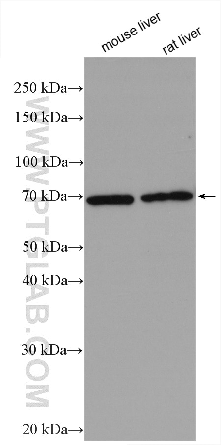
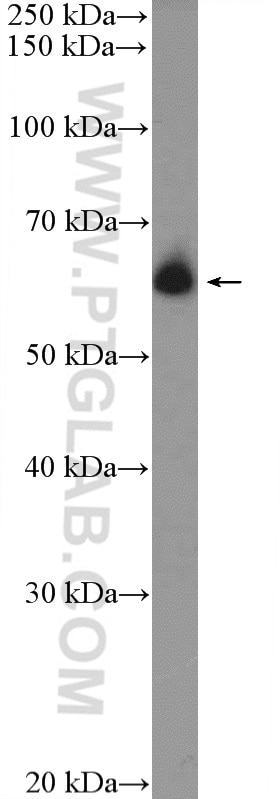
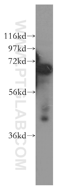
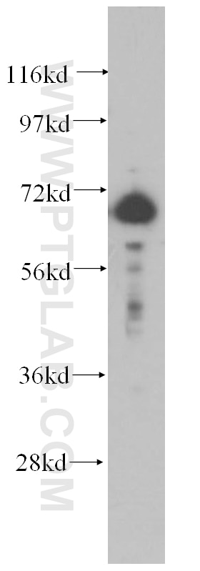
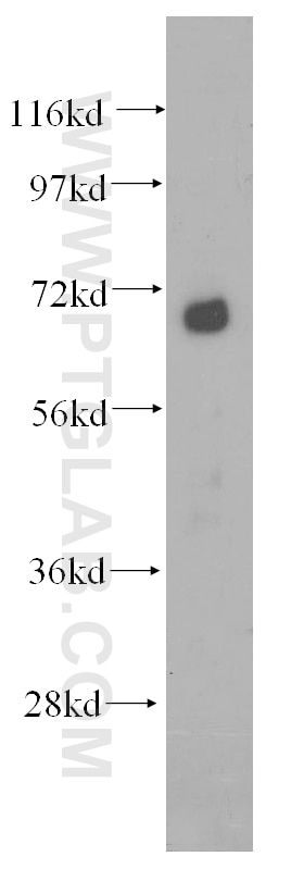
| Positive WB detected in | mouse liver tissue, HeLa cells, human liver tissue, Jurkat cells, rat liver tissue, rat spleen tissue |
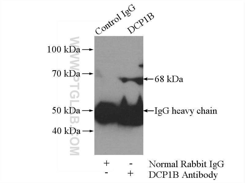
| Positive IP detected in | HeLa cells |
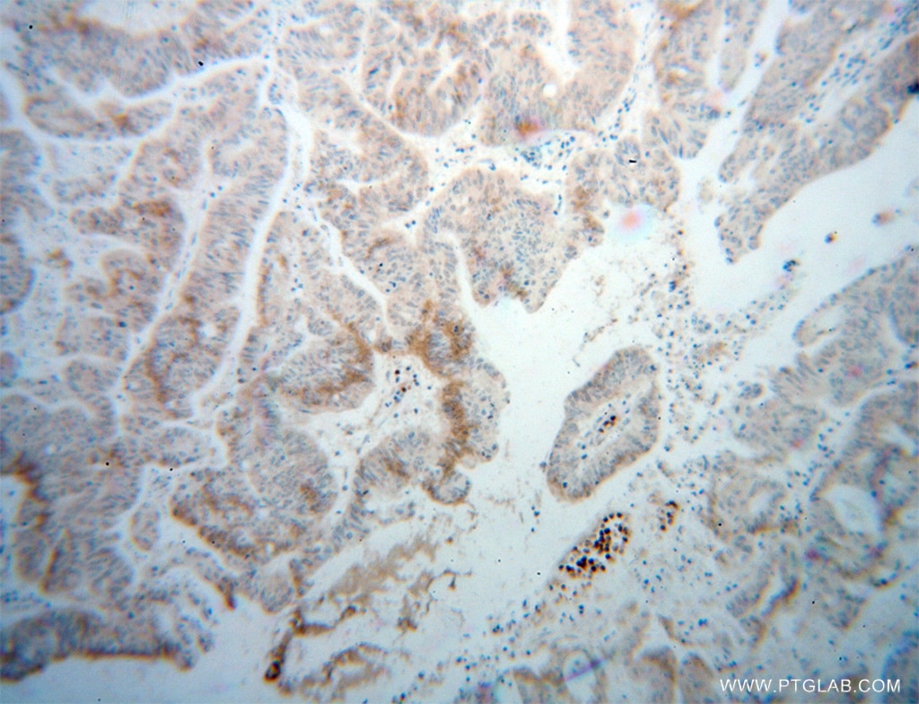
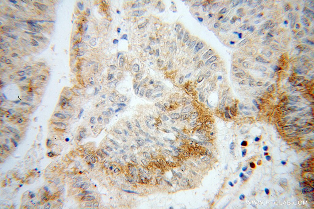
| Positive IHC detected in | human liver cancer tissue Note: suggested antigen retrieval with TE buffer pH 9.0; (*) Alternatively, antigen retrieval may be performed with citrate buffer pH 6.0 |
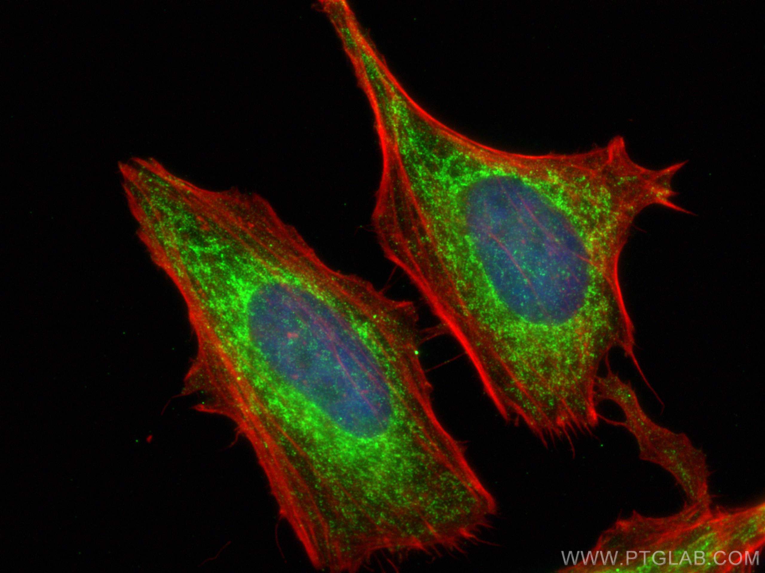
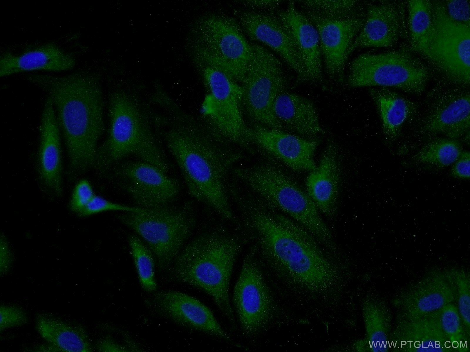
| Positive IF/ICC detected in | HeLa cells, A549 cells |
Recommended dilution
| Application | Dilution |
|---|---|
| Western Blot (WB) | WB : 1:1000-1:6000 |
| Immunoprecipitation (IP) | IP : 0.5-4.0 ug for 1.0-3.0 mg of total protein lysate |
| Immunohistochemistry (IHC) | IHC : 1:20-1:200 |
| Immunofluorescence (IF)/ICC | IF/ICC : 1:200-1:800 |
| It is recommended that this reagent should be titrated in each testing system to obtain optimal results. | |
| Sample-dependent, Check data in validation data gallery. | |
Product Information
13863-1-AP targets DCP1B in WB, IHC, IF/ICC, IP, ELISA, CUT&Tag applications and shows reactivity with human, mouse, rat samples.
| Tested Reactivity | human, mouse, rat |
| Cited Reactivity | human |
| Host / Isotype | Rabbit / IgG |
| Class | Polyclonal |
| Type | Antibody |
| Immunogen |
CatNo: Ag4845 Product name: Recombinant human DCP1B protein Source: e coli.-derived, PGEX-4T Tag: GST Domain: 269-618 aa of BC043437 Sequence: QGVVRSLSYEEPRRHSPPIEKQLCPAIQKLMVRSADLHPLSELPENRPCENGSTHSAGEFFTGPVQPGSPHNIGTSRGVQNASRTQNLFEKLQSTPGAANKCDPSTPAPASSAALNRSRAPTSVTPVAPGKGLAQPPQAYFNGSLPPQTVGHQAHGREQSTLPRQTLAISGSQTGSSGVISPQELLKKLQIVQQEQQLHASNRPALAAKFPVLAQSSGTGKPLESWINKTPNTEQQTPLFQVISPQRIPATAAPSLLMSPMVFAQPTSVPPKERESGLLPVGGQEPPAAATSLLLPIQSPEPSVITSSPLTKLQLQEALLYLIQNDDNFLNIIYEAYLFSMTQAAMKKTM Predict reactive species |
| Full Name | DCP1 decapping enzyme homolog B (S. cerevisiae) |
| Calculated Molecular Weight | 618 aa, 68 kDa |
| Observed Molecular Weight | 68 kDa |
| GenBank Accession Number | BC043437 |
| Gene Symbol | DCP1B |
| Gene ID (NCBI) | 196513 |
| RRID | AB_2090411 |
| Conjugate | Unconjugated |
| Form | Liquid |
| Purification Method | Antigen affinity purification |
| UNIPROT ID | Q8IZD4 |
| Storage Buffer | PBS with 0.02% sodium azide and 50% glycerol, pH 7.3. |
| Storage Conditions | Store at -20°C. Stable for one year after shipment. Aliquoting is unnecessary for -20oC storage. 20ul sizes contain 0.1% BSA. |
Background Information
DCP1B belongs to DCP1 family. DCP1B may play a role in the degradation of mRNAs, both in normal mRNA turnover and in nonsense-mediated mRNA decay. It may remove the 7-methyl guanine cap structure mRNA molecules, yielding a 5'-phosphorylated mRNA fragment and 7m-GDP. DCP1B has 2 isoforms with the molecular mass of 56 and 68 kDa. DCP1B is associated with DM-related atherogenesis (PMID:33195466).
Protocols
| Product Specific Protocols | |
|---|---|
| IF protocol for DCP1B antibody 13863-1-AP | Download protocol |
| IHC protocol for DCP1B antibody 13863-1-AP | Download protocol |
| IP protocol for DCP1B antibody 13863-1-AP | Download protocol |
| WB protocol for DCP1B antibody 13863-1-AP | Download protocol |
| Standard Protocols | |
|---|---|
| Click here to view our Standard Protocols |
Publications
What published studies show
The DCP1B Polyclonal antibody (Cat# 13863-1-AP) has 4 citations published in Cell, Nature Cancer, International Journal of Molecular Medicine, and Molecular Cell. Research fields span cancer biology (KRAS phase separation in colon tumors, lncRNA-encoded micropeptide in macrophage-mediated tumor growth), virology (Dengue virus RNA replication), and RNA biology/transcription (ECAS complex coupling transcription elongation and RNA processing). Applications cited include WB, IF, and CUT&Tag, all in human samples.
| Species | Application | Title |
|---|---|---|
Nat Cancer A micropeptide encoded by the lncRNA USP30-AS1 promotes tumor growth by attenuating cGAS-STING-type I IFN signaling in macrophages | ||
Int J Mol Med LSm1 binds to the Dengue virus RNA 3' UTR and is a positive regulator of Dengue virus replication. | ||
Reviews
What customers say
The single review rates this antibody 5/5 for immunohistochemistry on cortical slice cultures at a 1:100 dilution. The reviewer reports strong, clear signal: diffuse staining in unstressed tissue that becomes distinct granular structures after 90 minutes of 42°C heat shock. Bright staining was observed under low-power microscopy using a 488 secondary antibody. Overall, the antibody performed well at the concentration used.
The reviews below have been submitted by verified Proteintech customers who received an incentive for providing their feedback.
George (Verified Customer) (08-19-2019) | Shows up well under concentration used - diffuse in unstressed tissue, forms distinct granular structures following 90 minutes 42oC heat shock. Only tested using 488 secondary antibody but showed up brightly under low power microscopy.
|











