
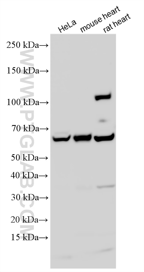



Tested Applications
| Positive WB detected in | HeLa cells, human heart tissue, mouse heart tissue, rat heart tissue |
| Positive IP detected in | mouse heart tissue |
| Positive IHC detected in | human breast cancer tissue, human normal colon, human oesophagus cancer tissue, human ovary tumor tissue Note: suggested antigen retrieval with TE buffer pH 9.0; (*) Alternatively, antigen retrieval may be performed with citrate buffer pH 6.0 |
Recommended dilution
| Application | Dilution |
|---|---|
| Western Blot (WB) | WB : 1:1000-1:6000 |
| Immunoprecipitation (IP) | IP : 0.5-4.0 ug for 1.0-3.0 mg of total protein lysate |
| Immunohistochemistry (IHC) | IHC : 1:2000-1:8000 |
| It is recommended that this reagent should be titrated in each testing system to obtain optimal results. | |
| Sample-dependent, Check data in validation data gallery. | |
Product Information
11440-1-AP targets EHD2 in WB, IHC, IF, IP, ELISA applications and shows reactivity with human, mouse, rat samples.
| Tested Reactivity | human, mouse, rat |
| Cited Reactivity | human, mouse, pig |
| Host / Isotype | Rabbit / IgG |
| Class | Polyclonal |
| Type | Antibody |
| Immunogen |
CatNo: Ag2002 Product name: Recombinant human EHD2 protein Source: e coli.-derived, PGEX-4T Tag: GST Domain: 194-543 aa of BC014445 Sequence: LEISDEFSEAIGALRGHEDKIRVVLNKADMVETQQLMRVYGALMWALGKVVGTPEVLRVYIGSFWSQPLLVPDNRRLFELEEQDLFRDIQGLPRHAALRKLNDLVKRARLVRVHAYIISYLKKEMPSVFGKENKKKQLILKLPVIFAKIQLEHHISPGDFPDCQKMQELLMAHDFTKFHSLKPKLLEALDEMLTHDIAKLMPLLRQEELESTEVGVQGGAFEGTHMGPFVERGPDEAMEDGEEGSDDEAEWVVTKDKSKYDEIFYNLAPADGKLSGSKAKTWMVGTKLPNSVLGRIWKLSDVDRDGMLDDEEFALASHLIEAKLEGHGLPANLPRRLVPPSKRRHKGSAE Predict reactive species |
| Full Name | EH-domain containing 2 |
| Calculated Molecular Weight | 543 aa, 61 kDa |
| Observed Molecular Weight | 61 kDa |
| GenBank Accession Number | BC014445 |
| Gene Symbol | EHD2 |
| Gene ID (NCBI) | 30846 |
| RRID | AB_2097325 |
| Conjugate | Unconjugated |
| Form | Liquid |
| Purification Method | Antigen affinity purification |
| UNIPROT ID | Q9NZN4 |
| Storage Buffer | PBS with 0.02% sodium azide and 50% glycerol, pH 7.3. |
| Storage Conditions | Store at -20°C. Stable for one year after shipment. Aliquoting is unnecessary for -20oC storage. 20ul sizes contain 0.1% BSA. |
Background Information
EH domain-containing protein 2 (EHD2) is a member of the EHD protein family. It is primarily located in the plasma membrane but can also be found in the cytoplasm and endosomes. EHD2 is also a nuclear-cytoplasmic shuttle protein, acting as a corepressor of transcription to inhibit gene transcription when it enters the cell nucleus. EHD2 is a dynamin-related ATPase that plays a key role in regulating a series of biological processes. As a crucial regulator of endocytic transport, it is involved in the formation and maintenance of endosomal tubules and vesicles, which are vital for intracellular transport of proteins and other substances. EHD2 has been implicated in several human cancers and has potential as a predictive biomarker of chemotherapy efficacy.
Protocols
| Product Specific Protocols | |
|---|---|
| IHC protocol for EHD2 antibody 11440-1-AP | Download protocol |
| IP protocol for EHD2 antibody 11440-1-AP | Download protocol |
| WB protocol for EHD2 antibody 11440-1-AP | Download protocol |
| Standard Protocols | |
|---|---|
| Click here to view our Standard Protocols |
Publications
What published studies show
The EHD2 Polyclonal antibody (Cat# 11440-1-AP) has 8 citations published in Nature, Nature Communications, Emerging Microbes & Infections, American Journal of Cancer Research, PLoS One, Cancer Investigation, and Journal of Molecular and Cellular Cardiology Plus. Research fields span beige fat biogenesis, adipocyte expansion, African swine fever virus inhibition, colon cancer progression, caveolae super-resolution imaging, and pulmonary arterial hypertension.
| Species | Application | Title |
|---|---|---|
Nat Commun Plasma membrane remodeling determines adipocyte expansion and mechanical adaptability | ||
Emerg Microbes Infect Identification of cepharanthine as an effective inhibitor of African swine fever virus replication
| ||
Am J Cancer Res Ese-3 contributes to colon cancer progression by downregulating EHD2 and transactivating INPP4B.
| ||
Cancer Invest Identification of Differentially Expressed Proteins of Normal and Cancerous Human Colorectal Tissues by Liquid Chromatograph-Mass Spectrometer Based on iTRAQ Approach. |
Reviews
What customers say
Both reviewers rated the antibody 5 stars. One user reported it works well for Western blot, detecting a specific band around 65 kDa, and also performs well for immunofluorescence on mouse and human cell lines. Another user confirmed good performance for Western blot on HUVEC samples. Overall, customers found the antibody reliable and effective for both WB and IF applications at a 1/1000 dilution.
The reviews below have been submitted by verified Proteintech customers who received an incentive for providing their feedback.
Laura (Verified Customer) (01-27-2022) | It works well for western blot with different bands but one specific around 65 kDa. It also works well for immunofluorescence of cells.
|
Kyosuke (Verified Customer) (06-12-2019) | It works well on HUVECs sample for Western blot.
|



