

Tested Applications
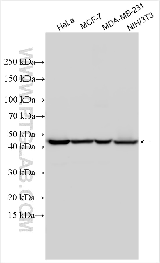
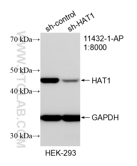
| Positive WB detected in | HeLa cells, HEK-293 cells, MCF-7 cells, MDA-MB-231 cells, NIH/3T3 cells |
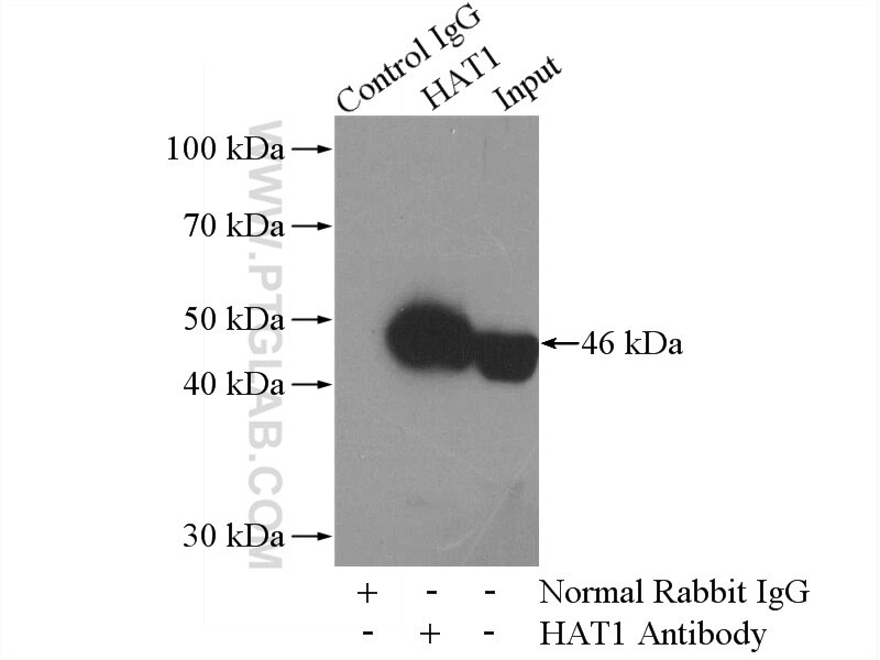
| Positive IP detected in | HepG2 cells |
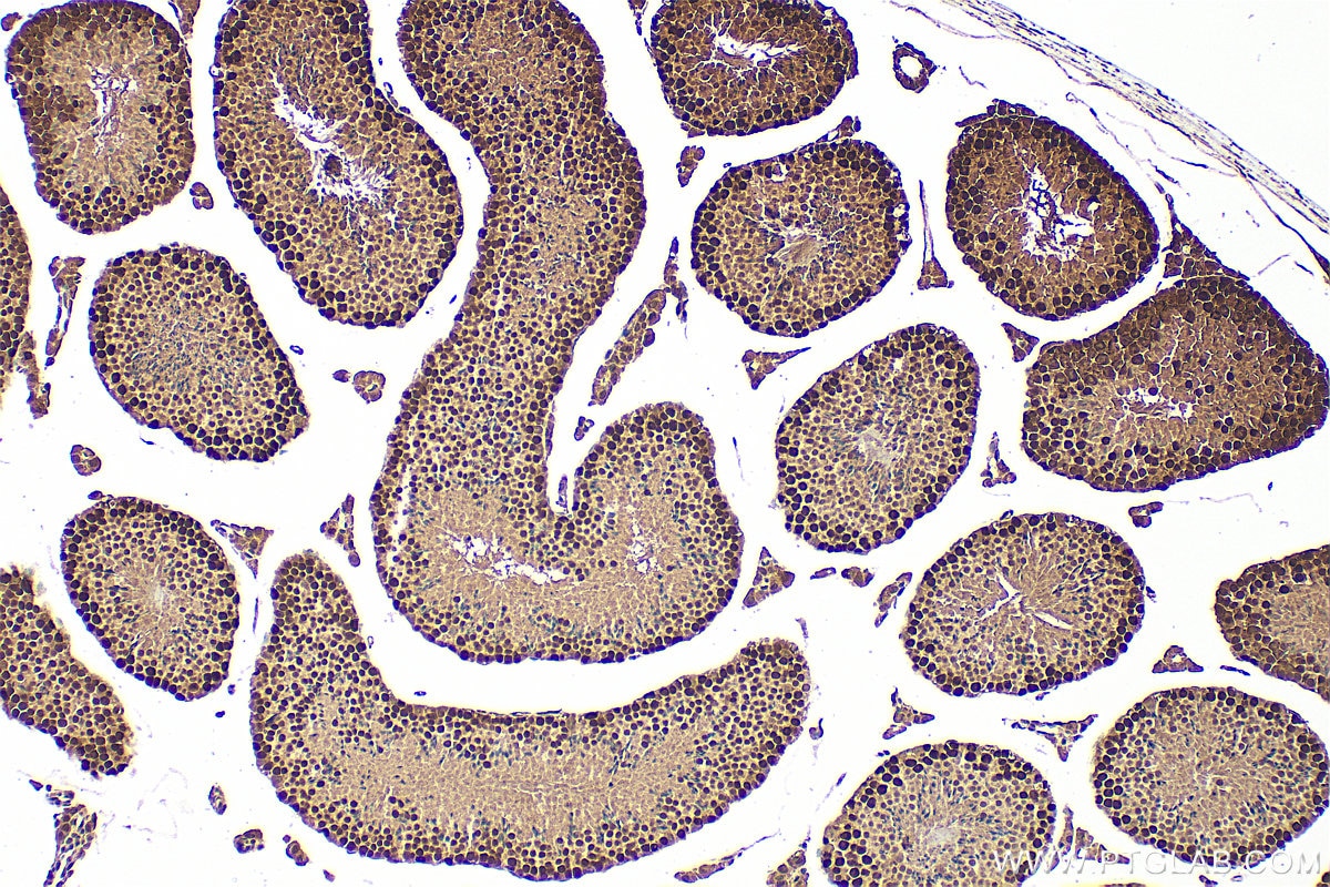
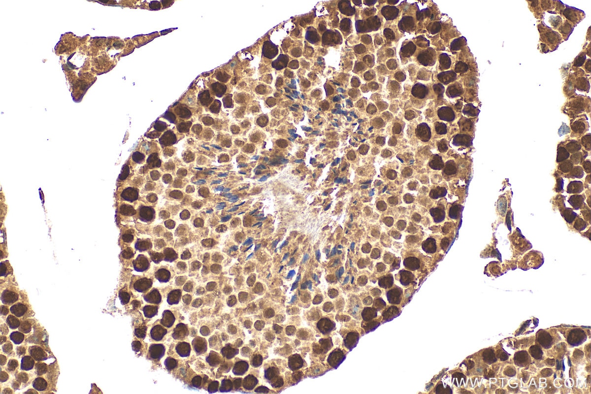
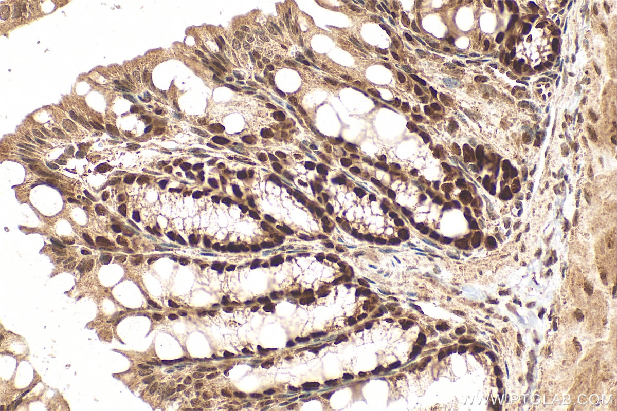
| Positive IHC detected in | mouse testis tissue, mouse colon tissue Note: suggested antigen retrieval with TE buffer pH 9.0; (*) Alternatively, antigen retrieval may be performed with citrate buffer pH 6.0 |
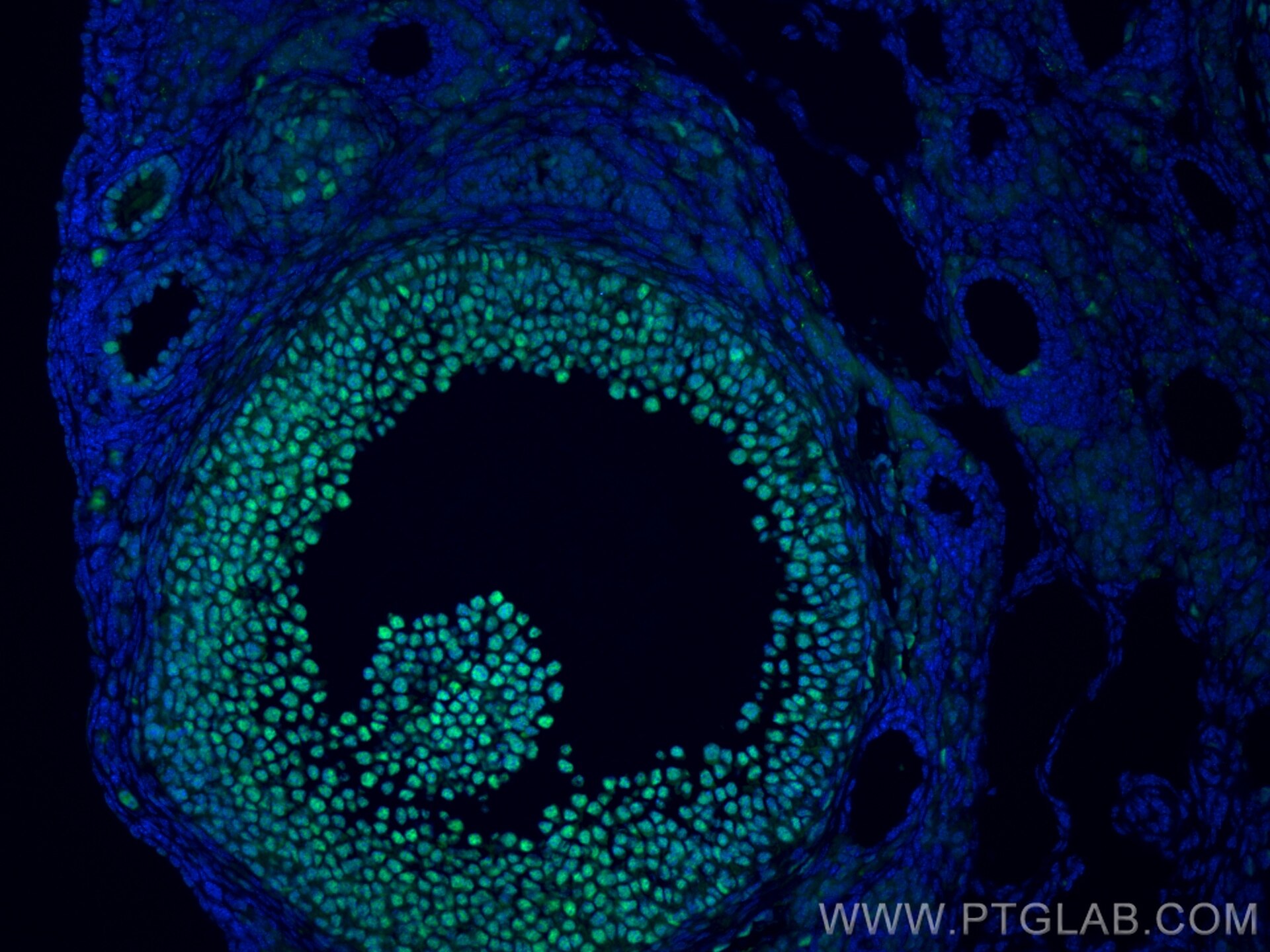
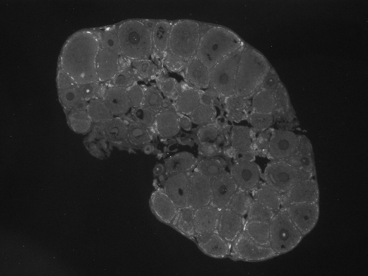
| Positive IF-P detected in | mouse ovary tissue |
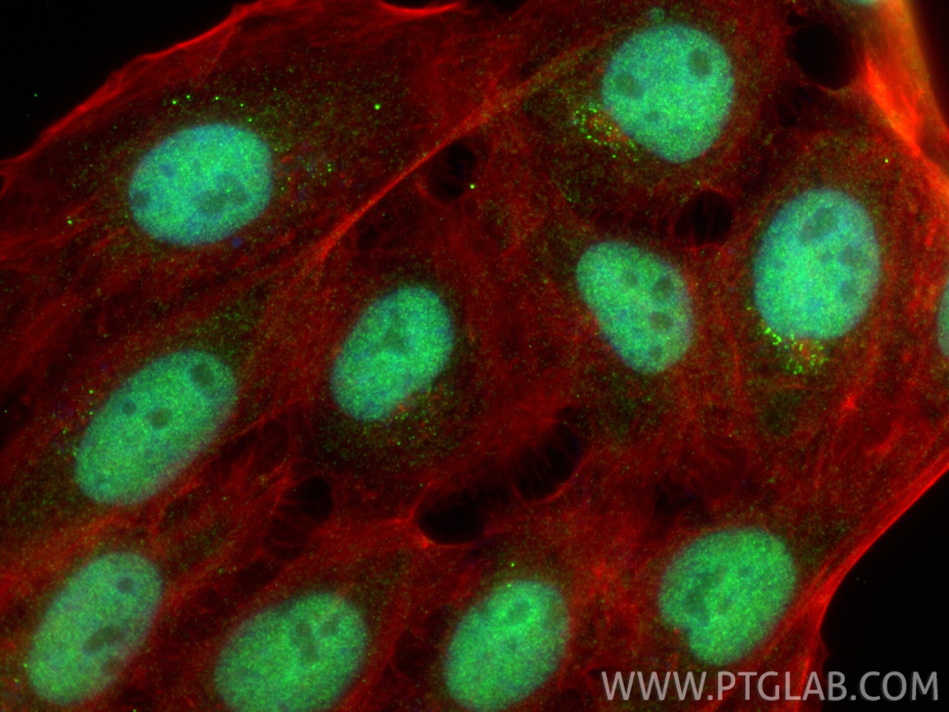
| Positive IF/ICC detected in | A431 cells |
Recommended dilution
| Application | Dilution |
|---|---|
| Western Blot (WB) | WB : 1:5000-1:50000 |
| Immunoprecipitation (IP) | IP : 0.5-4.0 ug for 1.0-3.0 mg of total protein lysate |
| Immunohistochemistry (IHC) | IHC : 1:250-1:1000 |
| Immunofluorescence (IF)-P | IF-P : 1:50-1:500 |
| Immunofluorescence (IF)/ICC | IF/ICC : 1:500-1:2000 |
| It is recommended that this reagent should be titrated in each testing system to obtain optimal results. | |
| Sample-dependent, Check data in validation data gallery. | |
Product Information
11432-1-AP targets HAT1 in WB, IHC, IF/ICC, IF-P, IP, ELISA applications and shows reactivity with human, mouse, rat samples.
| Tested Reactivity | human, mouse, rat |
| Cited Reactivity | human, mouse, rat |
| Host / Isotype | Rabbit / IgG |
| Class | Polyclonal |
| Type | Antibody |
| Immunogen |
CatNo: Ag1952 Product name: Recombinant human HAT1 protein Source: e coli.-derived, PGEX-4T Tag: GST Domain: 241-419 aa of BC018682 Sequence: MLILTPFQGQGHGAQLLETVHRYYTEFPTVLDITAEDPSKSYVKLRDFVLVKLCQDLPCFSREKLMQGFNEDMAIEAQQKFKINKQHARRVYEILRLLVTDMSDAEQYRSYRLDIKRRLISPYKKKQRDLAKMRKCLRPEELTNQMNQIEISMQHEQLEESFQELVEDYRRVIERLAQE Predict reactive species |
| Full Name | histone acetyltransferase 1 |
| Calculated Molecular Weight | 419 aa, 50 kDa |
| Observed Molecular Weight | 46 kDa |
| GenBank Accession Number | BC018682 |
| Gene Symbol | HAT1 |
| Gene ID (NCBI) | 8520 |
| RRID | AB_2116435 |
| Conjugate | Unconjugated |
| Form | Liquid |
| Purification Method | Antigen affinity purification |
| UNIPROT ID | O14929 |
| Storage Buffer | PBS with 0.02% sodium azide and 50% glycerol, pH 7.3. |
| Storage Conditions | Store at -20°C. Stable for one year after shipment. Aliquoting is unnecessary for -20oC storage. 20ul sizes contain 0.1% BSA. |
Protocols
| Product Specific Protocols | |
|---|---|
| IF protocol for HAT1 antibody 11432-1-AP | Download protocol |
| IHC protocol for HAT1 antibody 11432-1-AP | Download protocol |
| IP protocol for HAT1 antibody 11432-1-AP | Download protocol |
| WB protocol for HAT1 antibody 11432-1-AP | Download protocol |
| Standard Protocols | |
|---|---|
| Click here to view our Standard Protocols |
Publications
What published studies show
The HAT1 antibody (Cat# 11432-1-AP) has accumulated 21 citations published in high-impact journals including Nature, Nature Communications, Molecular Cell, Cell Death & Disease, EMBO Reports, eLife, and Cell Metabolism. Associated research fields encompass epigenetic regulation (histone acetylation, succinylation, and lactylation), oncology (pancreatic, lung, glioblastoma, ovarian, and liver cancers), ferroptosis, DNA repair, neurodegeneration, metabolic disorders, reproductive biology, and inflammatory/immune responses.
| Species | Application | Title |
|---|---|---|
Nat Commun Histone acetyltransferase 1 promotes postinfarction inflammatory response by regulation of monocyte histone succinylation.
| ||
Mol Cell Rate-limiting enzymes in nucleotide metabolism synchronize nucleotide biosynthesis and chromatin formation. | ||
Cell Death Dis Histone acetyltransferase 1 promotes gemcitabine resistance by regulating the PVT1/EZH2 complex in pancreatic cancer.
| ||
Cell Death Dis HAT1/HDAC2 mediated ACSL4 acetylation confers radiosensitivity by inducing ferroptosis in nasopharyngeal carcinoma | ||
Cell Death Dis HAT1 functions as a lactyltransferase and mediates RPA1 lactylation to promote DNA repair and radioresistance in lung adenocarcinoma.
|
Reviews
What customers say
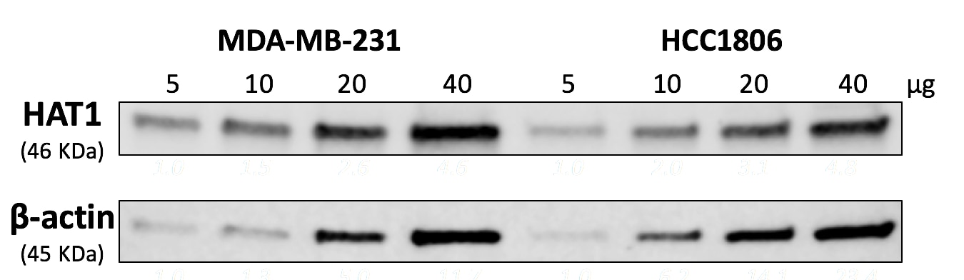
Both reviewers report positive results. One user (4/5) used the antibody at 1:100 for immunofluorescence on mouse ovary sections, noting it stained nuclei of developed oocytes very well. The other (5/5) used it at 1:1000 for Western blot on breast cancer cell lines (MDA-MB-231 and HCC1806), finding it performed well across a broad sample range of 5–40 µg and explicitly recommending the product. Overall, users are satisfied with its performance in both IF and WB applications.
The reviews below have been submitted by verified Proteintech customers who received an incentive for providing their feedback.
Kathryn (Verified Customer) (04-22-2022) | This antibody works very well to stain nuclei of developed oocytes in the mouse ovary.
 |
Leonardo (Verified Customer) (05-29-2019) | The antibody works well in a good range of sample (5-40 ug) in both cell lines I tested (MDA-MB-231 and HCC1806). I definitely recommend.
 |









