

Tested Applications
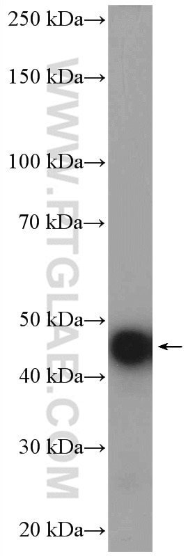
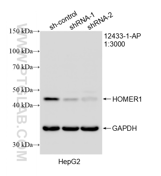
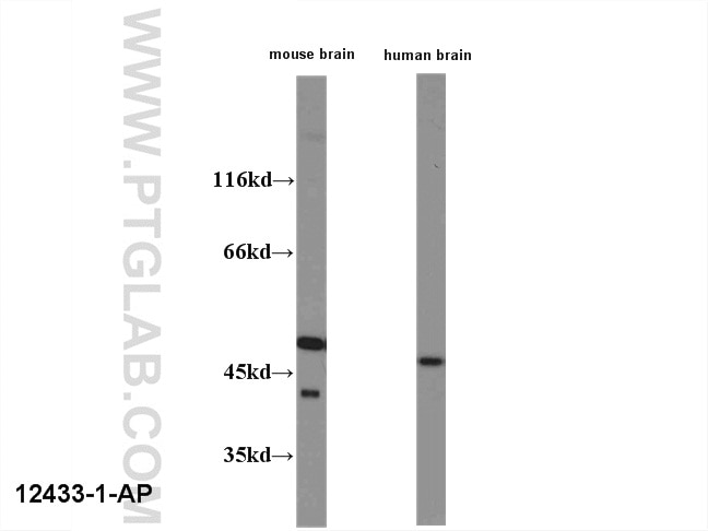
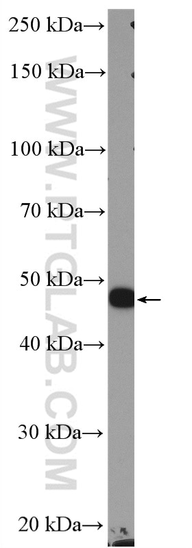
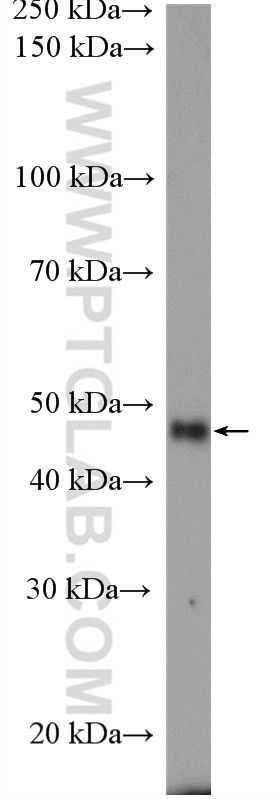
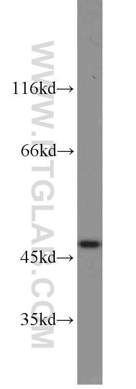
| Positive WB detected in | rat brain tissue, C2C12 cells, HepG2 cells, human brain tissue, human/mouse brain, NIH/3T3 cells |
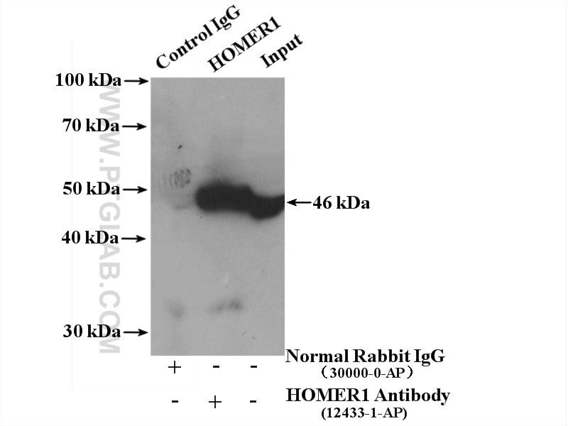
| Positive IP detected in | fetal human brain tissue |
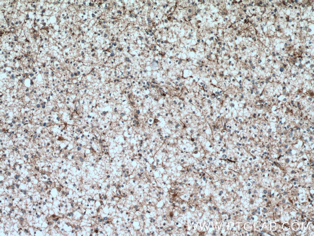
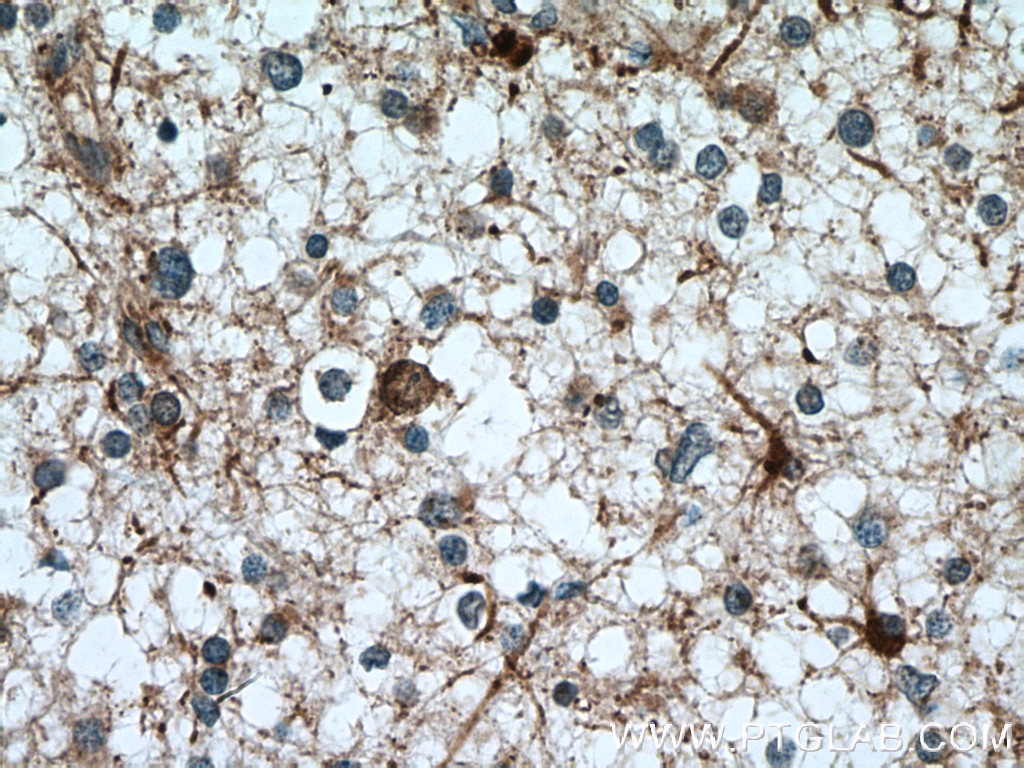
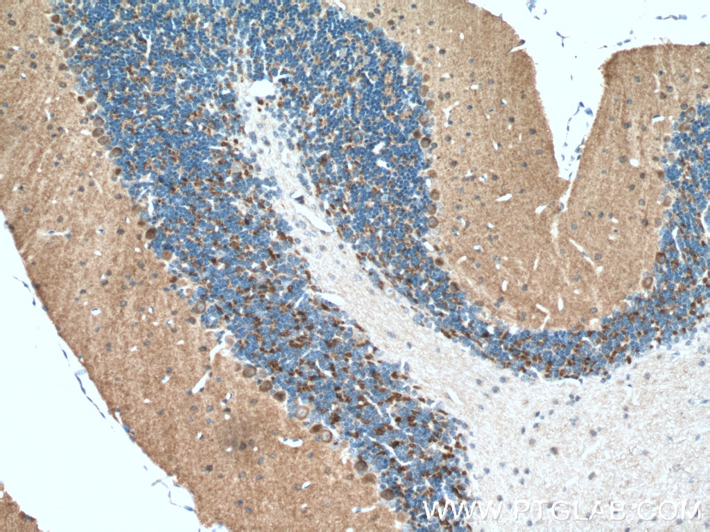
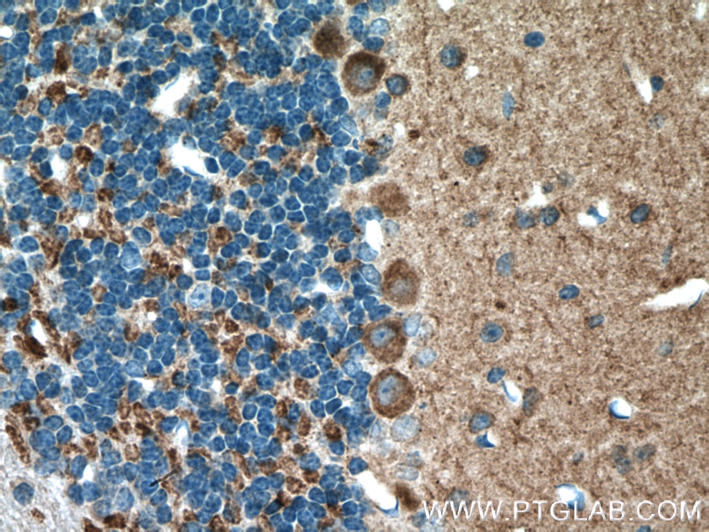
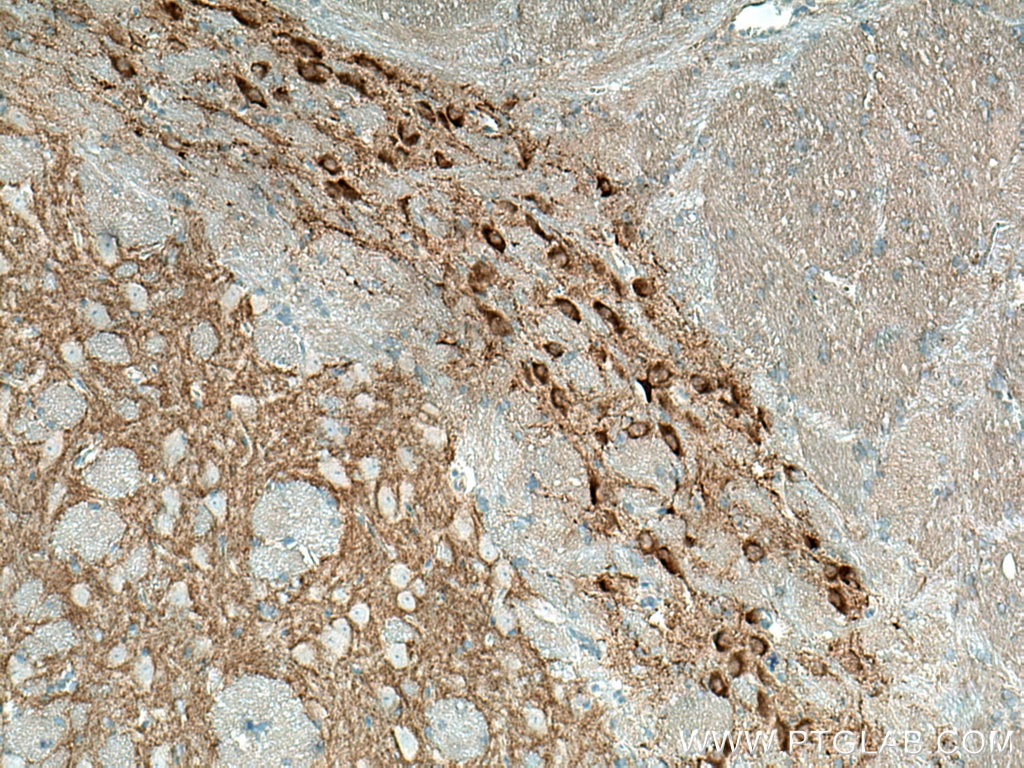
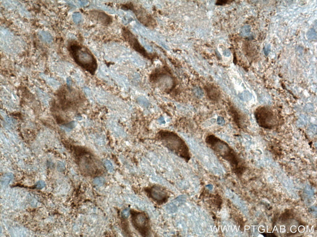
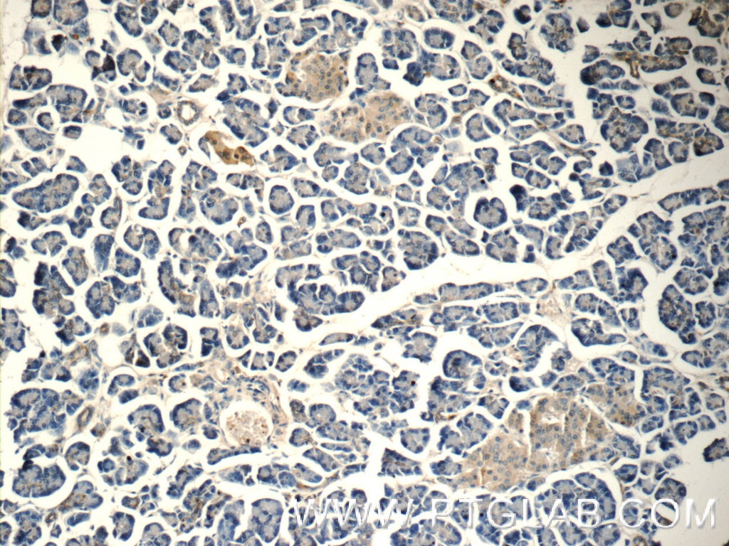
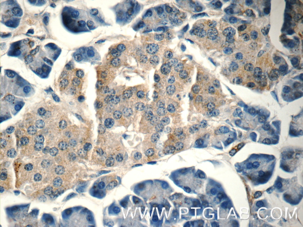
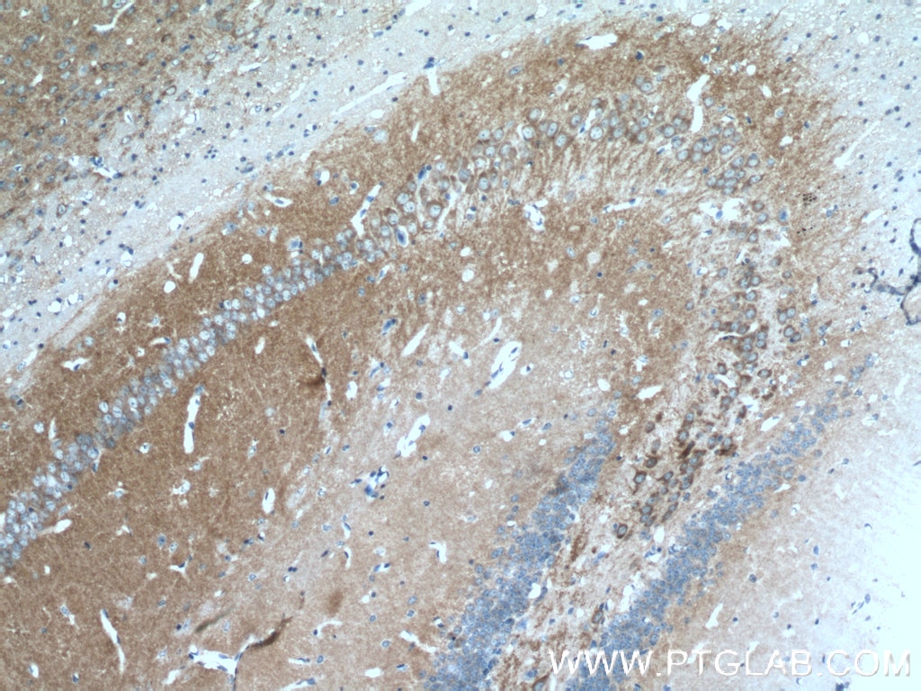
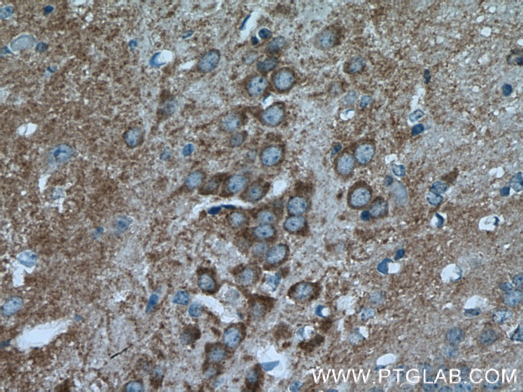
| Positive IHC detected in | human gliomas tissue, mouse brain tissue, rat brain tissue, human pancreas tissue Note: suggested antigen retrieval with TE buffer pH 9.0; (*) Alternatively, antigen retrieval may be performed with citrate buffer pH 6.0 |
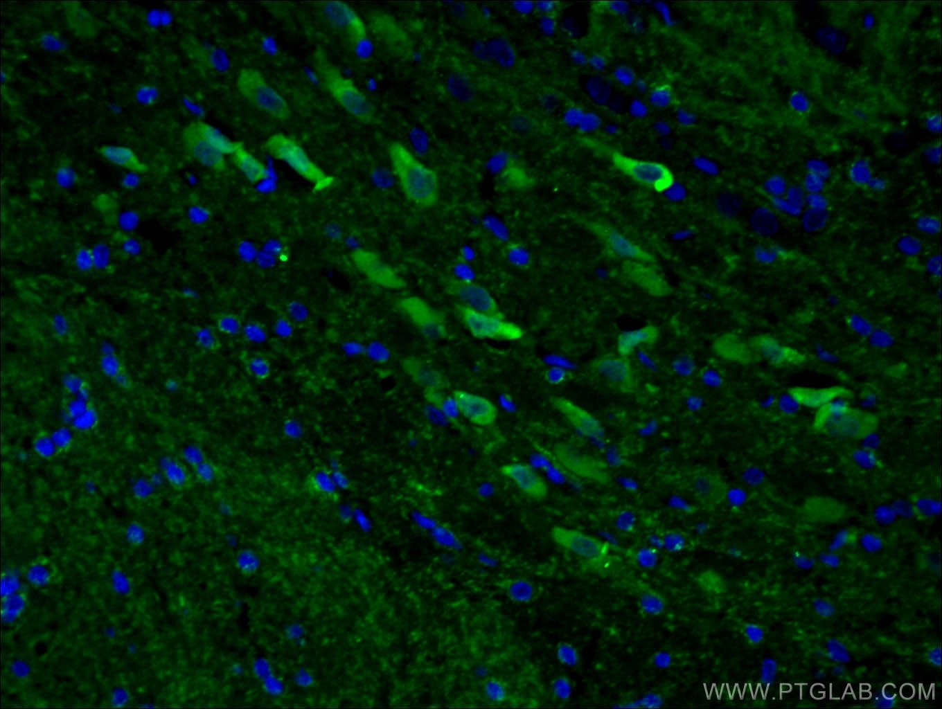
| Positive IF-P detected in | rat brain tissue |
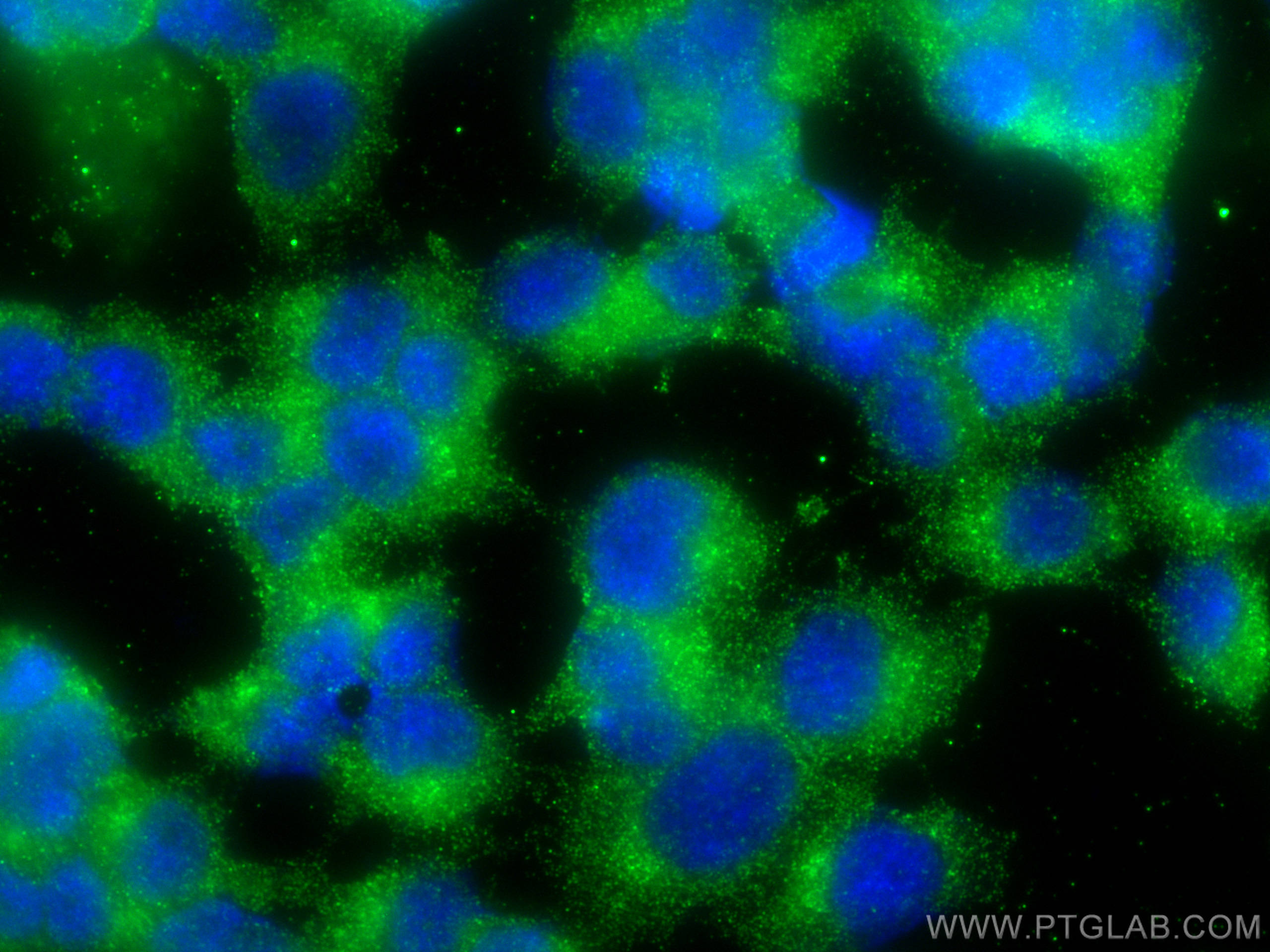
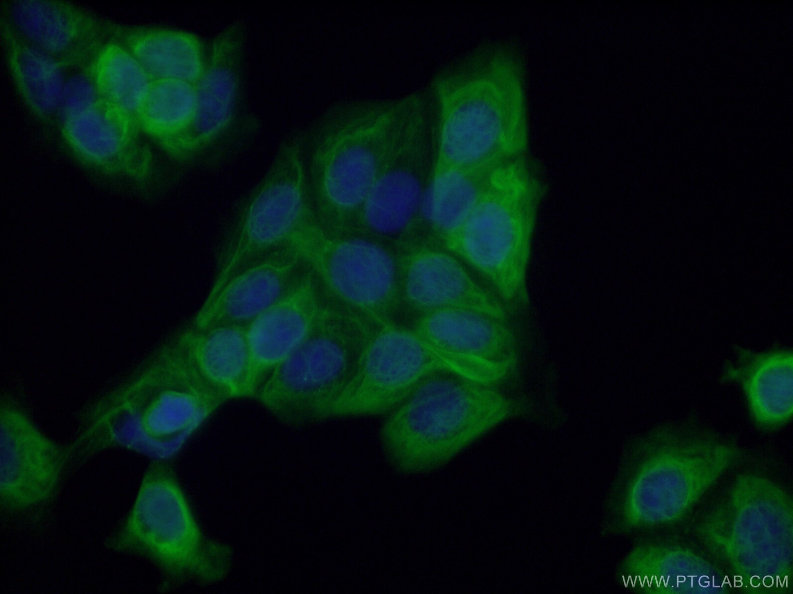
| Positive IF/ICC detected in | PC-12 cells, MCF-7 cells |
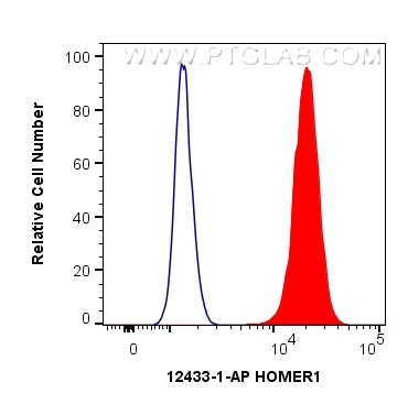
| Positive FC (Intra) detected in | SH-SY5Y cells |
Recommended dilution
| Application | Dilution |
|---|---|
| Western Blot (WB) | WB : 1:1000-1:5000 |
| Immunoprecipitation (IP) | IP : 0.5-4.0 ug for 1.0-3.0 mg of total protein lysate |
| Immunohistochemistry (IHC) | IHC : 1:50-1:500 |
| Immunofluorescence (IF)-P | IF-P : 1:50-1:500 |
| Immunofluorescence (IF)/ICC | IF/ICC : 1:200-1:800 |
| Flow Cytometry (FC) (INTRA) | FC (INTRA) : 0.40 ug per 10^6 cells in a 100 µl suspension |
| It is recommended that this reagent should be titrated in each testing system to obtain optimal results. | |
| Sample-dependent, Check data in validation data gallery. | |
Product Information
12433-1-AP targets HOMER1 in WB, IHC, IF/ICC, IF-P, FC (Intra), IP, ELISA applications and shows reactivity with human, mouse, rat samples.
| Tested Reactivity | human, mouse, rat |
| Cited Reactivity | human, mouse, rat, zebra finches |
| Host / Isotype | Rabbit / IgG |
| Class | Polyclonal |
| Type | Antibody |
| Immunogen |
CatNo: Ag3109 Product name: Recombinant human HOMER1 protein Source: e coli.-derived, PGEX-4T Tag: GST Domain: 7-354 aa of BC015502 Sequence: FSTRAHVFQIDPNTKKNWVPTSKHAVTVSYFYDSTRNVYRIISLDGSKAIINSTITPNMTFTKTSQKFGQWADSRANTVYGLGFSSEHHLSKFAEKFQEFKEAARLAKEKSQEKMELTSTPSQESAGGDLQSPLTPESINGTDDERTPDVTQNSEPRAEPTQNALPFSHSSAISKHWEAELATLKGNNAKLTAALLESTANVKQWKQQLAAYQEEAERLHKRVTELECVSSQANAVHTHKTELNQTIQELEETLKLKEEEIERLKQEIDNARELQEQRDSLTQKLQEVEIRNKDLEGQLSDLEQRLEKSQNEQEAFRNNLKTLLEILDGKIFELTELRDNLAKLLECS Predict reactive species |
| Full Name | homer homolog 1 (Drosophila) |
| Calculated Molecular Weight | 40 kDa |
| Observed Molecular Weight | 46-48 kDa |
| GenBank Accession Number | BC015502 |
| Gene Symbol | HOMER1 |
| Gene ID (NCBI) | 9456 |
| RRID | AB_2295573 |
| Conjugate | Unconjugated |
| Form | Liquid |
| Purification Method | Antigen affinity purification |
| UNIPROT ID | Q86YM7 |
| Storage Buffer | PBS with 0.02% sodium azide and 50% glycerol, pH 7.3. |
| Storage Conditions | Store at -20°C. Stable for one year after shipment. Aliquoting is unnecessary for -20oC storage. 20ul sizes contain 0.1% BSA. |
Background Information
Human HOMER1, also named as SYN47, belongs to the Homer family. Human HOMER1 has some isoforms: HOMER1a, HOMER1b, HOMER1c, HOMER1e and HOMER1h. Mouse Homer1 also named as Vesl-1 and it has some isoforms: Vesl-1l, Vesl-1m, Vesl-1s, Homer1b, and Homer1d. HOMER1 is a postsynaptic density scaffolding protein. It is a neuronal immediate-early gene (IEG) that is enriched at excitatory synapses and binds mGluRs. In WB, the MW of HOMER1 is about 46-50kd, 40-43kd and 20-26kd.
Protocols
| Product Specific Protocols | |
|---|---|
| FC protocol for HOMER1 antibody 12433-1-AP | Download protocol |
| IF protocol for HOMER1 antibody 12433-1-AP | Download protocol |
| IHC protocol for HOMER1 antibody 12433-1-AP | Download protocol |
| IP protocol for HOMER1 antibody 12433-1-AP | Download protocol |
| WB protocol for HOMER1 antibody 12433-1-AP | Download protocol |
| Standard Protocols | |
|---|---|
| Click here to view our Standard Protocols |
Publications
What published studies show
The anti-Homer1 antibody (Cat# 12433-1-AP) has accumulated 33 citations across journals including Nature Neuroscience, Cell Metabolism, Cell Stem Cell, Molecular Psychiatry, Journal of Neuroscience, CNS Neuroscience & Therapeutics, and eLife. Associated research fields encompass synaptic plasticity, autism spectrum disorder, neurodegeneration, hearing loss, pain, epilepsy, stroke, spinal cord injury, depression, cardiac protection, and brain development.
| Species | Application | Title |
|---|---|---|
Cell Metab Mitochondrial dysfunction reveals H(2)S-mediated synaptic sulfhydration as a potential mechanism for autism-associated social defects. | ||
Cell Stem Cell Molecular signature of primate astrocytes reveals pathways and regulatory changes contributing to human brain evolution | ||
Nat Neurosci Neuronal activity rapidly reprograms dendritic translation via eIF4G2:uORF binding | ||
Adv Sci (Weinh) Comparative Cochlear Transcriptomics in Echolocating Bats and Mouse Reveals Hras as Protector Against Noise-Induced Hearing Loss | ||
Mol Psychiatry A recurrent SHANK1 mutation implicated in autism spectrum disorder causes autistic-like core behaviors in mice via downregulation of mGluR1-IP3R1-calcium signaling. | ||
Neural Regen Res The interaction between KIF21A and KANK1 regulates dendritic morphology and synapse plasticity in neurons |
Reviews
What customers say
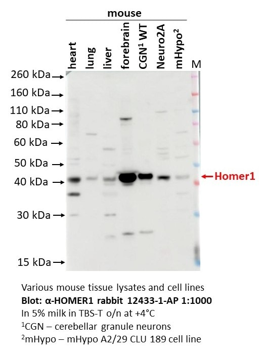
One reviewer (5/5 stars) used this antibody at 1:1000 dilution for Western Blot on mouse heart, lung, liver, forebrain, primary cerebellar granule neurons, and Neuro2A/mHypo cell lines. They reported a nice specific signal in mouse brain, primary neurons, and Neuro2A cells, but noted the presence of some additional non-specific bands. Overall, the antibody performed well with good specificity across multiple mouse tissues and cell types.
The reviews below have been submitted by verified Proteintech customers who received an incentive for providing their feedback.
Olga (Verified Customer) (09-12-2025) | Nice specific signal in mouse brain, primary neurons and Neuro2A cell line. However, some additional non-specific bands also present.
 |






















