
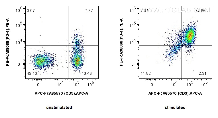

Tested Applications
| Positive FC detected in | PHA treated human PBMCs |
Recommended dilution
| Application | Dilution |
|---|---|
| Flow Cytometry (FC) | FC : 5 ul per 10^6 cells in 100 μl suspension |
| This reagent has been pre-titrated and tested for flow cytometric analysis. The suggested use of this reagent is 5 ul per 10^6 cells in a 100 µl suspension or 5 ul per 100 µl of whole blood. | |
| Sample-dependent, Check data in validation data gallery. | |
Product Information
PE-FcA98068 targets PD-1/CD279 in FC applications and shows reactivity with human samples.
| Tested Reactivity | human |
| Host / Isotype | Rabbit / IgG |
| Class | Recombinant |
| Type | Antibody |
| Immunogen |
CatNo: Eg0974 Product name: Recombinant Human PD-1/CD279 protein (His Tag) Source: mammalian cells-derived, pHZ-KIsec-C-6*HIS Tag: C-6*HIS Domain: 24-167 aa of BC074740 Sequence: FLDSPDRPWNPPTFSPALLVVTEGDNATFTCSFSNTSESFVLNWYRMSPSNQTDKLAAFPEDRSQPGQDCRFRVTQLPNGRDFHMSVVRARRNDSGTYLCGAISLAPKAQIKESLRAELRVTERRAEVPTAHPSPSPRPAGQFQ Predict reactive species |
| Full Name | programmed cell death 1 |
| Calculated Molecular Weight | 288 aa, 32 kDa |
| GenBank Accession Number | BC074740 |
| Gene Symbol | PD-1 |
| Gene ID (NCBI) | 5133 |
| RRID | AB_3749731 |
| Conjugate | PE Fluorescent Dye |
| Excitation/Emission Maxima Wavelengths | 496 nm, 565 nm / 578 nm |
| Excitation Laser | Blue Laser (488 nm), Green Laser (532 nm), Yellow-Green Laser (561 nm) |
| Form | Liquid |
| Purification Method | Protein A purification |
| UNIPROT ID | Q15116 |
| Storage Buffer | PBS with 0.09% sodium azide and 0.5% BSA, pH 7.3. |
| Storage Conditions | Store at 2-8°C. Avoid exposure to light. Stable for one year after shipment. |
Background Information
Programmed cell death 1 (PD-1, also known as CD279) is an immunoinhibitory receptor that belongs to the CD28/CTLA-4 subfamily of the Ig superfamily. It is a 288 amino acid (aa) type I transmembrane protein composed of one Ig superfamily domain, a stalk, a transmembrane domain, and an intracellular domain containing an immunoreceptor tyrosine-based inhibitory motif (ITIM) as well as an immunoreceptor tyrosine-based switch motif (ITSM) (PMID: 18173375). PD-1 is expressed during thymic development and is induced in a variety of hematopoietic cells in the periphery by antigen receptor signaling and cytokines (PMID: 20636820). Engagement of PD-1 by its ligands PD-L1 or PD-L2 transduces a signal that inhibits T-cell proliferation, cytokine production, and cytolytic function (PMID: 19426218). It is critical for the regulation of T cell function during immunity and tolerance. Blockade of PD-1 can overcome immune resistance and also has been shown to have antitumor activity (PMID: 22658127; 23169436). It has been reported that PD-1 is heavily glycosylated and migrates with an apparent molecular mass of 47-55 kDa on SDS-PAGE , which is larger than its predicted mass of 32 kDa (PMID: 8671665; 17640856; 17003438).
Protocols
| Product Specific Protocols | |
|---|---|
| FC protocol for PE PD-1/CD279 antibody PE-FcA98068 | Download protocol |
| Standard Protocols | |
|---|---|
| Click here to view our Standard Protocols |
Publications
What published studies show
PE-FcA98068 is the PE-conjugated FcZero-rAb® Anti-Human PD-1/CD279 Rabbit Recombinant Antibody (clone 240724G11). It has a total of 1 citation. The citation appears in Clinical and Translational Medicine (IF 7.9), published in 2026. The study investigates how TALDO1 promotes lipid metabolic reprogramming and immunosuppressive microenvironment remodeling in hepatocellular carcinoma, placing the research in the fields of liver cancer, lipid metabolism, and tumor immunology. The antibody was used in flow cytometry (FC) in this publication.
| Species | Application | Title |
|---|---|---|
Clin Transl Med TALDO1 promotes lipid metabolic reprogramming and immunosuppressive microenvironment remodelling in hepatocellular carcinoma |


