

Tested Applications
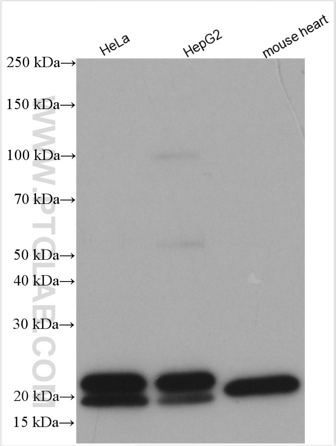
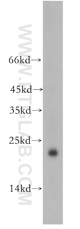
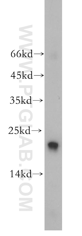
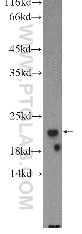
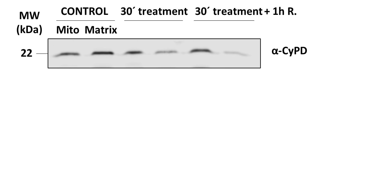
| Positive WB detected in | HeLa cells, HepG2 cells, human heart tissue, rat heart tissue, mouse heart tissue |
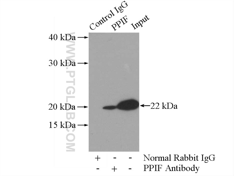
| Positive IP detected in | mouse heart tissue |
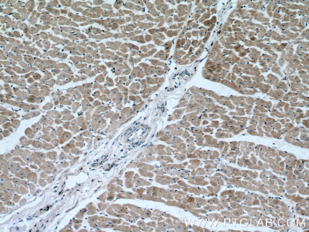
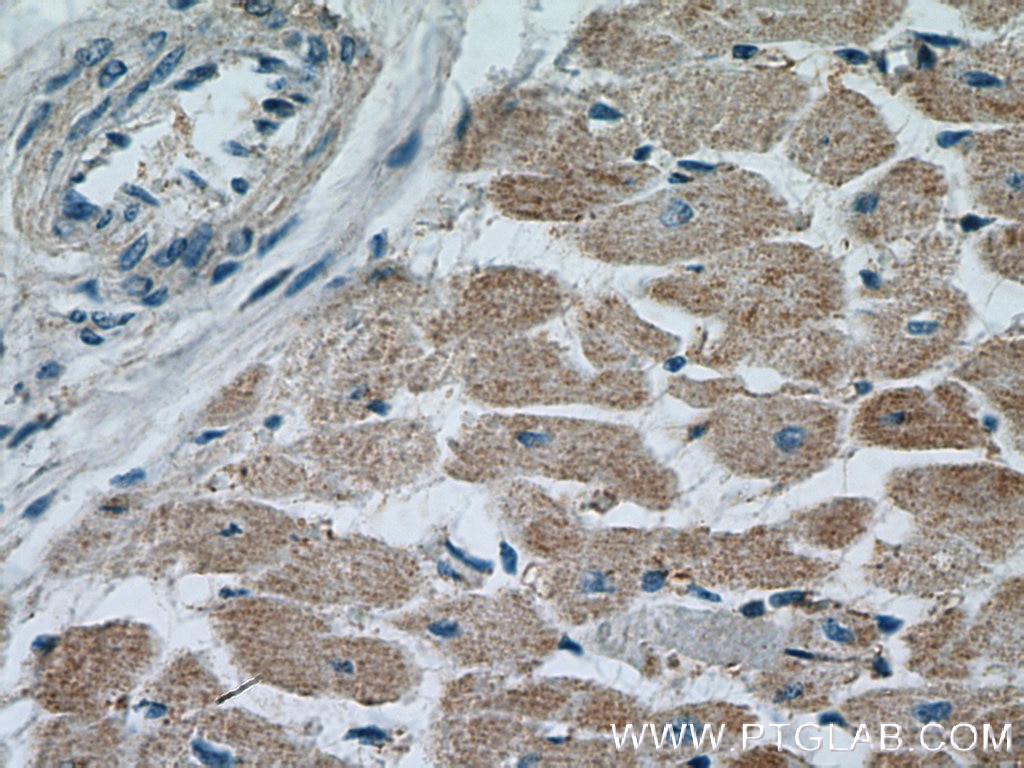
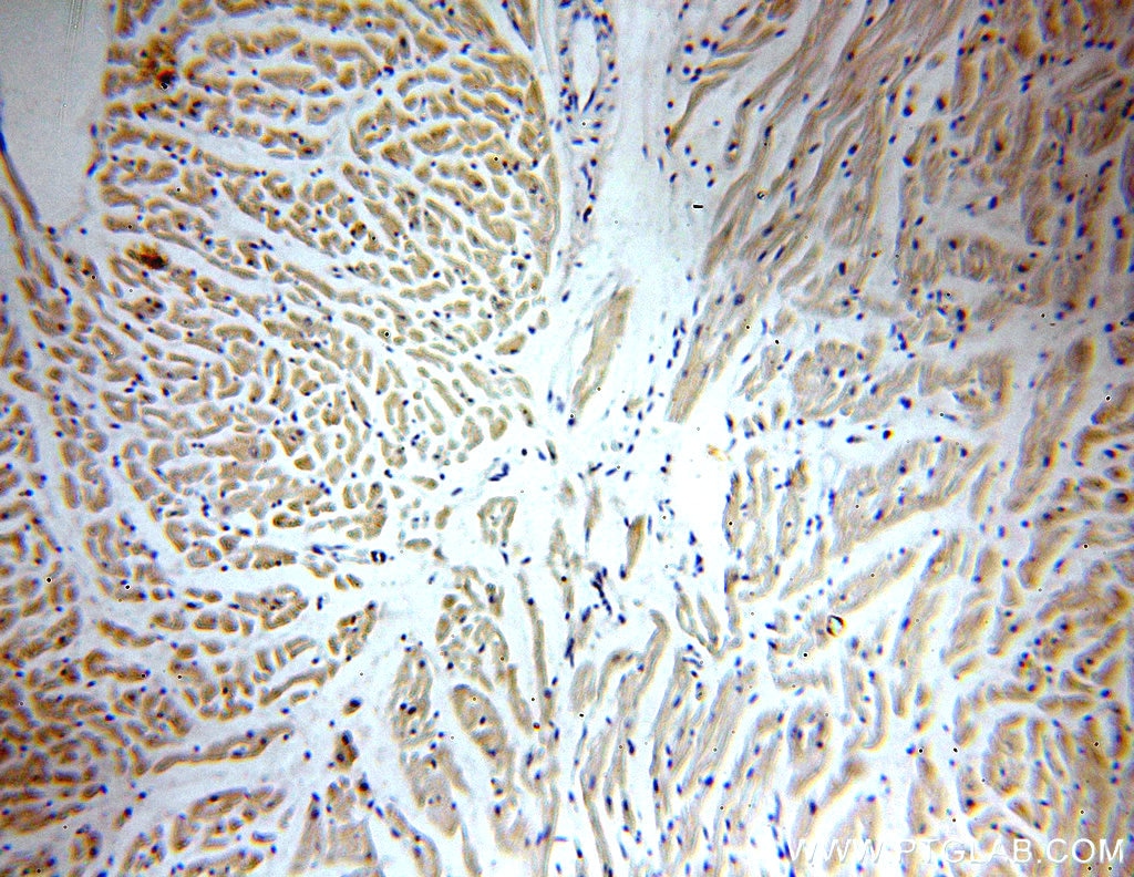
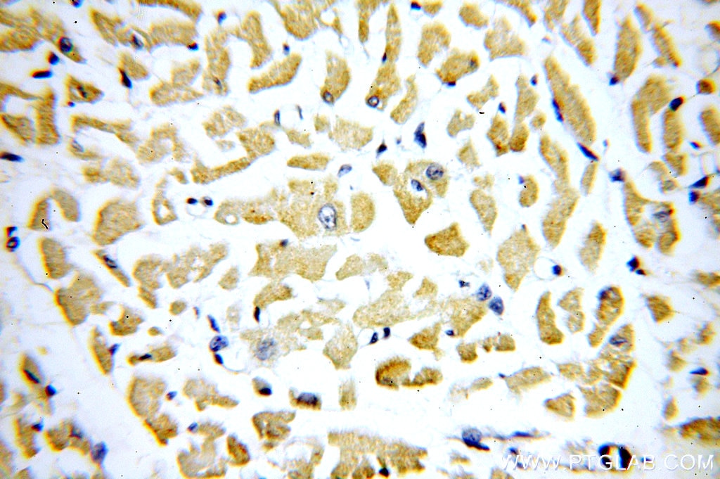
| Positive IHC detected in | human heart tissue Note: suggested antigen retrieval with TE buffer pH 9.0; (*) Alternatively, antigen retrieval may be performed with citrate buffer pH 6.0 |
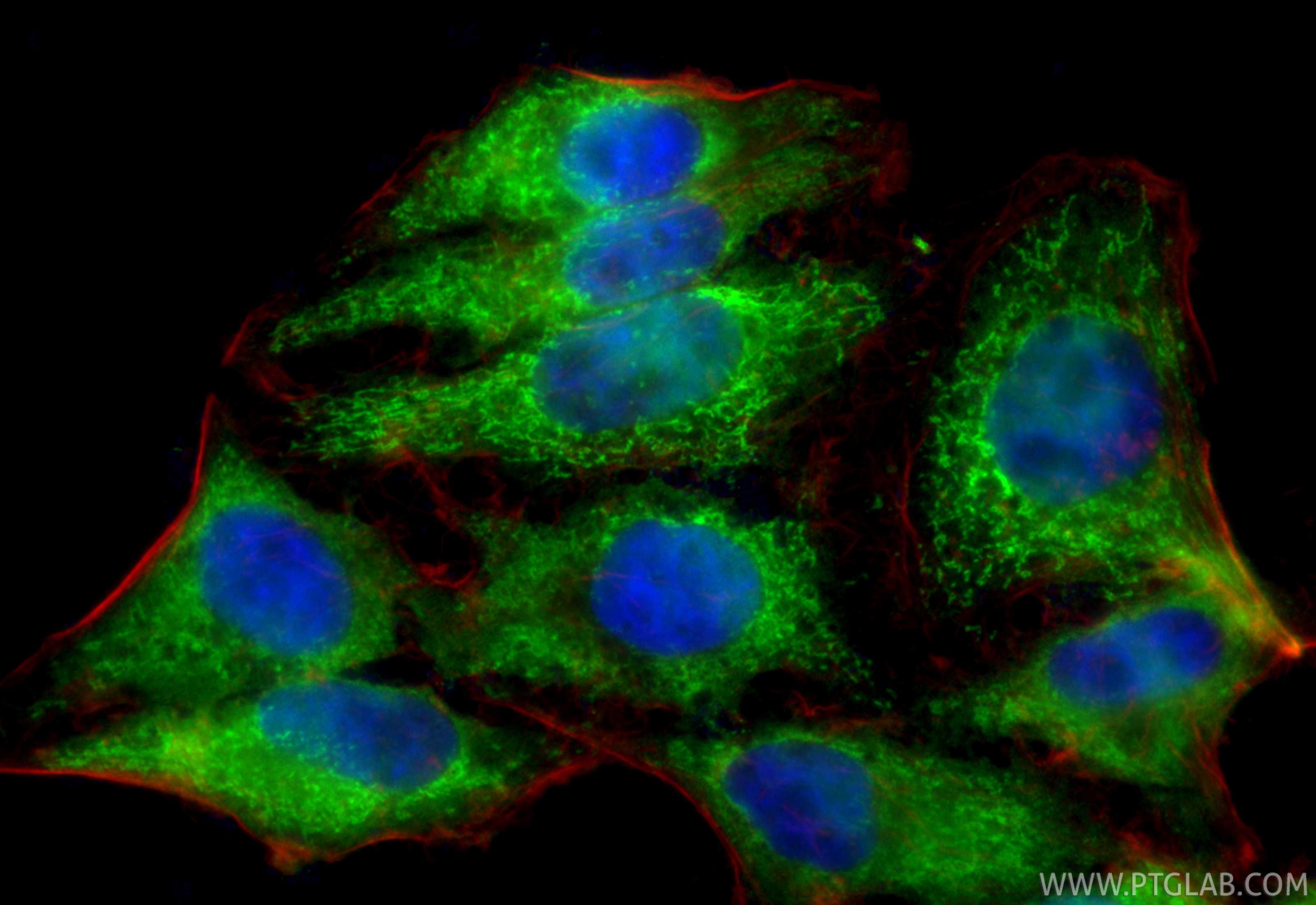
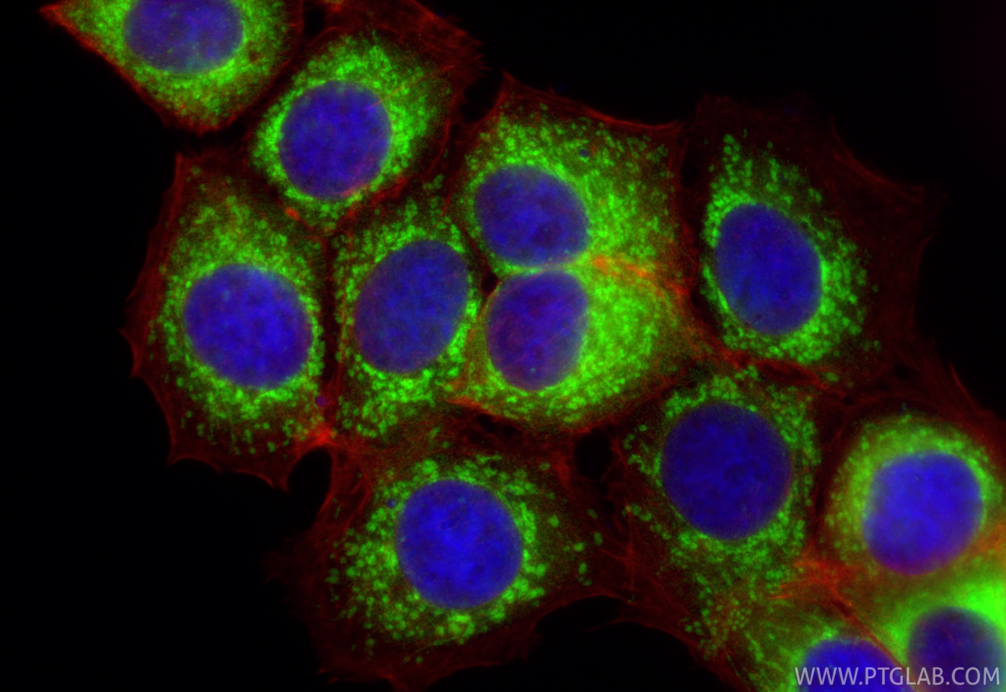
| Positive IF/ICC detected in | HepG2 cells, MCF-7 cells |
Recommended dilution
| Application | Dilution |
|---|---|
| Western Blot (WB) | WB : 1:1000-1:4000 |
| Immunoprecipitation (IP) | IP : 0.5-4.0 ug for 1.0-3.0 mg of total protein lysate |
| Immunohistochemistry (IHC) | IHC : 1:100-1:400 |
| Immunofluorescence (IF)/ICC | IF/ICC : 1:200-1:800 |
| It is recommended that this reagent should be titrated in each testing system to obtain optimal results. | |
| Sample-dependent, Check data in validation data gallery. | |
Product Information
18466-1-AP targets PPIF in WB, IHC, IF/ICC, IP, CoIP, ELISA applications and shows reactivity with human, mouse, rat samples.
| Tested Reactivity | human, mouse, rat |
| Cited Reactivity | human, mouse, rat |
| Host / Isotype | Rabbit / IgG |
| Class | Polyclonal |
| Type | Antibody |
| Immunogen |
CatNo: Ag13151 Product name: Recombinant human PPIF-Specific protein Source: e coli.-derived, PET28a Tag: 6*His Domain: 1-207 aa of BC005020 Sequence: MLALRCGSRWLGLLSVPRSVPLRLPAARACSKGSGDPSSSSSSGNPLVYLDVDANGKPLGRVVLELKADVVPKTAENFRALCTGEKGFGYKGSTFHRVIPSFMCQAGDFTNHNGTGGKSIYGSRFPDENFTLKHVGPGVLSMANAGPNTNGSQFFICTIKTDWLDGKHVVFGHVKEGMDVVKKIESFGSKSGRTSKKIVITDCGQLS Predict reactive species |
| Full Name | peptidylprolyl isomerase F |
| Calculated Molecular Weight | 22 kDa |
| Observed Molecular Weight | 16~22 kDa |
| GenBank Accession Number | BC005020 |
| Gene Symbol | PPIF |
| Gene ID (NCBI) | 10105 |
| RRID | AB_2169273 |
| Conjugate | Unconjugated |
| Form | Liquid |
| Purification Method | Antigen affinity purification |
| UNIPROT ID | P30405 |
| Storage Buffer | PBS with 0.02% sodium azide and 50% glycerol, pH 7.3. |
| Storage Conditions | Store at -20°C. Stable for one year after shipment. Aliquoting is unnecessary for -20oC storage. 20ul sizes contain 0.1% BSA. |
Background Information
PPIF, also named as Cyclophilin D (CypD), CYP3, PPIase F and Rotamase F, belongs to the cyclophilin-type PPIase family. PPIases accelerate the folding of proteins. It catalyzes the cis-trans isomerization of proline imidic peptide bonds in oligopeptides. PPIF is a mitochondrial matrix protein involved in the induction of necrotic and apoptotic cell death through the activation of the mitochondrial permeability transition (mPT) pore. This antibody is immunized with full length gene PPIF and absorbed by PPIA. So this antibody is specific to PPIF.
Protocols
| Product Specific Protocols | |
|---|---|
| IF protocol for PPIF antibody 18466-1-AP | Download protocol |
| IHC protocol for PPIF antibody 18466-1-AP | Download protocol |
| IP protocol for PPIF antibody 18466-1-AP | Download protocol |
| WB protocol for PPIF antibody 18466-1-AP | Download protocol |
| Standard Protocols | |
|---|---|
| Click here to view our Standard Protocols |
Publications
What published studies show
Anti-PPIF (Peptidylprolyl Isomerase F) antibody (Cat# 18466-1-AP) has 21 citations published in journals including Nature Nanotechnology, Nature Communications, Advanced Science, Science Advances, EMBO Journal, Cell Signaling, Journal of Biological Chemistry, FEBS Letters, and eLife. Research fields span mitochondrial biology (mtDNA release, mPTP, OPA1), ferroptosis, cancer (colorectal, endometrial, prostate, breast, osteosarcoma, glioblastoma), inflammation, oxidative stress, and gene editing/nanotechnology.
| Species | Application | Title |
|---|---|---|
Nat Nanotechnol An orally administered gene editing nanoparticle boosts chemo-immunotherapy in colorectal cancer | ||
Nat Commun The mitochondrial fusion protein OPA1 is dispensable in the liver and its absence induces mitohormesis to protect liver from drug-induced injury | ||
Adv Sci (Weinh) CypD Dependent mPTP Opening Is Crucial for Oxidized Mitochondrial DNA Release in Ferroptosis.
| ||
Adv Sci (Weinh) LPS-Induced Mitochondrial Damage via SLC41A1-Mediated Magnesium Ion Efflux Leads to the Pyroptosis of Dental Stem Cells | ||
Sci Adv Apolipoprotein E drives microglia activation in the development of autoimmune uveitis through up-regulation of peptidyl prolyl isomerase F. | ||
Cell Commun Signal Mitochondrial DNA release via mPTP and BAX/BAK drives inflammatory injury in intestinal ischemia reperfusion. |
Reviews
What customers say
Both reviewers gave 5-star ratings. One used the antibody for Western Blot and IHC on human cells at 1:1000, praising sharp bands, minimal non-specific binding, and superior performance compared to competitors at a lower dilution. The antibody was reusable across four WB experiments with consistent quality, and IHC signal intensity was rated above average. The other reviewer used it for WB on HeLa cells at 1:2000, reporting clear bands with no aspecific signal. Both highly recommend the product for reliable results.
The reviews below have been submitted by verified Proteintech customers who received an incentive for providing their feedback.
Samuel (Verified Customer) (10-29-2025) | After having used an anti-PPIF primary antibody from two competing vendors for Western Blot analyses, both at a dilution of 1:750, I can say with confidence that the Proteintech anti-PPIF antibody works a lot better when taking into consideration the sharpness of the protein bands observed during Western Blot imaging, the relative lack of non-specific binding, and the fact that all of this was achieved at a primary antibody dilution of 1:1000 rather than 1:750, making the product not only higher quality when compared to competitors, but also more cost effective in the long run. I have been able to re-use the same primary antibody solution for four different Western Blot experiments and there is little to no reduction in band quality. I have also used this specific antibody for one IHC experiment, which yielded results that were very much to my liking, primarily as a result of the signal intensity, which I would rate above average compared to competitors. All this being said, I would highly recommend this specific antibody for anyone who is considering using it for great results, at least from a Western Blot and IHC perspective.
|
Veronica (Verified Customer) (01-30-2024) | Really satisfied with the result in WB. No aspecific signal, clear band. Reccomended! Conditions: 12% acrylammide gel Ab’: 1:2000 in 5% milk O/N
 |












