
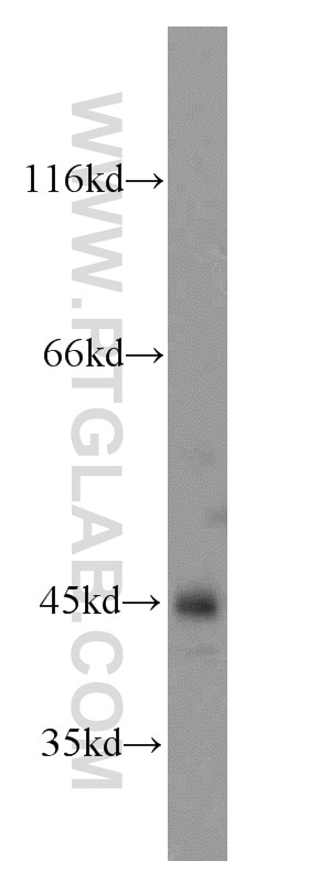
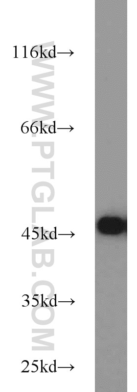
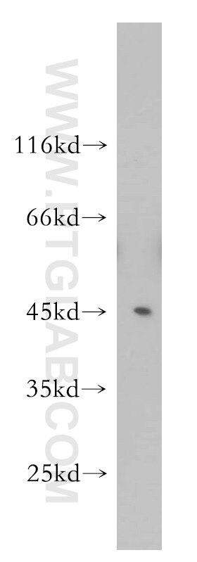
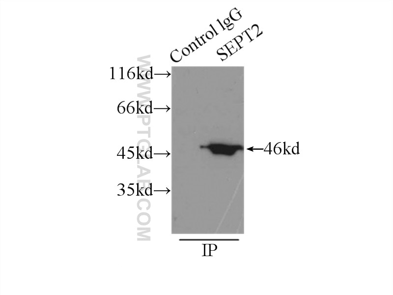

Tested Applications
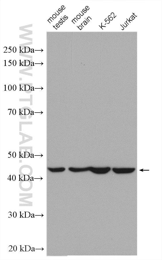
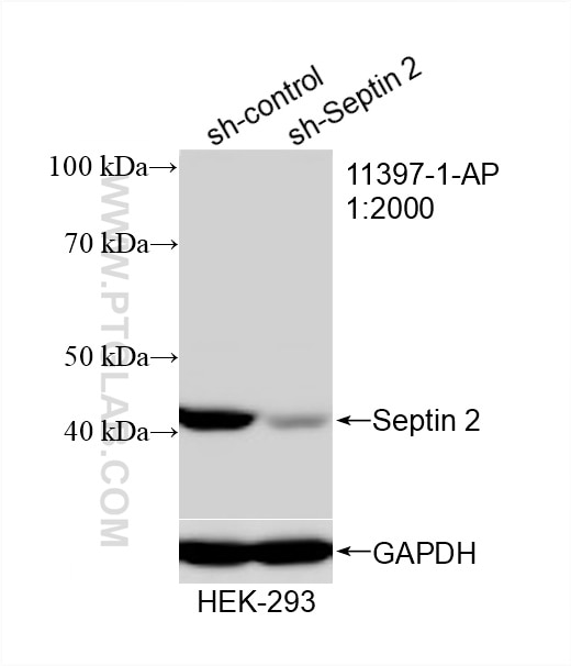
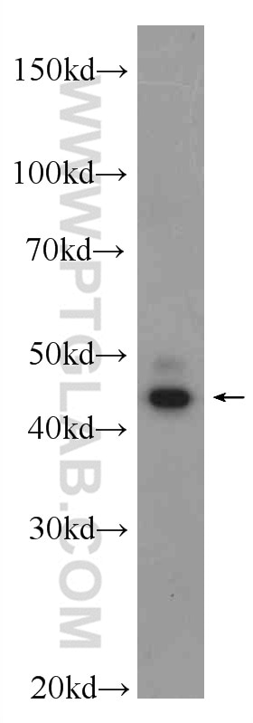
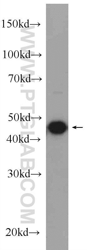
| Positive WB detected in | mouse brain tissue, mouse heart tissue, HeLa cells, human skeletal muscle tissue, rat brain tissue, C6 cells, HEK-293 cells, K-562 cells, Jurkat cells |
| Positive IP detected in | mouse brain tissue |
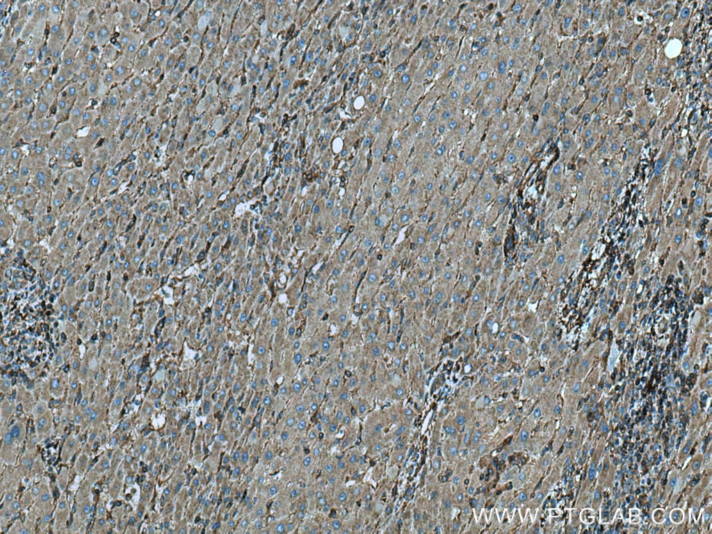
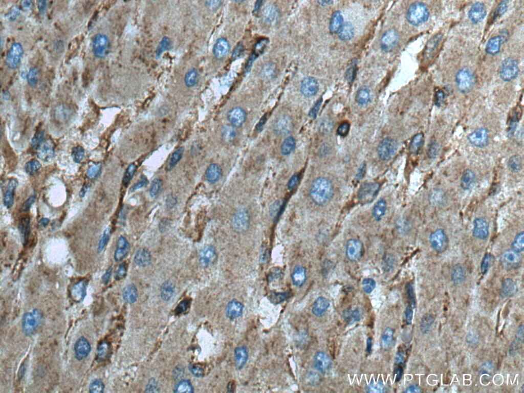
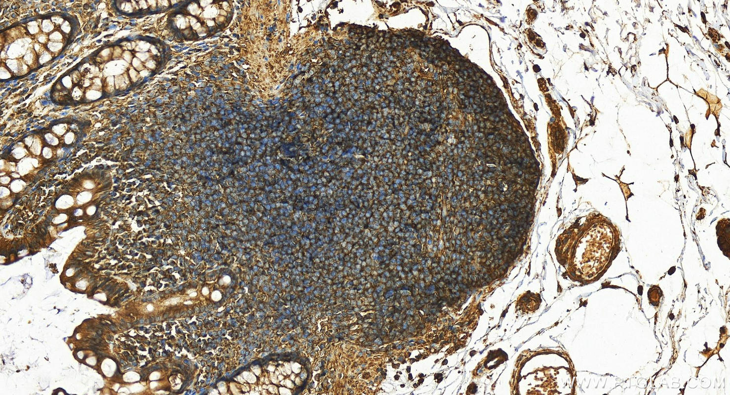
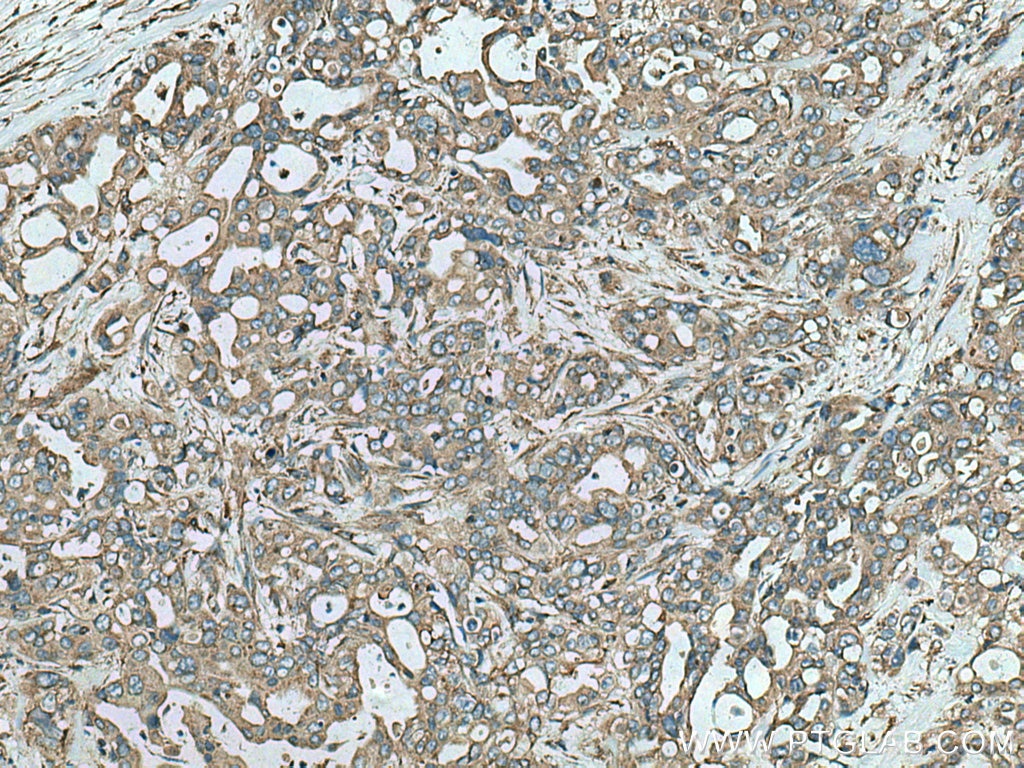
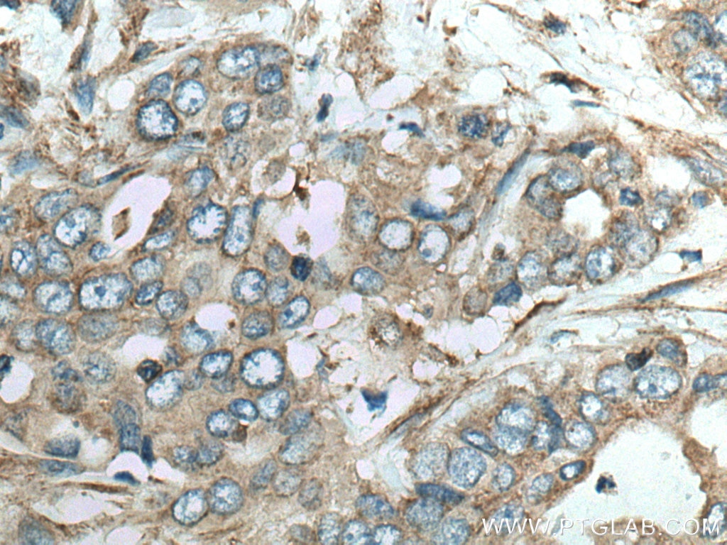
| Positive IHC detected in | human liver cancer tissue, human colon tissue, human pancreas cancer tissue Note: suggested antigen retrieval with TE buffer pH 9.0; (*) Alternatively, antigen retrieval may be performed with citrate buffer pH 6.0 |
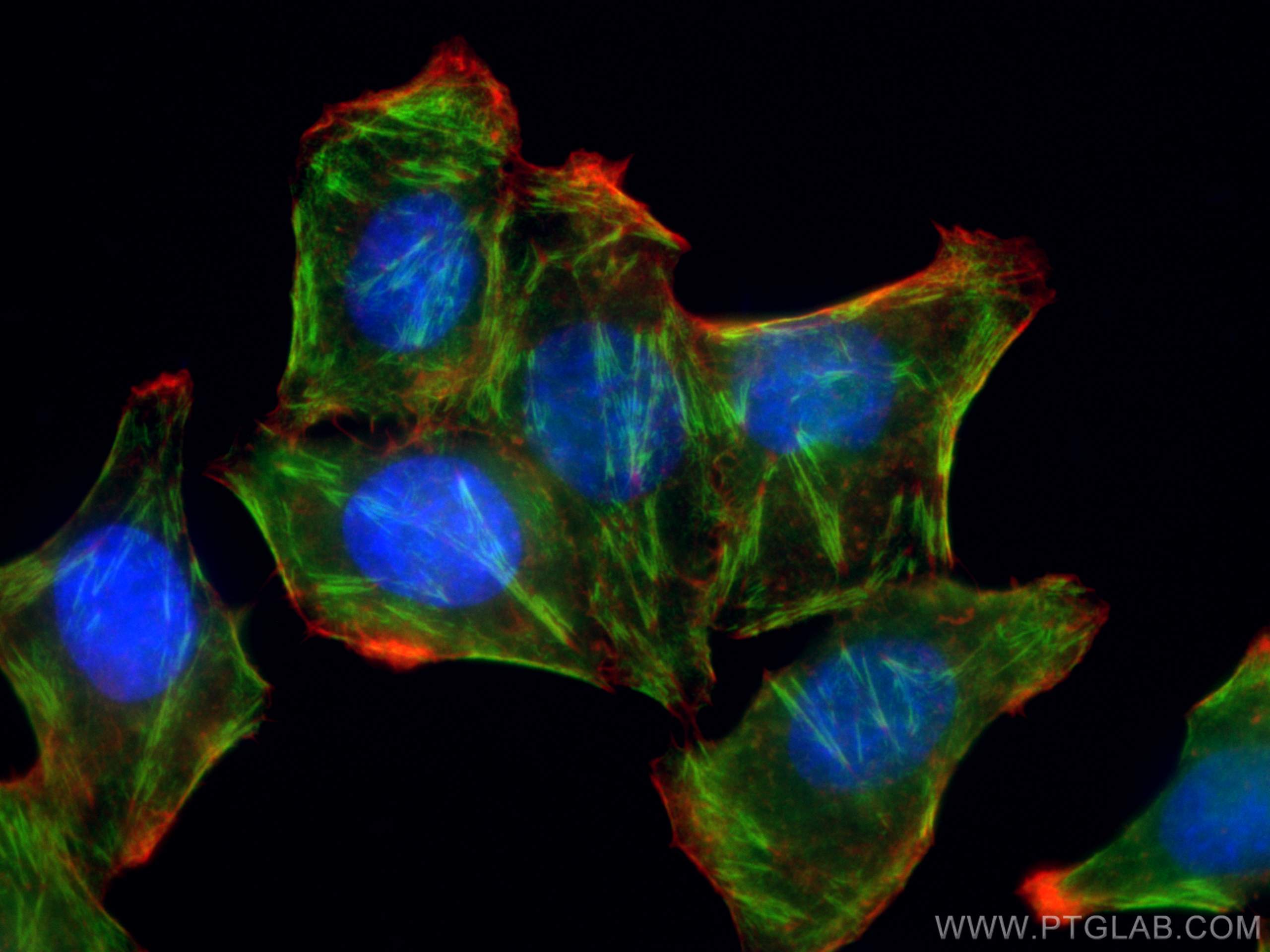
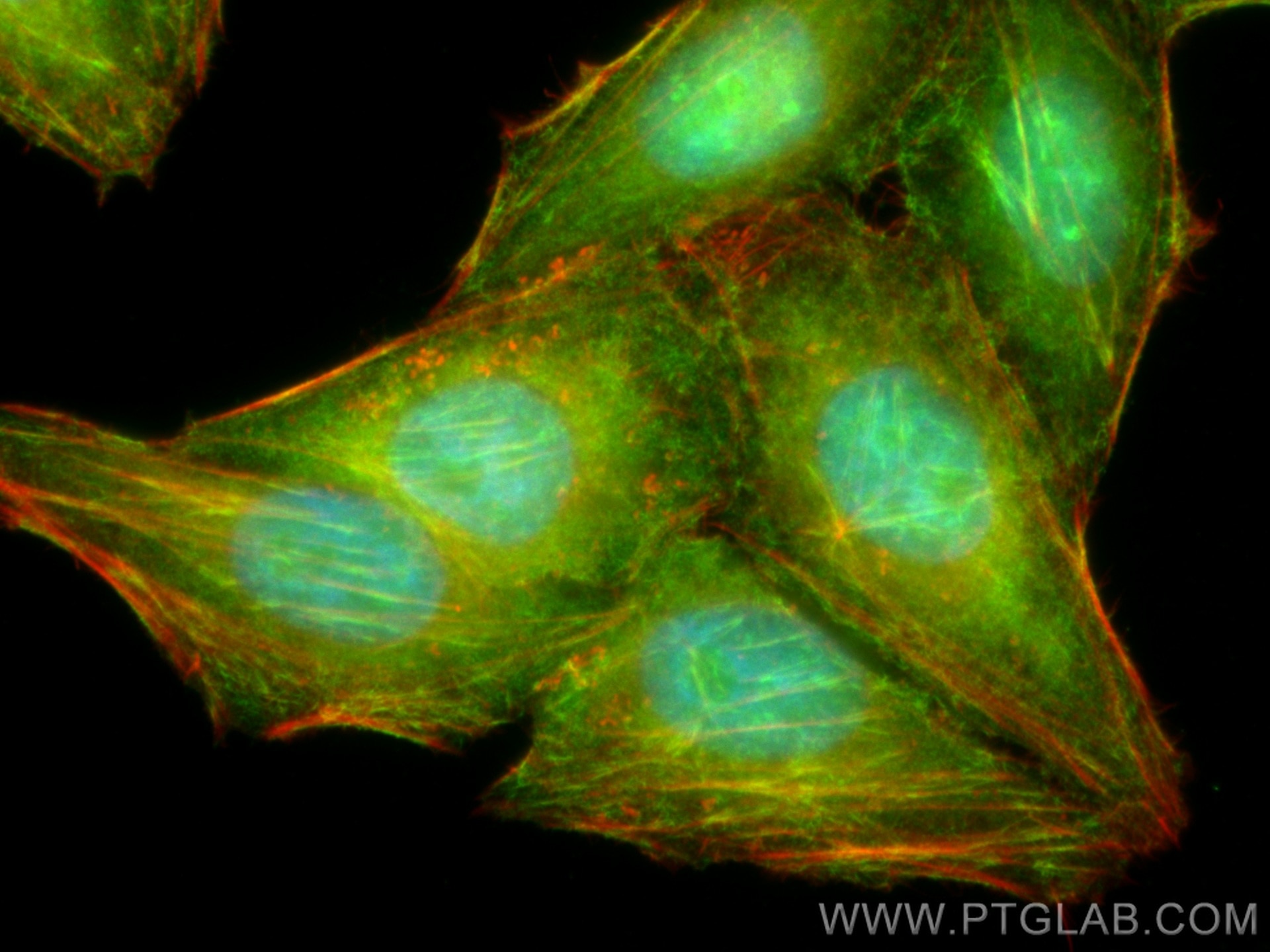
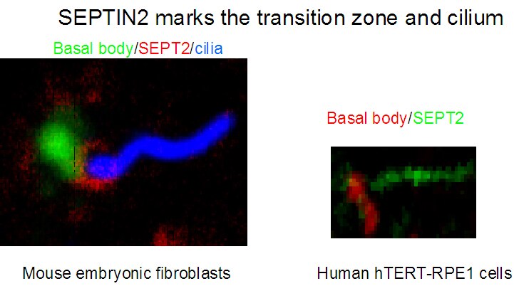
| Positive IF/ICC detected in | HepG2 cells, hTERT-RPE1 cells and Mouse embryonic fibroblasts |
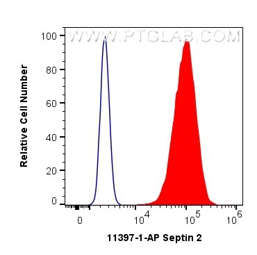
| Positive FC (Intra) detected in | MCF-7 cells |
Recommended dilution
| Application | Dilution |
|---|---|
| Western Blot (WB) | WB : 1:1000-1:4000 |
| Immunoprecipitation (IP) | IP : 0.5-4.0 ug for 1.0-3.0 mg of total protein lysate |
| Immunohistochemistry (IHC) | IHC : 1:50-1:500 |
| Immunofluorescence (IF)/ICC | IF/ICC : 1:50-1:500 |
| Flow Cytometry (FC) (INTRA) | FC (INTRA) : 0.40 ug per 10^6 cells in a 100 µl suspension |
| It is recommended that this reagent should be titrated in each testing system to obtain optimal results. | |
| Sample-dependent, Check data in validation data gallery. | |
Product Information
11397-1-AP targets Septin 2 in WB, IHC, IF/ICC, FC (Intra), IP, ELISA applications and shows reactivity with human, mouse, rat samples.
| Tested Reactivity | human, mouse, rat |
| Cited Reactivity | human, mouse, rat, canine |
| Host / Isotype | Rabbit / IgG |
| Class | Polyclonal |
| Type | Antibody |
| Immunogen |
CatNo: Ag1946 Product name: Recombinant human SEPT2 protein Source: e coli.-derived, PGEX-4T Tag: GST Domain: 1-361 aa of BC014455 Sequence: MSKQQPTQFINPETPGYVGFANLPNQVHRKSVKKGFEFTLMVVGESGLGKSTLINSLFLTDLYPERVISGAAEKIERTVQIEASTVEIEERGVKLRLTVVDTPGYGDAINCRDCFKTIISYIDEQFERYLHDESGLNRRHIIDNRVHCCFYFISPFGHGLKPLDVAFMKAIHNKVNIVPVIAKADTLTLKERERLKKRILDEIEEHNIKIYHLPDAESDEDEDFKEQTRLLKASIPFSVVGSNQLIEAKGKKVRGRLYPWGVVEVENPEHNDFLKLRTMLITHMQDLQEVTQDLHYENFRSERLKRGGRKVENEDMNKDQILLEKEAELRRMQEMIARMQAQMQMQMQGGDGDGGALGHHV Predict reactive species |
| Full Name | septin 2 |
| Calculated Molecular Weight | 41 kDa |
| Observed Molecular Weight | 45 kDa |
| GenBank Accession Number | BC014455 |
| Gene Symbol | Septin 2 |
| Gene ID (NCBI) | 4735 |
| RRID | AB_2239110 |
| Conjugate | Unconjugated |
| Form | Liquid |
| Purification Method | Antigen affinity purification |
| UNIPROT ID | Q15019 |
| Storage Buffer | PBS with 0.02% sodium azide and 50% glycerol, pH 7.3. |
| Storage Conditions | Store at -20°C. Stable for one year after shipment. Aliquoting is unnecessary for -20oC storage. 20ul sizes contain 0.1% BSA. |
Background Information
SEPT2 belongs to a family of septins, GTP-binding proteins that are a component of the cytoskeleton. Septins can assemble into intracellular structures of various shapes and are known to influence membrane stability, forming diffusion barriers and serving as scaffolds.
What is the molecular weight of SEPT2? Is SEPT2 post-translationally modified?
The molecular weight of SEPT2 is 45 kDa. In a cellular context, SEPT2 forms hetero-hexamers and hetero-octamers with other septins. These hexamers and octamers further assemble into high-order structures such as filaments and rings. SEPT2 can be phosphorylated on Ser218 by casein kinase 2 (PMID: 19165576) and can also be SUMOylated (PMID: 29051266).
What is the subcellular localization of SEPT2?
SEPT2 forms a diffusion barrier and localizes to the primary cilium; in some cell types, it has been found at the transition zone/basal body (PMID: 20558667), while in others localized to the axoneme (PMID: 23572511). During cell division, SEPT2 accumulates near the contractile ring from anaphase through telophase, and finally condenses into the midbody (PMID: 9203580). In interphase cells, SEPT2 was found to associate with polyglutamylated microtubules (PMID: 18209106) and can affect microtubule dynamics via interaction with MAP4.
What is the tissue expression pattern of SEPT2?
While some septins have tissue-specific expression profiles, SEPT2 is ubiquitously expressed (PMID: 22314400).
Protocols
| Product Specific Protocols | |
|---|---|
| FC protocol for Septin 2 antibody 11397-1-AP | Download protocol |
| IF protocol for Septin 2 antibody 11397-1-AP | Download protocol |
| IHC protocol for Septin 2 antibody 11397-1-AP | Download protocol |
| IP protocol for Septin 2 antibody 11397-1-AP | Download protocol |
| WB protocol for Septin 2 antibody 11397-1-AP | Download protocol |
| Standard Protocols | |
|---|---|
| Click here to view our Standard Protocols |
Publications
What published studies show
The Septin 2 (SEPT2) Polyclonal antibody, catalog number 11397-1-AP, has 36 citations. These appear in journals including Nature Communications, Cell Death & Disease, Cell Chemical Biology, Cell Reports, Journal of Cell Biology, Glia, mBio, PLoS Genetics, Molecular Biology of the Cell, Development, eLife, and Oncotarget. Research fields span septin cytoskeleton biology, cytokinesis, CNS myelination, sperm formation and male fertility, cancer, ciliogenesis, and host-pathogen interactions.
| Species | Application | Title |
|---|---|---|
Nat Commun Wiskott-Aldrich syndrome protein regulates autophagy and inflammasome activity in innate immune cells. | ||
Nat Commun Shigella flexneri evades septin-mediated cell-autonomous immunity via protein ADP-riboxanation. | ||
Cell Death Dis JMJD2C-mediated long non-coding RNA MALAT1/microRNA-503-5p/SEPT2 axis worsens non-small cell lung cancer. | ||
Cell Death Dis TMEM232 is required for the formation of sperm flagellum and male fertility in mice | ||
Pharmacol Res BET degrader exhibits lower antiproliferative activity than its inhibitor via EGR1 recruiting septins to promote E2F1-3 transcription in triple-negative breast cancer
|
Reviews
What customers say
Users report mixed results. For Western blot at 1:1000 on HEK293T lysates, one reviewer observed a strong band just below 47 kDa (expected Septin 2 size) plus a fainter band above it, rating the product 5 stars. For immunofluorescence at 1:200 on testis tissue, another reviewer noted staining at the annulus and acrosome but complained of high background even when dilution was reduced, giving 3 stars. Overall, the antibody performs well for WB but may require optimization for IF applications to reduce background.
The reviews below have been submitted by verified Proteintech customers who received an incentive for providing their feedback.
Christine (Verified Customer) (01-27-2023) | Incubation for a few hours at room temperature on HEK293T lysates. Detection of a very strong signal just below the 47 kDa marker (expected size of Septin 2) and a fainter signal just above the 47 kDa marker.
|
Mohammed (Verified Customer) (08-19-2020) | Some staining at the annulus, there seems to be staining in the acrosome. Overall, really high background (even when the concentration is lowered).
|


















