

Tested Applications
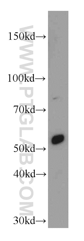
| Positive WB detected in | mouse pancreas tissue |
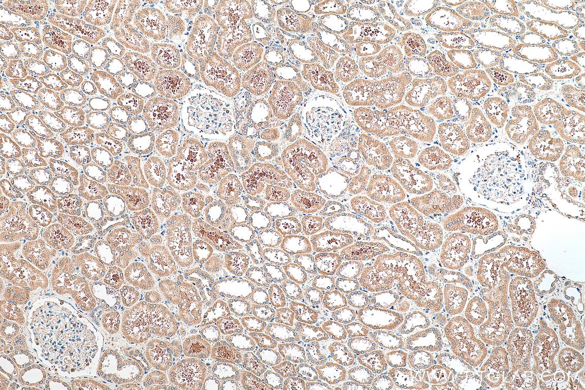
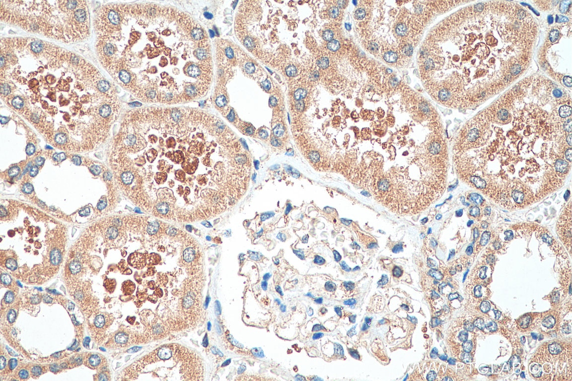
| Positive IHC detected in | human kidney tissue Note: suggested antigen retrieval with TE buffer pH 9.0; (*) Alternatively, antigen retrieval may be performed with citrate buffer pH 6.0 |
Recommended dilution
| Application | Dilution |
|---|---|
| Western Blot (WB) | WB : 1:200-1:1000 |
| Immunohistochemistry (IHC) | IHC : 1:50-1:500 |
| It is recommended that this reagent should be titrated in each testing system to obtain optimal results. | |
| Sample-dependent, Check data in validation data gallery. | |
Product Information
20267-1-AP targets SERINC3 in WB, IHC, ELISA applications and shows reactivity with human, mouse, rat samples.
| Tested Reactivity | human, mouse, rat |
| Cited Reactivity | human |
| Host / Isotype | Rabbit / IgG |
| Class | Polyclonal |
| Type | Antibody |
| Immunogen |
Peptide Predict reactive species |
| Full Name | serine incorporator 3 |
| Calculated Molecular Weight | 53 kDa |
| Observed Molecular Weight | 53 kDa |
| GenBank Accession Number | NM_006811 |
| Gene Symbol | SERINC3 |
| Gene ID (NCBI) | 10955 |
| RRID | AB_10693614 |
| Conjugate | Unconjugated |
| Form | Liquid |
| Purification Method | Antigen affinity purification |
| UNIPROT ID | Q13530 |
| Storage Buffer | PBS with 0.02% sodium azide and 50% glycerol, pH 7.3. |
| Storage Conditions | Store at -20°C. Stable for one year after shipment. Aliquoting is unnecessary for -20oC storage. 20ul sizes contain 0.1% BSA. |
Background Information
SERINC, also named as DIFF33 and TDE1, belongs to the TDE1 family. SERINC3 may be involved in cellular transformation. The antibody is specific to SERINC3.
Protocols
| Product Specific Protocols | |
|---|---|
| IHC protocol for SERINC3 antibody 20267-1-AP | Download protocol |
| WB protocol for SERINC3 antibody 20267-1-AP | Download protocol |
| Standard Protocols | |
|---|---|
| Click here to view our Standard Protocols |
Publications
What published studies show
The SERINC3 Polyclonal antibody (Cat# 20267-1-AP) has 2 citations. One appears in Cell Reports (2026), investigating how genome-bound cellular proteins are incorporated into HIV-1 particles to regulate viral infection. The other is in Frontiers in Immunology (2026), describing a machine-learning-based cell death and senescence index for prognosis and immune landscape characterization in head and neck squamous cell carcinoma. Research fields span virology/HIV biology and oncology/immunology.
| Species | Application | Title |
|---|---|---|
Cell Rep Incorporation of genome-bound cellular proteins into HIV-1 particles regulates viral infection. | ||
Front Immunol CDCSI: a machine learning-based interpretable cell death and cellular senescence index for prognosis improvement, immune landscape characterization, and therapeutic response prediction in head and neck squamous cell carcinoma. |
Reviews
What customers say
The single review reports unsatisfactory performance. In Western blot, the antibody detected a band around 42 kD rather than the expected ~53 kD, and this 42 kD band persisted even after knockdown with smart-pool RNAs, suggesting it may not be the intended target. In immunofluorescence, the expected Golgi localization described in the literature was not observed. Overall, the reviewer rated the product 1 out of 5, indicating it did not meet expectations for either application.
The reviews below have been submitted by verified Proteintech customers who received an incentive for providing their feedback.
Boyan (Verified Customer) (01-25-2019) | By WB, I saw a band around 42 kD, but the expected band is around 53 kD. The 42 kD band did not disappear when I knock it down with smart-pool RNAs.By IF, I didn't see its Golgi localisation, which is shown in literature.
|




