

Tested Applications
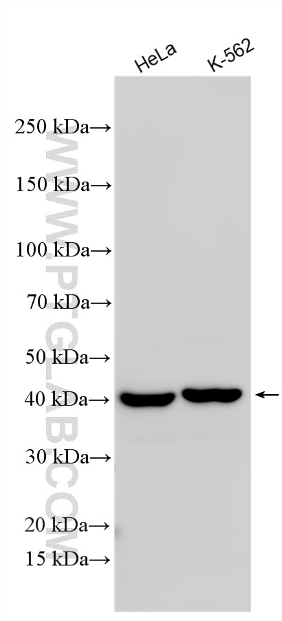
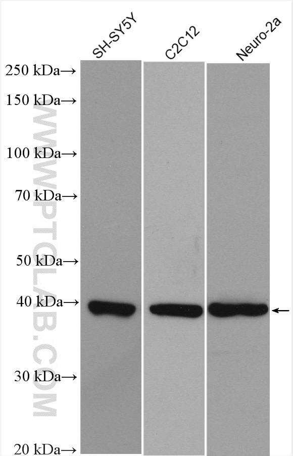
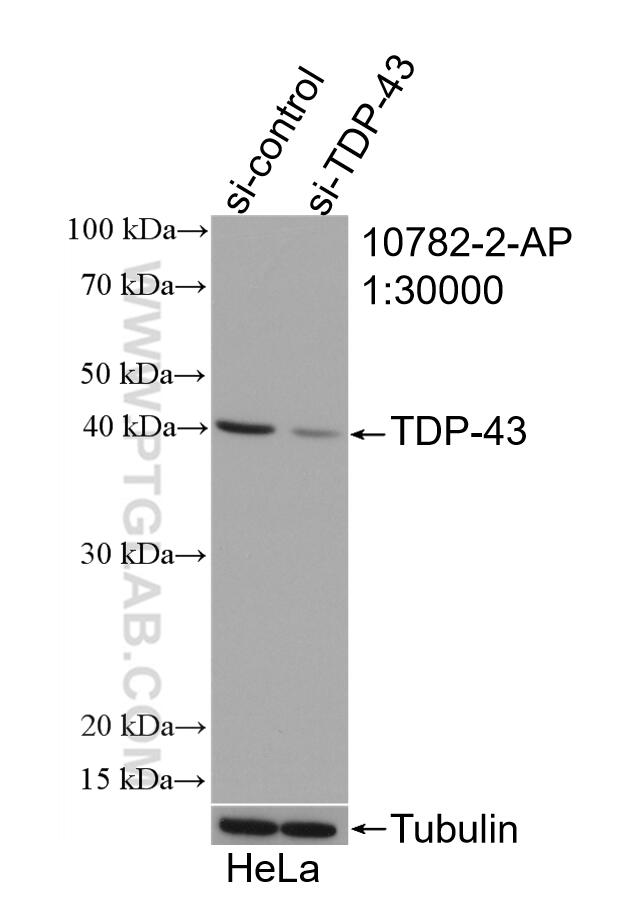
| Positive WB detected in | HeLa cells, SH-SY5Y cells, C2C12 cells, Neuro-2a cells |
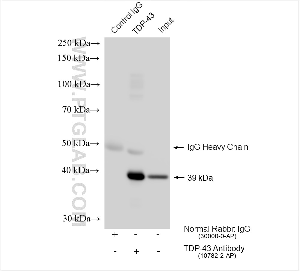
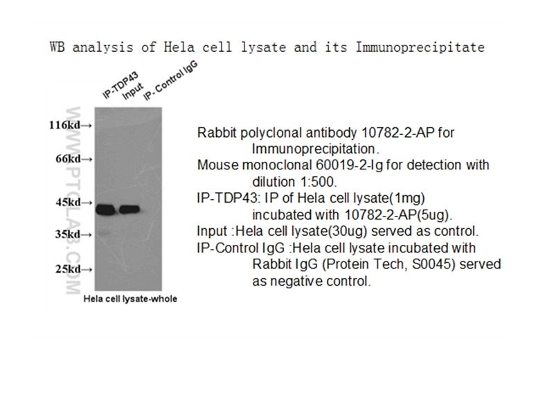
| Positive IP detected in | HeLa cells |
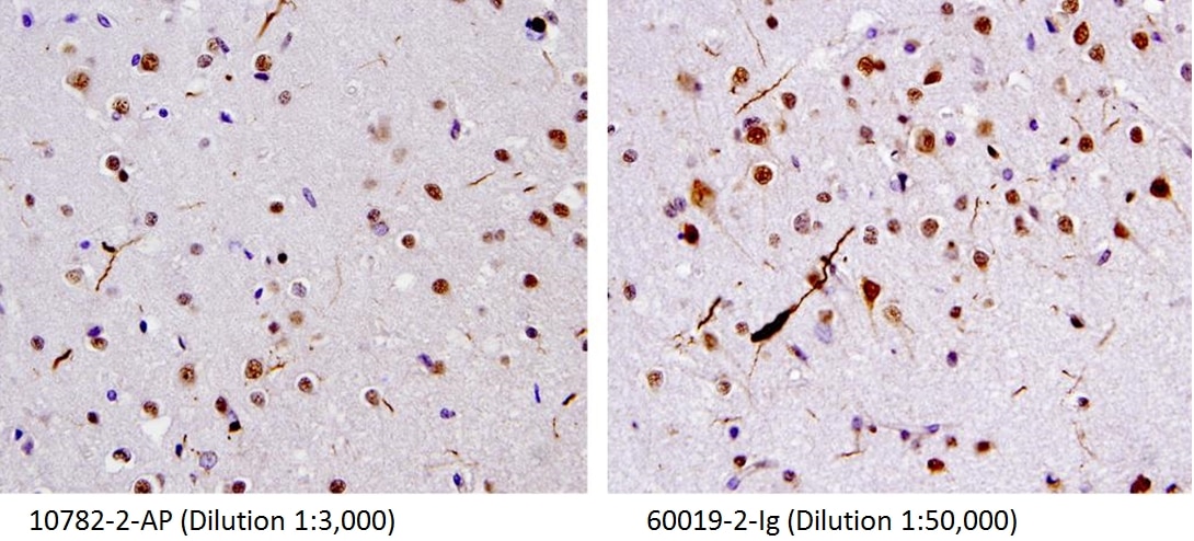
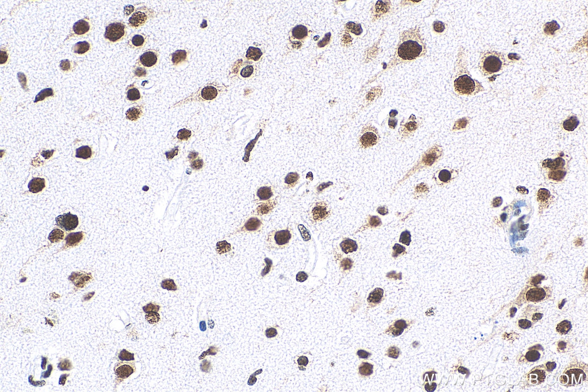
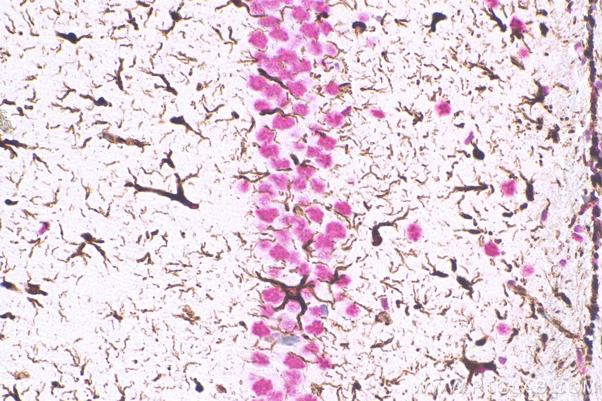
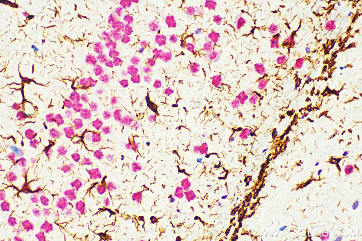
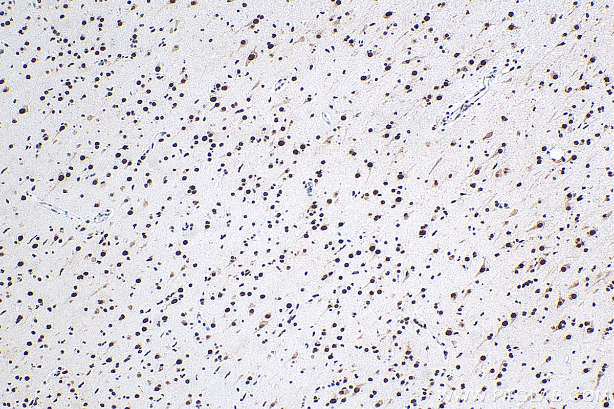
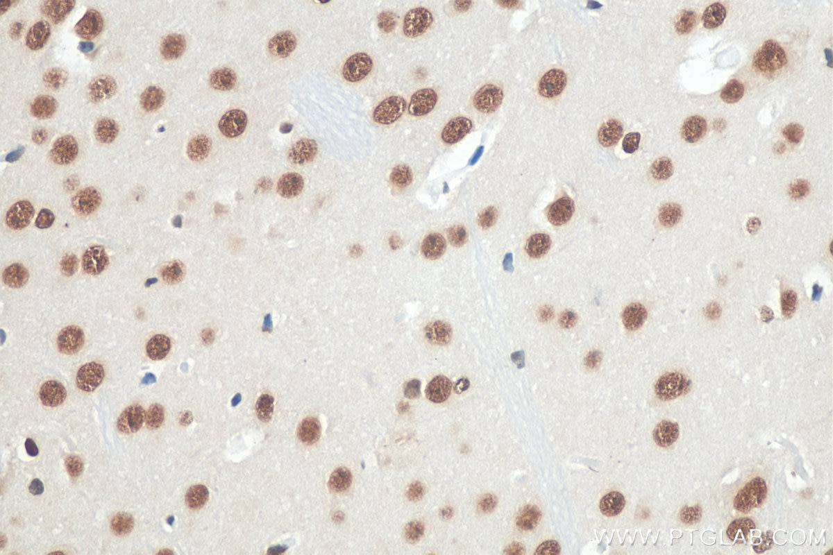
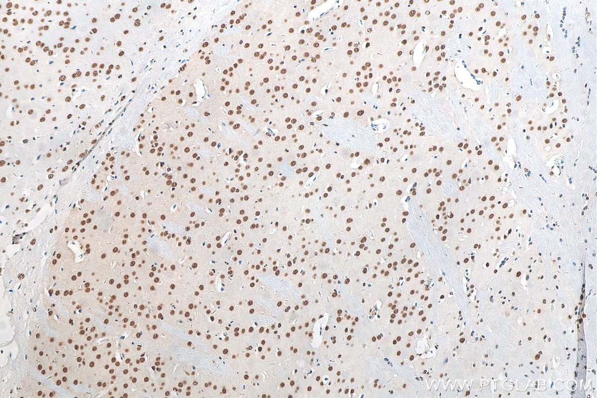
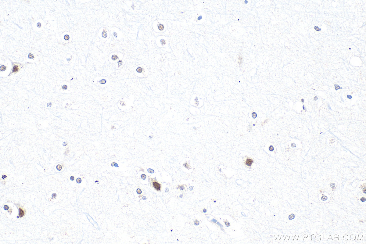
| Positive IHC detected in | human gliomas tissue, human brain tissue, human brain (FTLD-U) tissue, mouse brain tissue Note: suggested antigen retrieval with TE buffer pH 9.0; (*) Alternatively, antigen retrieval may be performed with citrate buffer pH 6.0 |
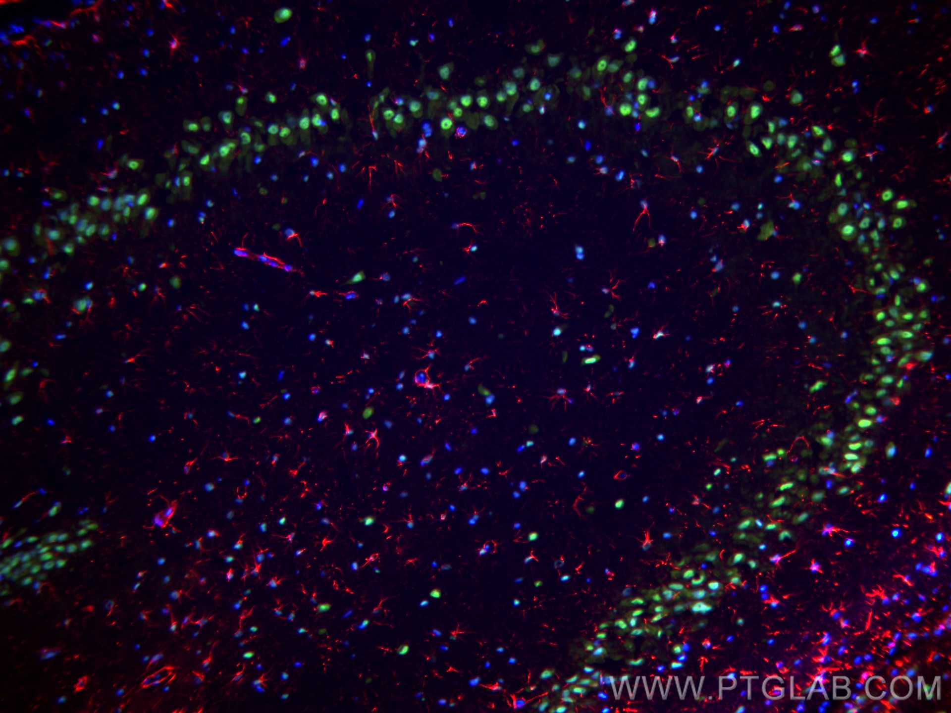
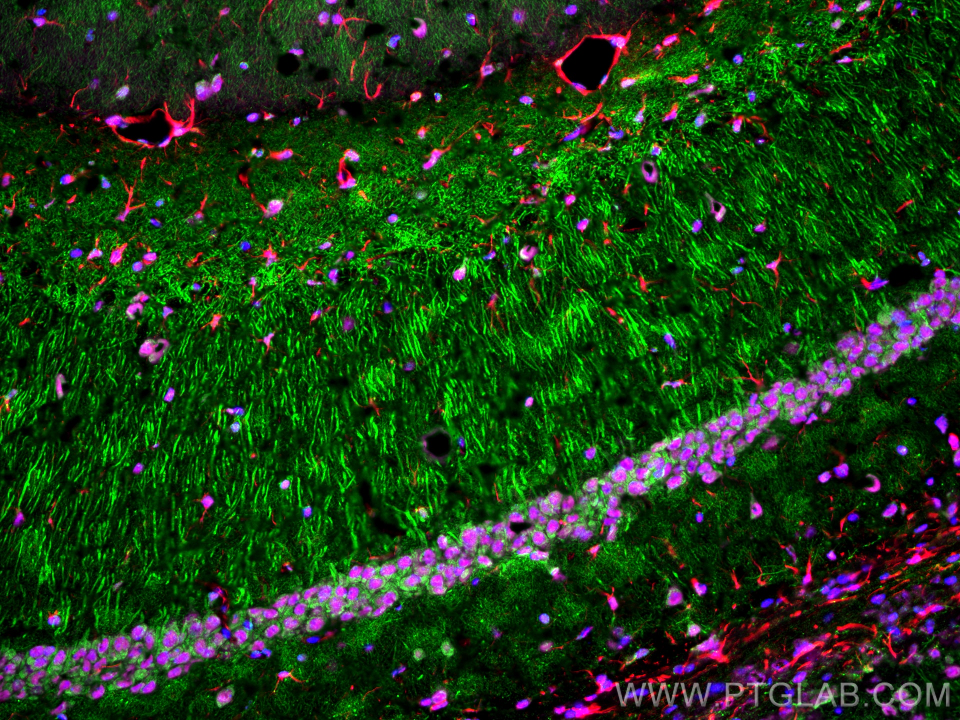
| Positive IF-Fro detected in | rat brain tissue, mouse brain tissue |
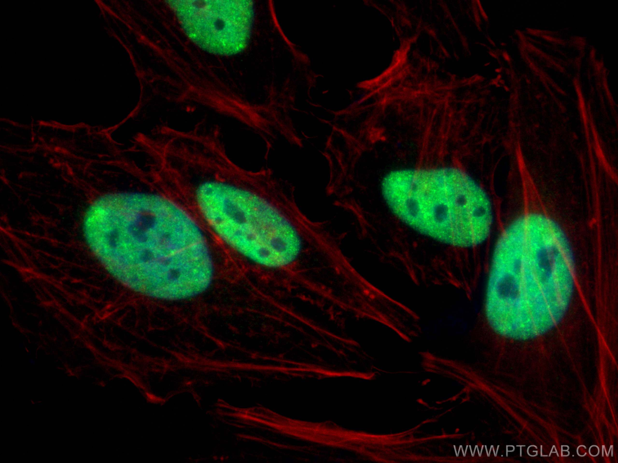
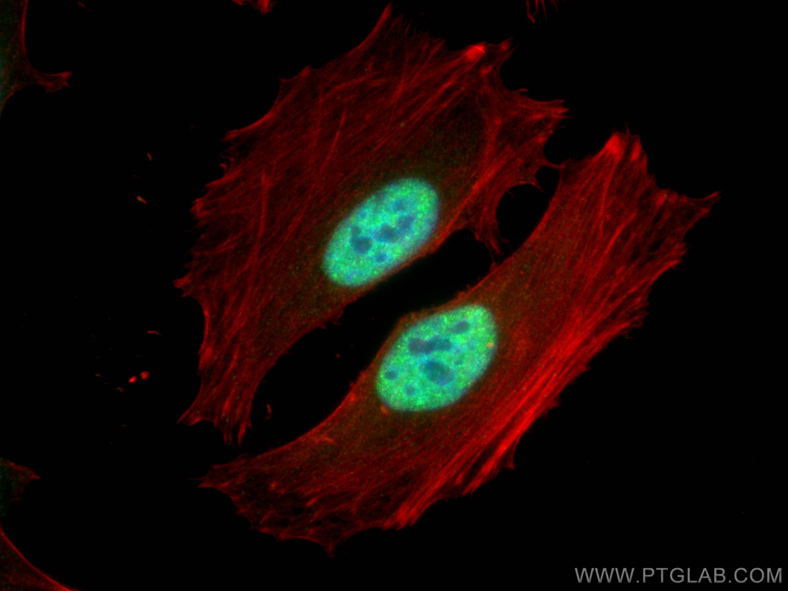
| Positive IF/ICC detected in | HeLa cells |
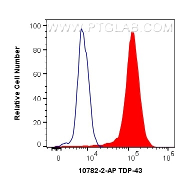
| Positive FC (Intra) detected in | HeLa cells |
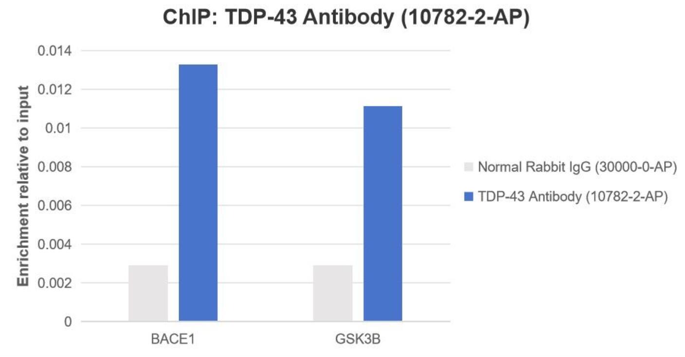
| Positive ChIP detected in | HeLa cells |
Recommended dilution
| Application | Dilution |
|---|---|
| Western Blot (WB) | WB : 1:20000-1:100000 |
| Immunoprecipitation (IP) | IP : 0.5-4.0 ug for 1.0-3.0 mg of total protein lysate |
| Immunohistochemistry (IHC) | IHC : 1:2000-1:8000 |
| Immunofluorescence (IF)-FRO | IF-FRO : 1:1000-1:4000 |
| Immunofluorescence (IF)/ICC | IF/ICC : 1:3000-1:12000 |
| Flow Cytometry (FC) (INTRA) | FC (INTRA) : 0.40 ug per 10^6 cells in a 100 µl suspension |
| Chromatin immunoprecipitation (ChIP) | CHIP : 1:10-1:100 |
| It is recommended that this reagent should be titrated in each testing system to obtain optimal results. | |
| Sample-dependent, Check data in validation data gallery. | |
Product Information
10782-2-AP targets TDP-43 in WB, IHC, IF/ICC, IF-Fro, FC (Intra), IP, CoIP, ChIP, RIP, ELISA, IEM applications and shows reactivity with human, mouse, rat, zebrafish samples.
| Tested Reactivity | human, mouse, rat, zebrafish |
| Cited Reactivity | human, mouse, rat, monkey, chicken, zebrafish, yeast, horse, seal |
| Host / Isotype | Rabbit / IgG |
| Class | Polyclonal |
| Type | Antibody |
| Immunogen |
Recombinant protein Predict reactive species |
| Full Name | TAR DNA binding protein |
| Calculated Molecular Weight | 43 kDa |
| Observed Molecular Weight | 44 kDa |
| GenBank Accession Number | BC001487 |
| Gene Symbol | TDP-43 |
| Gene ID (NCBI) | 23435 |
| RRID | AB_615042 |
| Conjugate | Unconjugated |
| Form | Liquid |
| Purification Method | Antigen affinity purification |
| UNIPROT ID | Q13148 |
| Storage Buffer | PBS with 0.02% sodium azide and 50% glycerol, pH 7.3. |
| Storage Conditions | Store at -20°C. Stable for one year after shipment. Aliquoting is unnecessary for -20oC storage. 20ul sizes contain 0.1% BSA. |
Background Information
The TARDBP gene encodes the TDP-43 protein, initially found to repress HIV-1 transcription by binding TAR DNA. TDP-43 has since been shown to bind RNA as well as DNA, and have multiple functions in transcriptional repression, translational regulation and pre-mRNA splicing. For instance, it is reported to regulate alternate splicing of the CTFR gene. In 2006 Neumann et al. found that hyperphosphorylated, ubiquitinated and/or cleaved forms of TDP-43, collectively known as pathological TDP-43, play a major role in the disease mechanisms of ubiquitin-positive, tau- and alpha-synuclein-negative frontotemporal dementia (FTLD-U) and in amyotrophic lateral sclerosis (ALS). Proteintech's 10782-2-AP antibody is a rabbit polyclonal antibody recognizing N-terminal TDP-43. It recognizes the intact 43 kDa protein as well as all posttranslationally modified and truncated forms in multiple applications. Various forms of TDP-43 exist, including 18-35 kDa of cleaved C-terminal fragments, 45-50 kDa phospho-protein, 55 kDa glycosylated form, 75 kDa hyperphosphorylated form, and 90-300 kDa cross-linked form. (17023659, 19823856, 21666678, 22193176) Recently TDP-43 has been reported to be overexpressed in triple negative breast cancer (TNBC) and it may be a potential target for TNBC diagnosis and drug design. (29581274)
Protocols
| Product Specific Protocols | |
|---|---|
| FC protocol for TDP-43 antibody 10782-2-AP | Download protocol |
| IF protocol for TDP-43 antibody 10782-2-AP | Download protocol |
| IHC protocol for TDP-43 antibody 10782-2-AP | Download protocol |
| IP protocol for TDP-43 antibody 10782-2-AP | Download protocol |
| WB protocol for TDP-43 antibody 10782-2-AP | Download protocol |
| Standard Protocols | |
|---|---|
| Click here to view our Standard Protocols |
Publications
What published studies show
Anti-TDP-43 (Cat# 10782-2-AP) has 1,795 total citations. Citations appear in high-impact journals including Nature, Science, Cell, Neuron, Nature Neuroscience, Nature Communications, Molecular Cell, Molecular Neurodegeneration, Nature Medicine, Cell Stem Cell, Autophagy, and Nucleic Acids Research. Research fields span neurodegeneration (ALS, FTD, Alzheimer's disease), RNA biology, stress granules, phase separation, autophagy, and protein aggregation.
| Species | Application | Title |
|---|---|---|
Reviews
What customers say
Out of 37 reviews, the vast majority are 5-star ratings. Users consistently report a clear single band at the expected ~45 kDa by Western blot and strong, low-background staining by immunofluorescence and immunohistochemistry. Common applications include WB (1:1000–1:5000), IF (1:100–500), and IHC (1:1000–8000), with tissues ranging from brain sections to iPSC-derived neurons and fibroblasts. Several researchers highlight its reliability in ALS studies. A small number of negative reports mention high background or low signal in specific ICC conditions, and one user noted it was unsuccessful for immunoprecipitation.
The reviews below have been submitted by verified Proteintech customers who received an incentive for providing their feedback.
Nicole (Verified Customer) (02-20-2026) | Worked great for our application!
|
Yuanmin (Verified Customer) (10-06-2025) | It works really nice for the IF, nice picture with less background
|
Emilie (Verified Customer) (09-24-2025) | I used it at 1:1000 for WB and at 1:200 for IF, and it worked as expected.
|
Manon (Verified Customer) (09-23-2025) | The antibody shows very nice staining by IF and also works well by WB
|
Nikita (Verified Customer) (08-14-2025) | Used this for checking our in house TDP production quality on WB. We did a lot of tests with other Ab and we trust this one here.
|
Cynthia (Verified Customer) (07-28-2025) | This antibody worked well for our lab.
|
Paloma (Verified Customer) (03-13-2025) | We love the results of this antibody
|
Jose (Verified Customer) (02-24-2025) | N/A
|
Kevin (Verified Customer) (01-31-2025) | Worked well for Opal staining of TDP-43 in human brain tissue sections and visualization on confocal
|
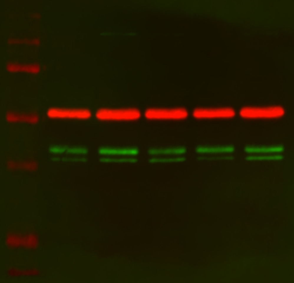
Scott (Verified Customer) (10-22-2024) | 10µg of protein was loaded and antibody was incubated overnight at 4oC following a total protein stain. The band appeared at the expected size but with two bands, blue bars, with Alpha-tubulin internal control (red band - 66031-1-Ig). Precision plus protein standard ladder #1610373.
 |
Makenna (Verified Customer) (07-16-2024) | Didn't work well for western blot, had high background
|
Paloma (Verified Customer) (03-26-2024) | We love this item since we start to use it.
|
MANOHAR (Verified Customer) (03-06-2024) |
|
David (Verified Customer) (01-02-2024) | Top notch antibody, single band at predicted molecular weight. Gold standard.
|
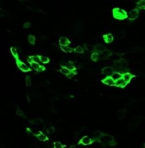
Parijat (Verified Customer) (09-09-2023) | Good antibody for IF
 |
Florencia (Verified Customer) (07-17-2023) | This antibody is one of the most important ones we use in the lab for studying ALS! It works very well and we will continue to use it.
|
Pauline (Verified Customer) (02-23-2022) | everything ok
|
Xin (Verified Customer) (01-23-2022) | Very good in WB with a band of around 45 kD.
|
Jessica (Verified Customer) (06-10-2021) | good for WB and IF
|
Ana (Verified Customer) (03-14-2021) | TDP43 in human primary fibroblasts. 10 ug of total protein. Primary ab incubation 16h 4ºC 1:1000 in BSA 3% in PBST. Secondary ab Goat anti-rabbit HRP 1:5000 1h RT incubation.
 |
Yasuyo (Verified Customer) (01-20-2021) | Works good for ICC in fibroblasts. High nuclear signal. Cells fixed with 4% PFA 15 min. TDP43 ab dilution 1:200 in PBST O/N 4ºC. Donkey anti-rabbit Alexafluor 568 1:500 for 1h RT.
|
MEIMEI (Verified Customer) (11-12-2020) | the antibody work prefect for my staining.
|
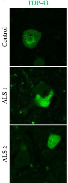
Stephane (Verified Customer) (10-31-2020) | Good product for staining.
 |
Marina (Verified Customer) (10-25-2020) | TDP43 in human primary fibroblasts. Loading: 10 ug in LB with DTT. TGX 4-15% gel. Blocking: 1h 3% BSA in PBST (0.1% tween). Primary ab incubation 16h 4ºC 1:1000 in BSA 3% in PBST (0.1% Tween) Detection: Goat anti-rabbit HRP + ECL+.
 |
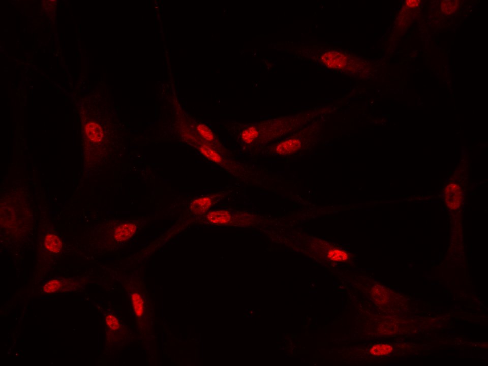
Uxoa (Verified Customer) (02-18-2020) | Good ICC in human primary fibroblats. Small amount of signal in cytoplasm in UT and control cells. High nuclear signal. Fibroblats fixed with 4% PFA 15 min. TDP43 ab dilution 1:100 in PBST (0.1% triton) + 10% DS O/N 4ºC. Donkey anti-rabbit alexa fluor 555 1:500 in PBST+10%DS 1h RT.
 |
Paul (Verified Customer) (01-15-2020) | Produces good IF images with minimal background.
|
Laura (Verified Customer) (01-10-2020) | Works very well
|
Benjamin (Verified Customer) (01-07-2020) | Works well in both western blotting and Immunofluorescence. Nice clear images obtained with IF.
|
Apoorva (Verified Customer) (12-18-2019) | It has great specificity over wide range of species.
|
Tilly (Verified Customer) (11-27-2019) | Great N-terminal TDP-43 antibody, works really well for immunohistochemistry, immunofluorescence and western blot from mouse brain tissue. Unfortunately, I have not been able to get it to work for precipitation in mouse brain.
|
Azita (Verified Customer) (10-04-2019) | I used it for ICC and it had low signal and high background with following (1/500, 1/300, 1/100,1/50) dilutions. Cell were fixed by 4% PFA.
|
Francisco (Verified Customer) (09-28-2019) | Antibody worked well. My lab has used the antibody in various experiments for quantifications, many of which were included in published works.
|
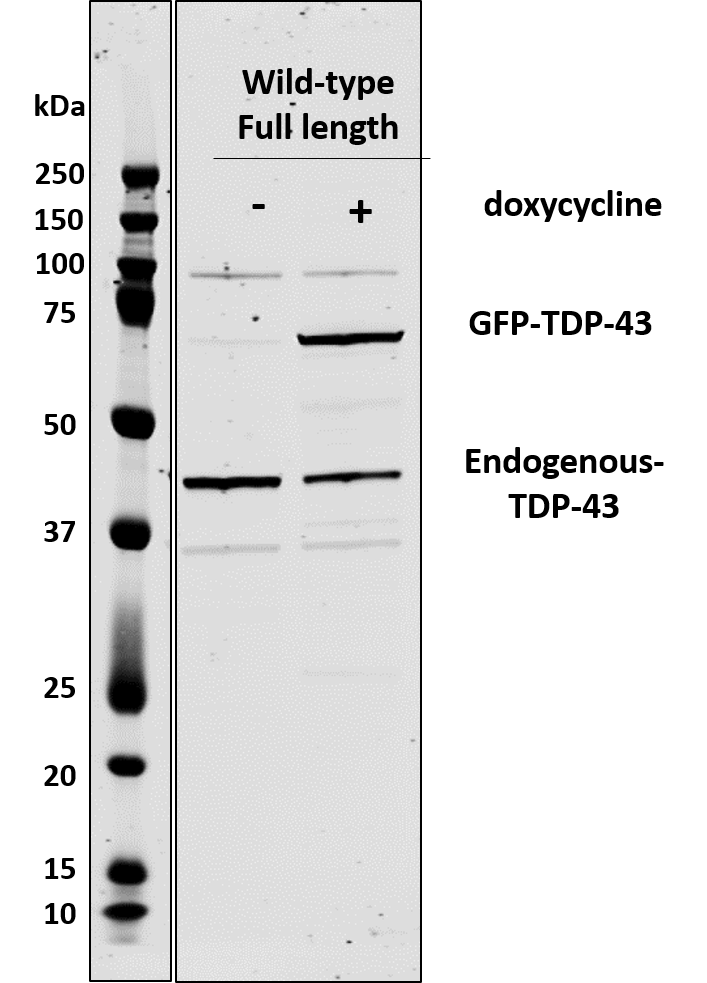
Alinda (Verified Customer) (09-11-2019) | Doxycycline-inducible SHSY5Y show GFP-TDP-43 expression upon dox addition.
 |
David (Verified Customer) (07-18-2019) | Excellent for both immunoblot and immunocytochemistry. Strong signal and low background in both cases. Single band in control cells for immunoblot.
|
George (Verified Customer) (07-07-2019) | Good antibody to show mislocalisation in transgenic ALS disease mouse model tissue culture. Needs a high concentration to show up for microscopy but very strong and clear signal.
|
Elena (Verified Customer) (08-17-2018) |
|
Petra (Verified Customer) (03-06-2018) |
|




















