

Tested Applications
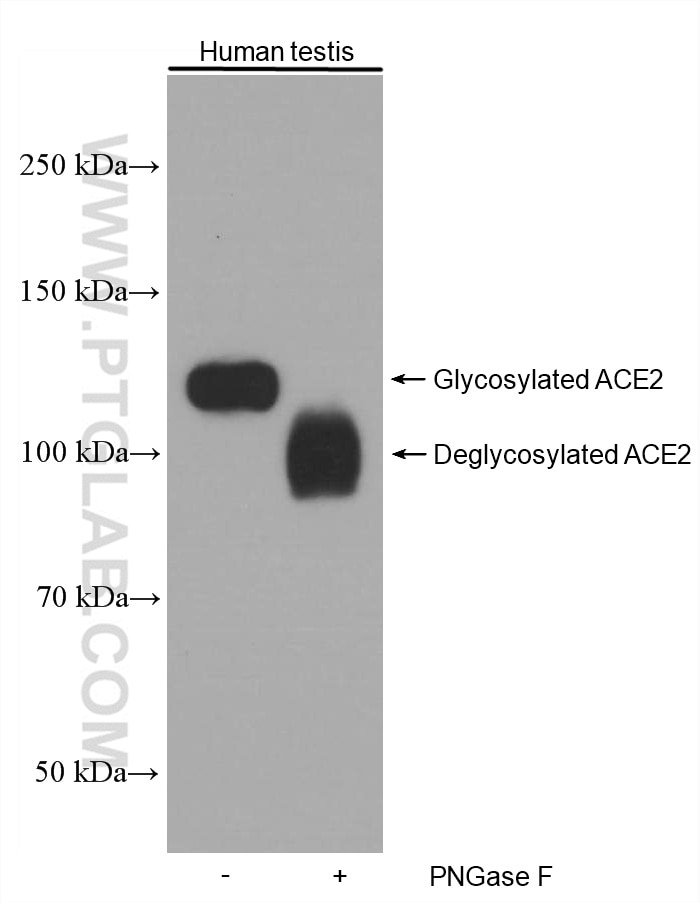
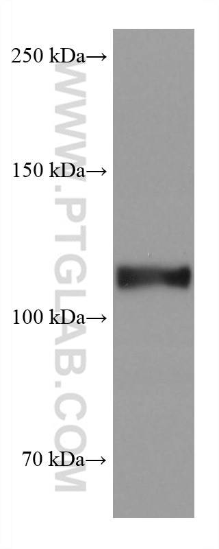
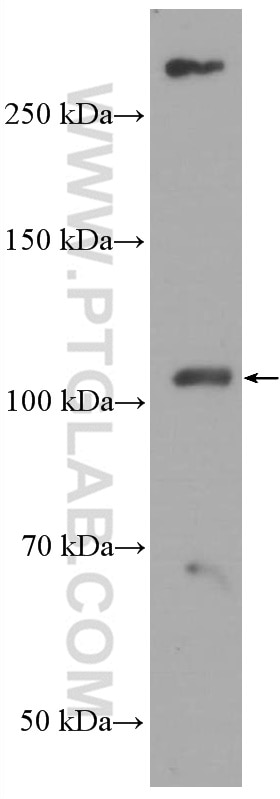
| Positive WB detected in | human testis tissue, human skeletal muscle tissue |
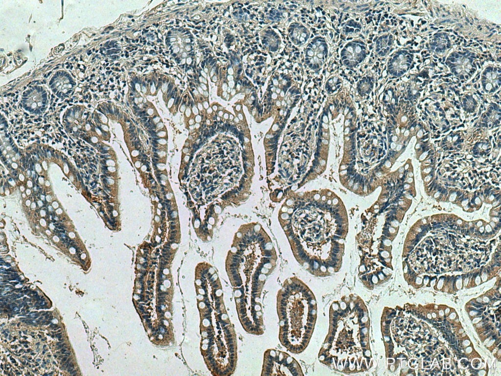
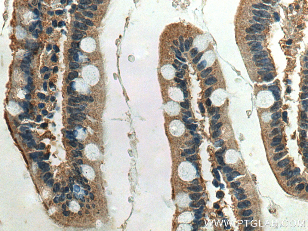
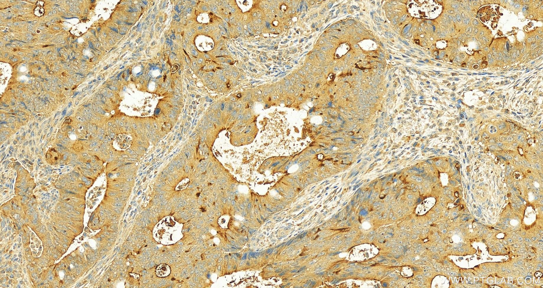
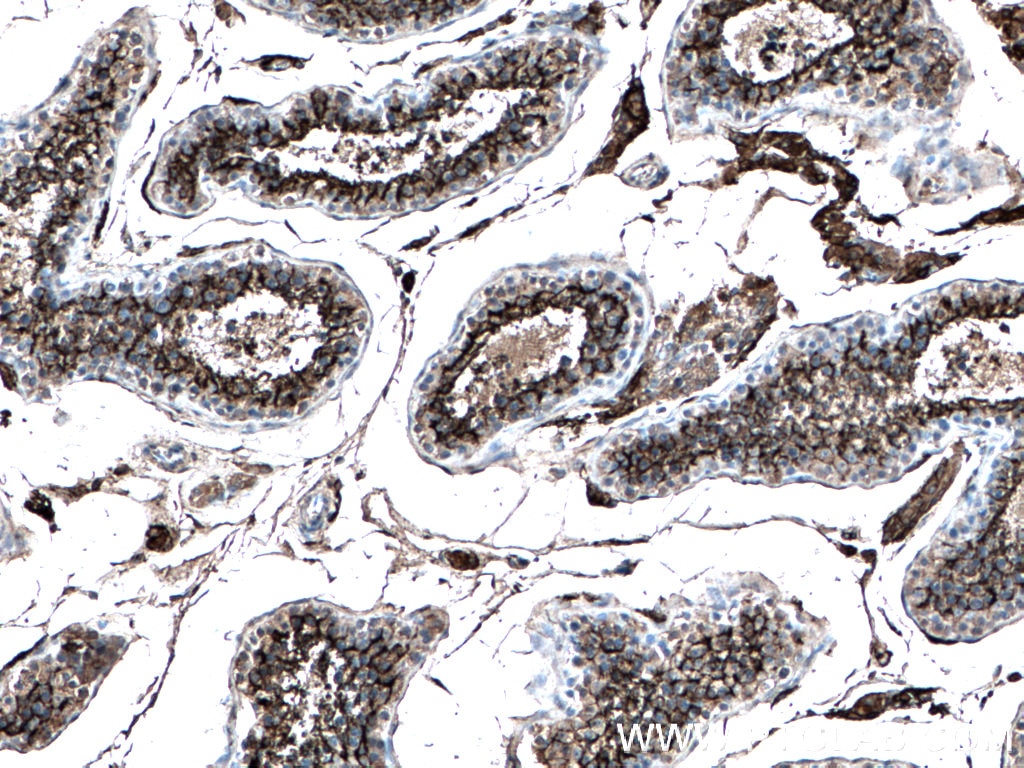
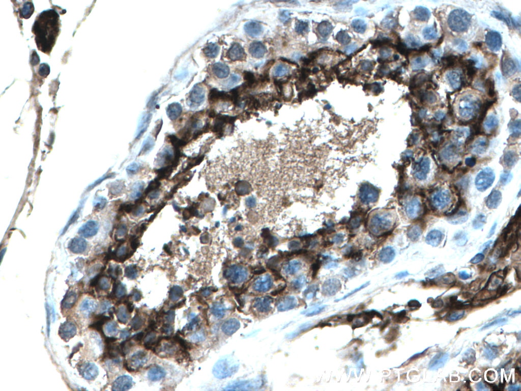
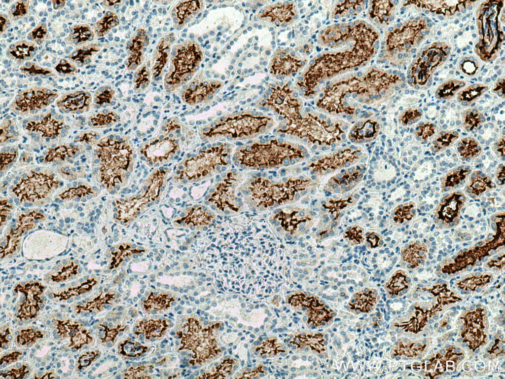
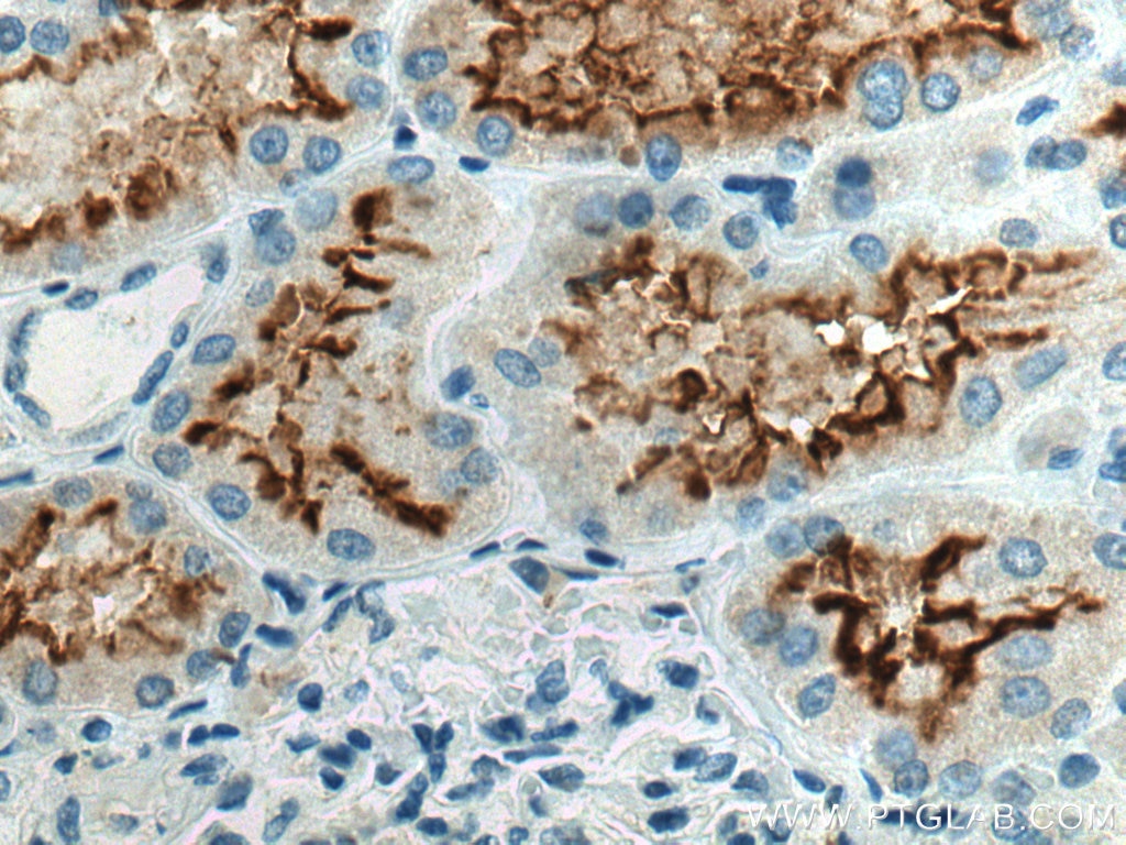
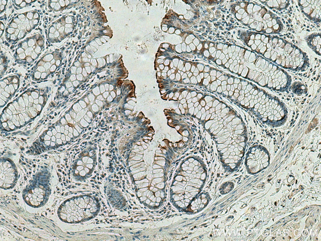
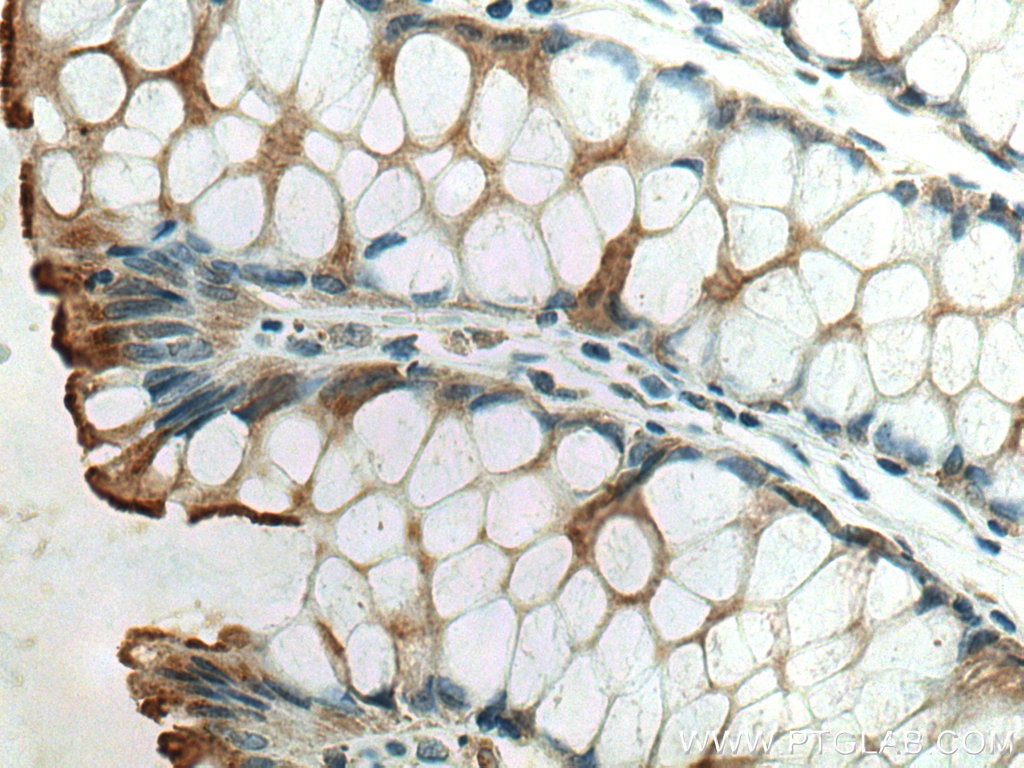
| Positive IHC detected in | human small intestine tissue, human colon tissue, human colon cancer tissue, human kidney tissue, human testis tissue Note: suggested antigen retrieval with TE buffer pH 9.0; (*) Alternatively, antigen retrieval may be performed with citrate buffer pH 6.0 |
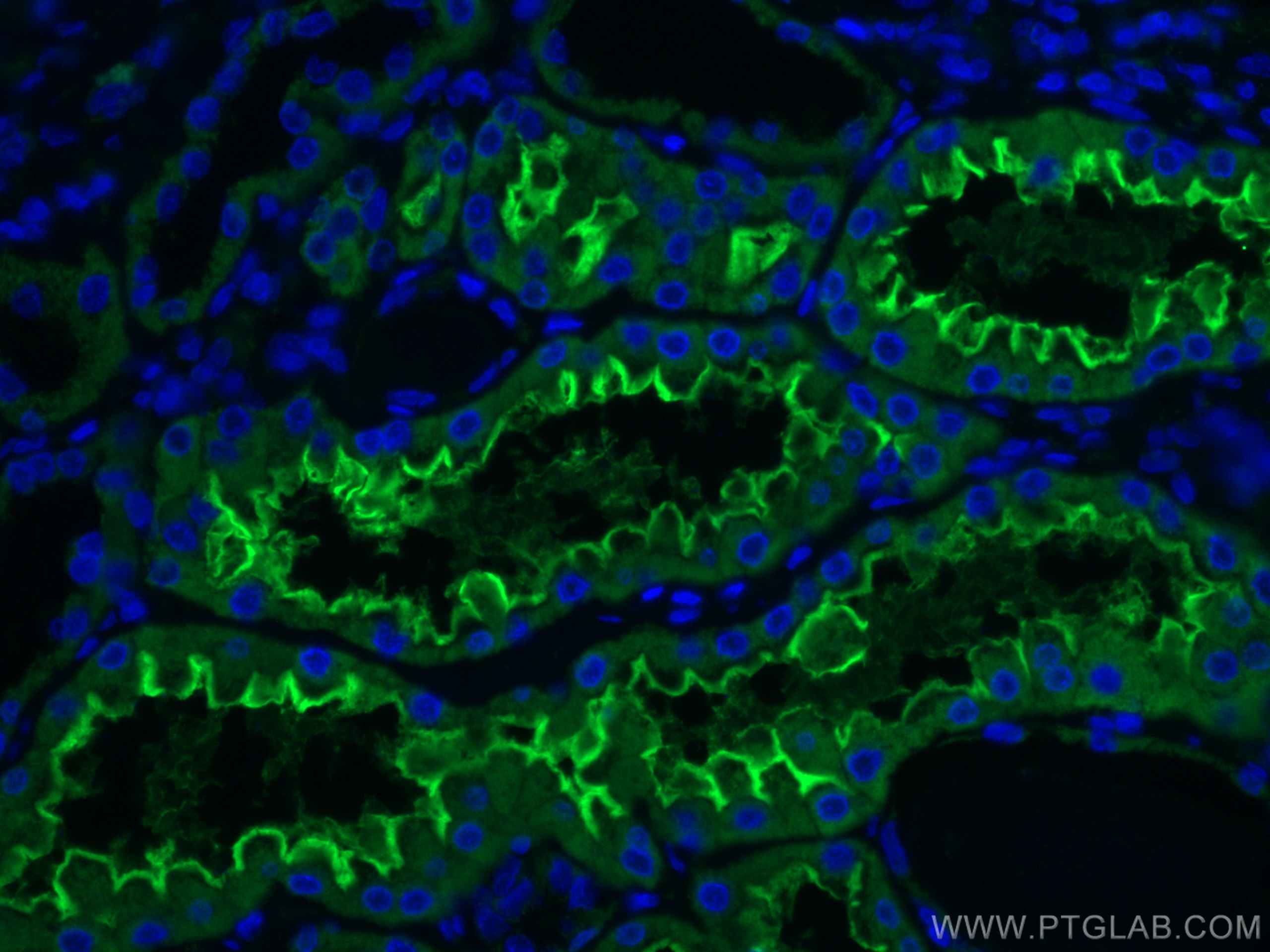
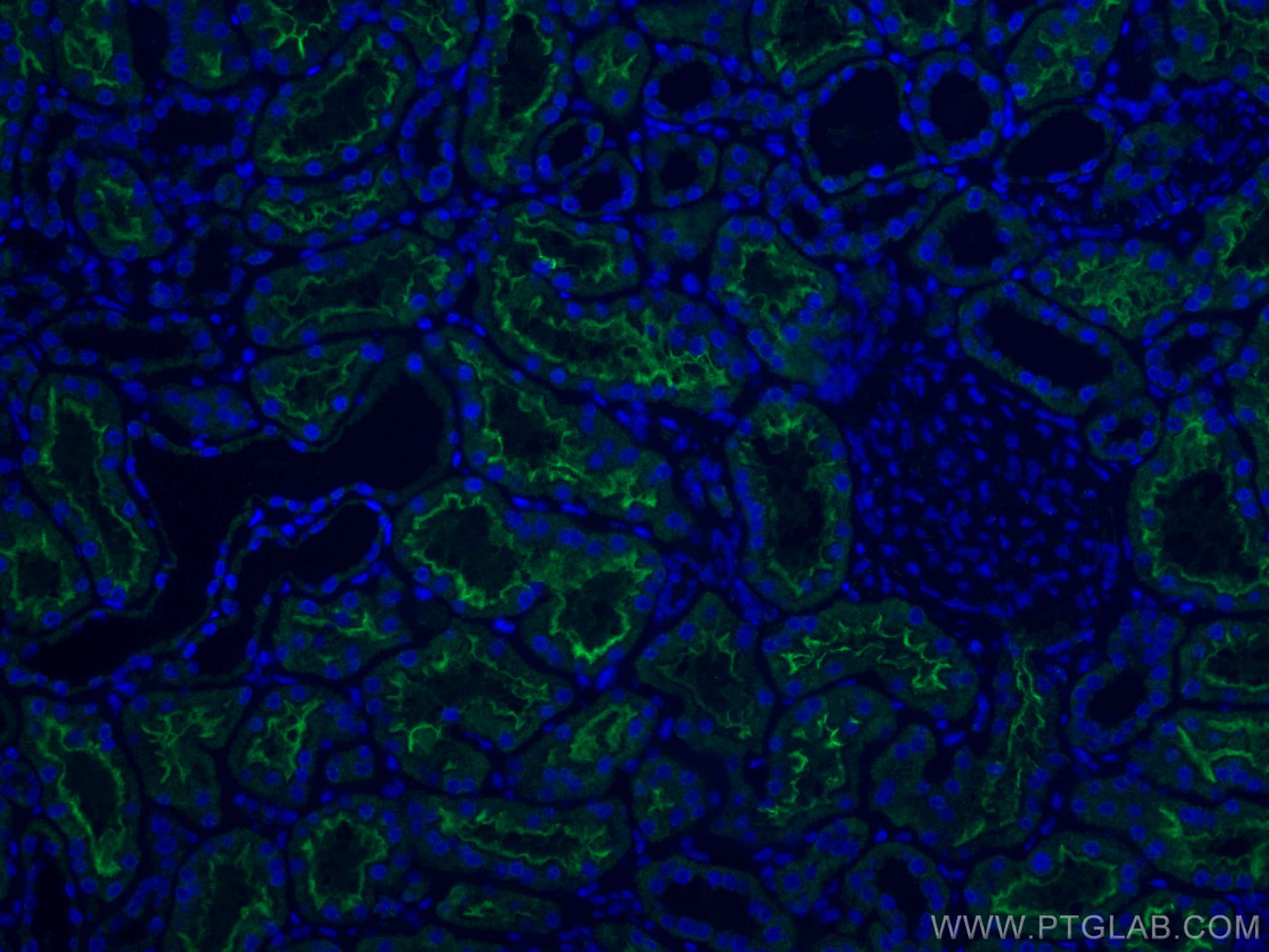
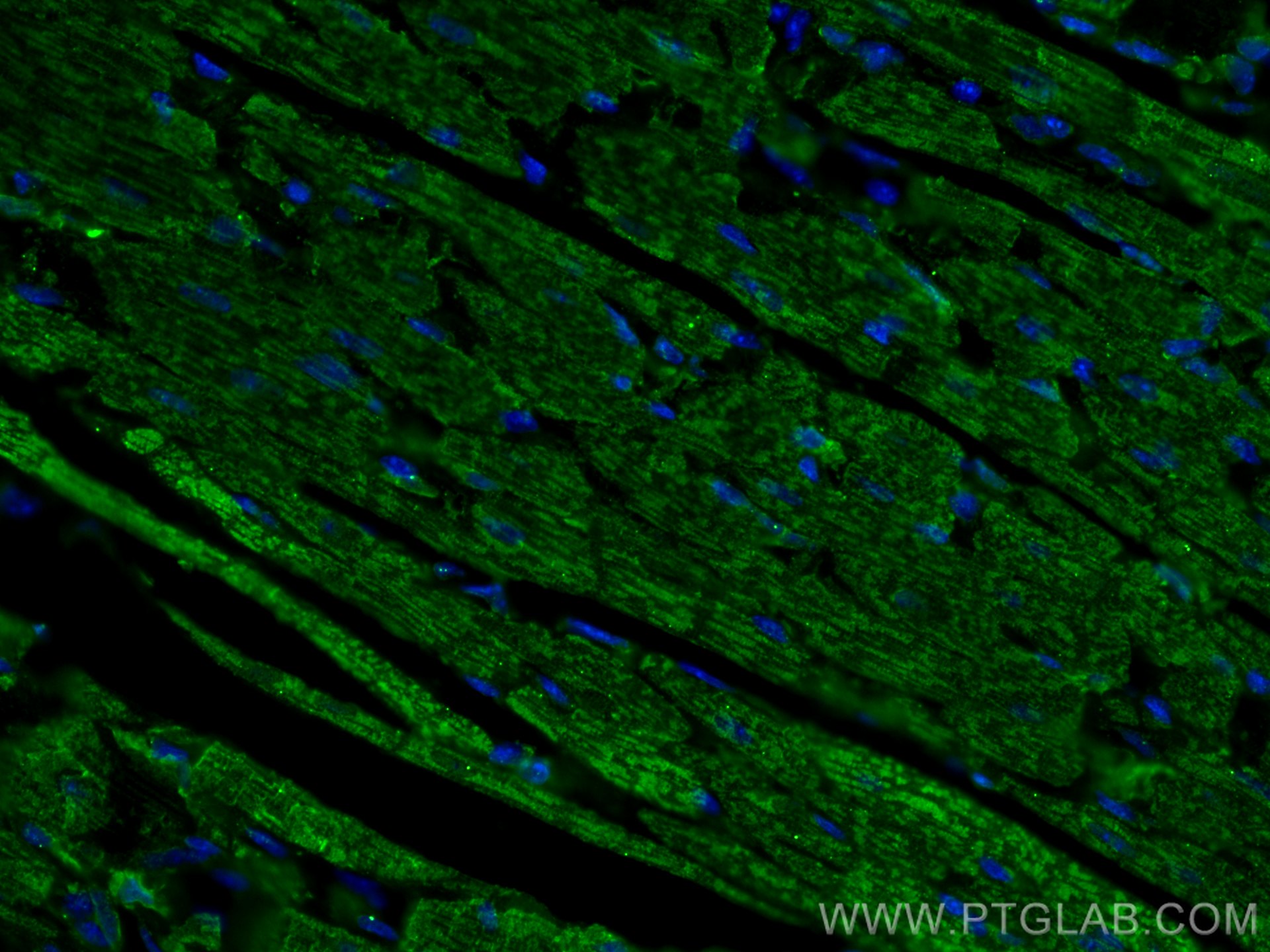
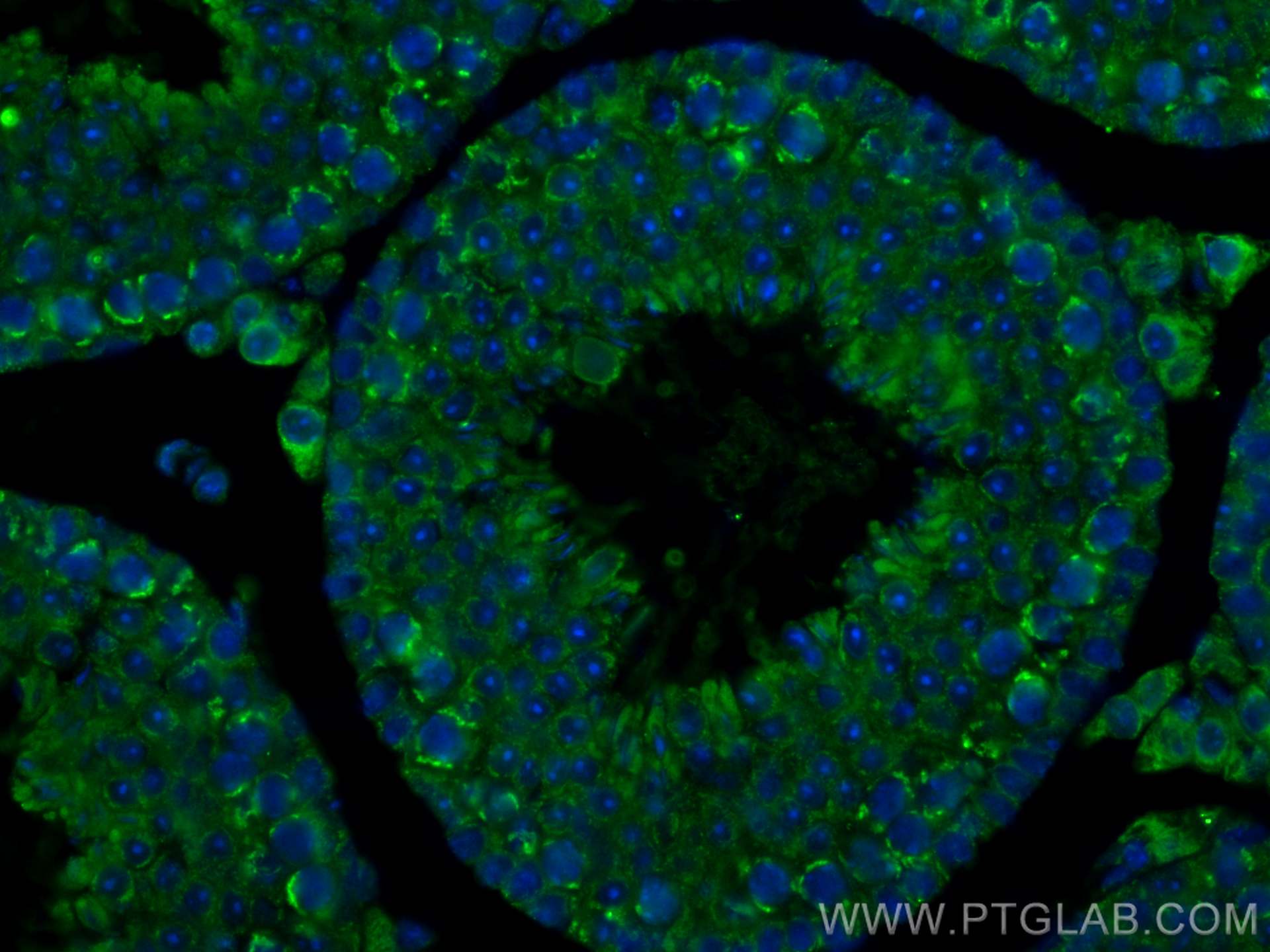
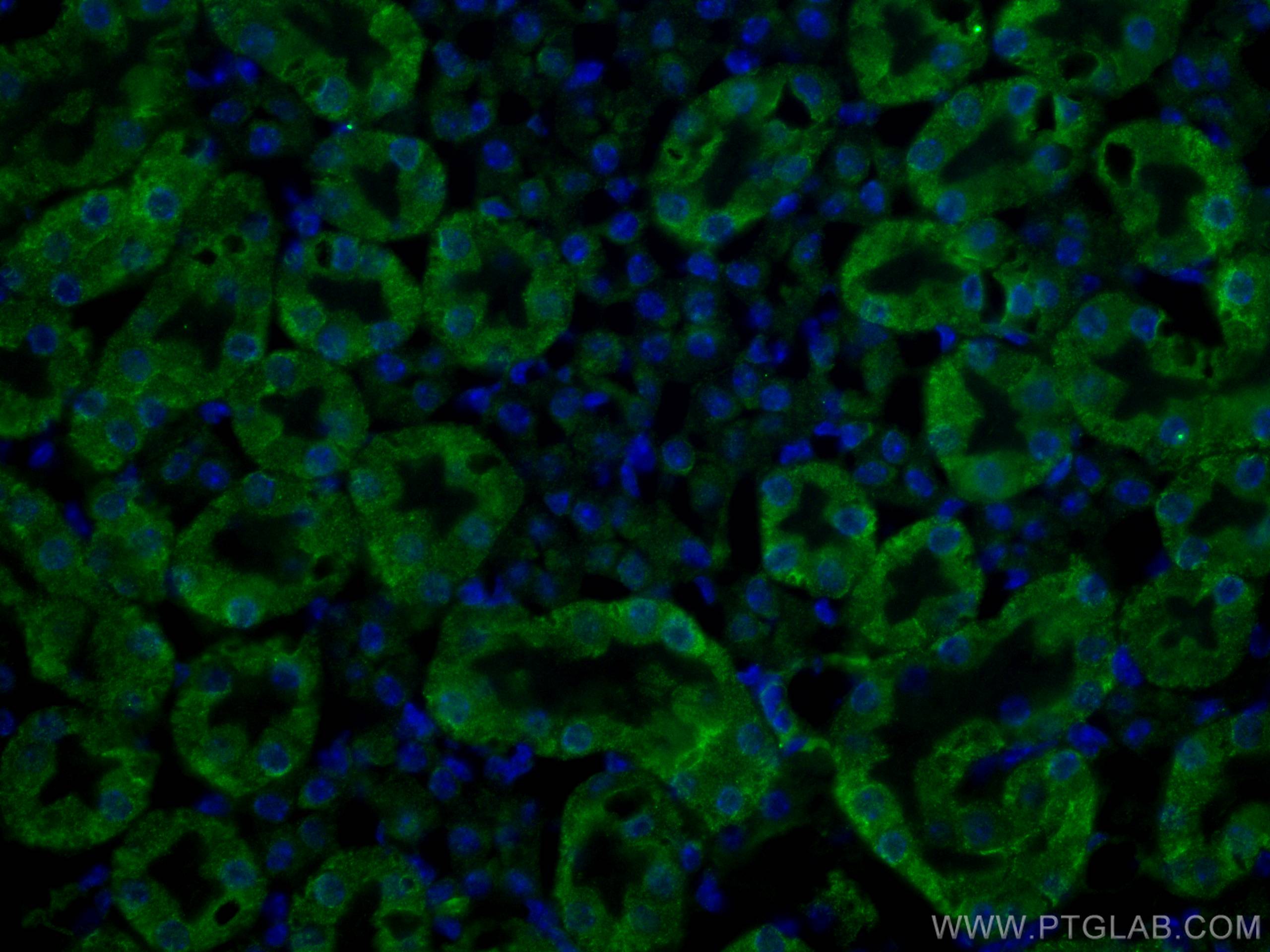
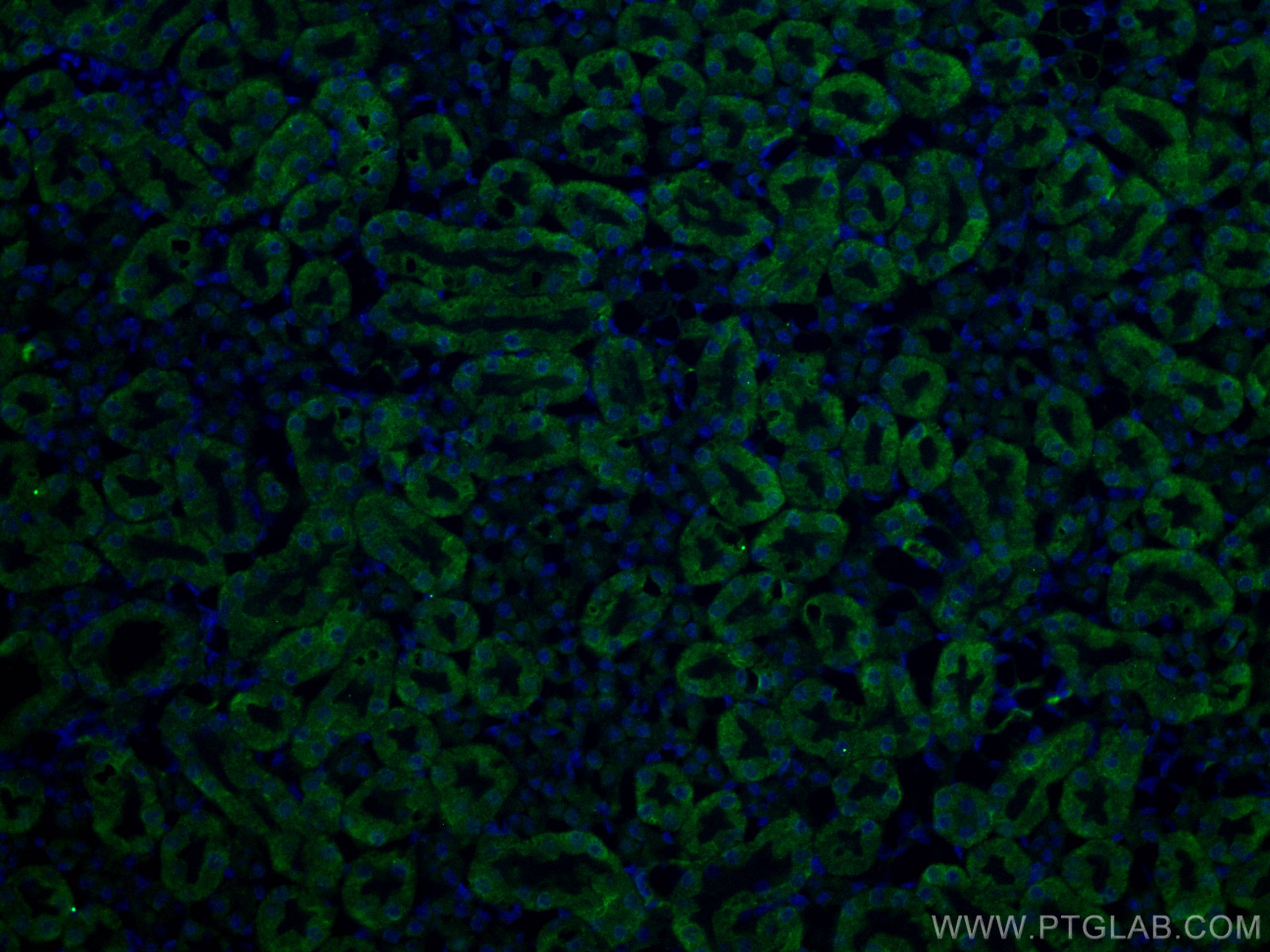
| Positive IF-P detected in | human kidney tissue, mouse testis tissue, mouse heart tissue |
Recommended dilution
| Application | Dilution |
|---|---|
| Western Blot (WB) | WB : 1:2000-1:6000 |
| Immunohistochemistry (IHC) | IHC : 1:1000-1:4000 |
| Immunofluorescence (IF)-P | IF-P : 1:200-1:800 |
| It is recommended that this reagent should be titrated in each testing system to obtain optimal results. | |
| Sample-dependent, Check data in validation data gallery. | |
Product Information
66699-1-Ig targets ACE2 in WB, IHC, IF-P, IP, RIP, ELISA applications and shows reactivity with human, mouse samples.
| Tested Reactivity | human, mouse |
| Cited Reactivity | human, mouse, monkey, goat |
| Host / Isotype | Mouse / IgG1 |
| Class | Monoclonal |
| Type | Antibody |
| Immunogen |
CatNo: Ag15554 Product name: Recombinant human ACE2 protein Source: e coli.-derived, PET28a Tag: 6*His Domain: 392-744 aa of BC048094 Sequence: LRNGANEGFHEAVGEIMSLSAATPKHLKSIGLLSPDFQEDNETEINFLLKQALTIVGTLPFTYMLEKWRWMVFKGEIPKDQWMKKWWEMKREIVGVVEPVPHDETYCDPASLFHVSNDYSFIRYYTRTLYQFQFQEALCQAAKHEGPLHKCDISNSTEAGQKLFNMLRLGKSEPWTLALENVVGAKNMNVRPLLNYFEPLFTWLKDQNKNSFVGWSTDWSPYADQSIKVRISLKSALGDKAYEWNDNEMYLFRSSVAYAMRQYFLKVKNQMILFGEEDVRVANLKPRISFNFFVTAPKNVSDIIPRTEVEKAIRMSRSRINDAFRLNDNSLEFLGIQPTLGPPNQPPVSIWLI Predict reactive species |
| Full Name | angiotensin I converting enzyme (peptidyl-dipeptidase A) 2 |
| Calculated Molecular Weight | 805 aa, 92 kDa |
| Observed Molecular Weight | 120 kDa, 92 kDa |
| GenBank Accession Number | BC048094 |
| Gene Symbol | ACE2 |
| Gene ID (NCBI) | 59272 |
| RRID | AB_2882052 |
| Conjugate | Unconjugated |
| Form | Liquid |
| Purification Method | Protein G purification |
| UNIPROT ID | Q9BYF1 |
| Storage Buffer | PBS with 0.02% sodium azide and 50% glycerol, pH 7.3. |
| Storage Conditions | Store at -20°C. Stable for one year after shipment. Aliquoting is unnecessary for -20oC storage. 20ul sizes contain 0.1% BSA. |
Background Information
ACE2 (Angiotensin-converting enzyme 2), also named as ACEH, is a zinc metalloprotease of the ACE family and a critical regulator of the reninangiotensin system. ACE2 has a more restricted tissue distribution than ACE, being found predominantly in the heart, kidneys, and testes although low levels have been detected in a variety of tissues (PMID:15983030). ACE2 has been shown to be a functional receptor of the human coronaviruses SARS-CoV and SARS-CoV-2 (PMID: 32142651). The expression level and expression pattern of human ACE2 in different tissues might be critical for the susceptibility, symptoms, and outcome of 2019-nCoV/SARS-CoV-2 infection (PMID: 32133153). It can be used as a potential therapeutic target of SARS-CoV-2 (PMID: 32125455). The calculated molecular weight of ACE2 is 92kDa but it migrates to 120kDa due to N-glycosylation (PMID:16166094). Sometimes, several cleaved fragments can also be detected as 75kDa, 50 kDa or 37kDa (PMID: 29561187, 22009550, 30759273). It has 2 isoforms produced by alternative splicing. This antibody is specific to ACE2. The location of ACE2 is membrane and cytoplasm, however it accumulates in the nucleus during the mitosis (PMID: 1730413/PMID: 18292088).
Protocols
| Product Specific Protocols | |
|---|---|
| IF protocol for ACE2 antibody 66699-1-Ig | Download protocol |
| IHC protocol for ACE2 antibody 66699-1-Ig | Download protocol |
| WB protocol for ACE2 antibody 66699-1-Ig | Download protocol |
| Standard Protocols | |
|---|---|
| Click here to view our Standard Protocols |
Publications
What published studies show
The anti-ACE2 antibody (Cat# 66699-1-Ig) has 48 total citations published in journals including Nature Medicine, Science, Nature Aging, Cell Stem Cell, Nature Communications, EBioMedicine, EMBO Reports, PLoS Pathogens, mBio, and Molecular Therapy. Research fields encompass SARS-CoV-2/COVID-19 viral entry and receptor biology, neurodegeneration (Alzheimer's, Parkinson's), cardiovascular dysfunction, inflammation, ferroptosis, glycosylation, and endocytosis. Primary applications are Western blot, immunofluorescence, and immunohistochemistry across human, mouse, monkey, and goat species.
| Species | Application | Title |
|---|---|---|
Nat Aging Brain-wide alterations revealed by spatial transcriptomics and proteomics in COVID-19 infection | ||
Cell Stem Cell Androgen Signaling Regulates SARS-CoV-2 Receptor Levels and Is Associated with Severe COVID-19 Symptoms in Men. | ||
J Adv Res Key β1-4 galactosylated glycan receptors of SARS-CoV-2 and its inhibitor from the galactosylated glycoproteins of bovine milk |
Reviews
What customers say
Users rate this ACE2 mouse monoclonal antibody highly across applications. One reviewer achieved clear, specific Western Blot results on D407 cells using chemiluminescence. Another used it successfully for IHC and IF on human lung (FFPE) and mouse lung (frozen) sections at 1:1000 dilution, noting that 1:500 works best for FFPE IF. A third reviewer confirmed strong IF performance with detailed optimization notes including Tris-EDTA antigen retrieval at 95°C for 30 minutes and 5% normal serum blocking. Overall, users report excellent specificity and reliable staining across multiple tissue types and formats.
The reviews below have been submitted by verified Proteintech customers who received an incentive for providing their feedback.
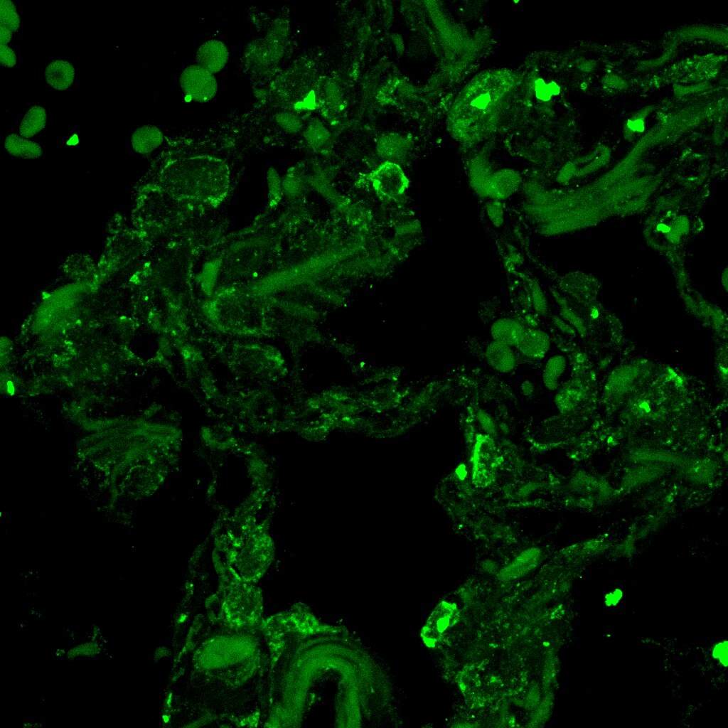
Alexandra (Verified Customer) (08-26-2026) | The Ace 2 Mouse McAb worked perfectly for western blot. It was very specific and the staining was easily visible with chemiluminescence.
|
Lindsey (Verified Customer) (05-24-2021) | Great antibody---looking forward to trying the RabPolyAb as well
 |
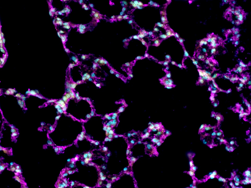
Lindsey (Verified Customer) (09-21-2020) | Perfomed IF analysis on human FFPE and mouse frozen tissue sections; for FFPE tissue, antigen retrieval was performed for 30 min at 95C using Tris EDTA Buffer (pH9.0); Permeabilization was not performed; Blocking was performed using 5% Normal Serum and 0.3% TritonX-100 for 30. minutes at room temperature. Mouse lung sections were costained with ARL13B (Proteintech #1711-1-AP) to visualize primary cilia of the lung epithelia; all sections were counterstained with DAPI Fluormount (Abcam); image attached is of mouse lung frozen tissue; due to size restrictions I am limited to this image only but am happy to provide additional images (human tissue etc.) upon request
 |



















