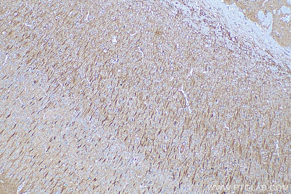
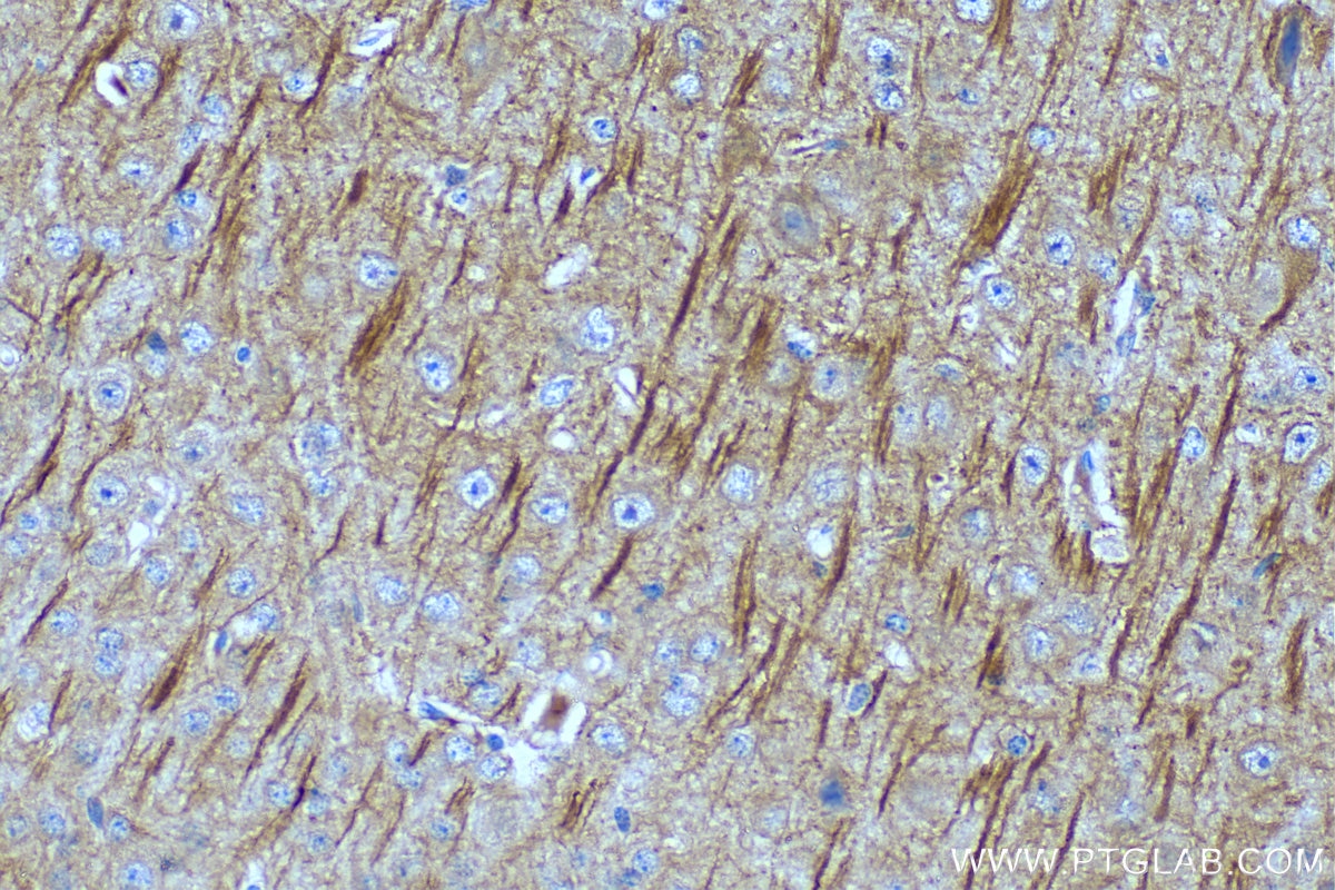
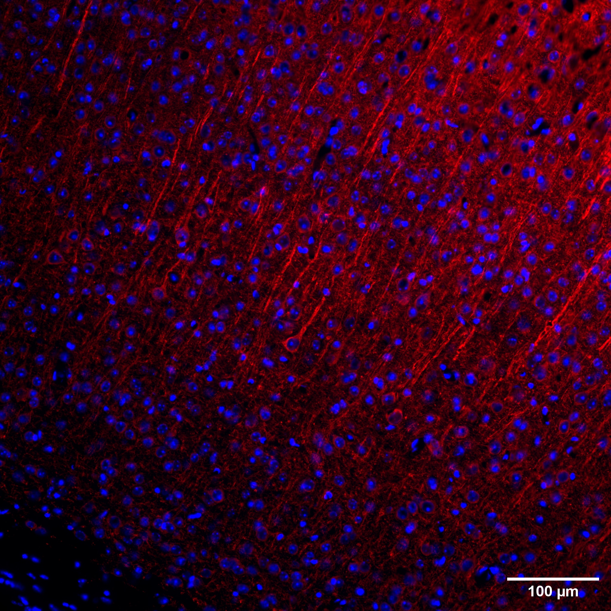
staining of mouse brain tissue using MAP2 Monoclonal antibody (67015-1-Ig)")
 staining of mouse brain tissue using MAP2 Monoclonal antibody (67015-1-Ig)")
 staining of mouse brain tissue using MAP2 Monoclonal antibody (67015-1-Ig)")
 staining of mouse brain tissue using MAP2 Monoclonal antibody (67015-1-Ig)")
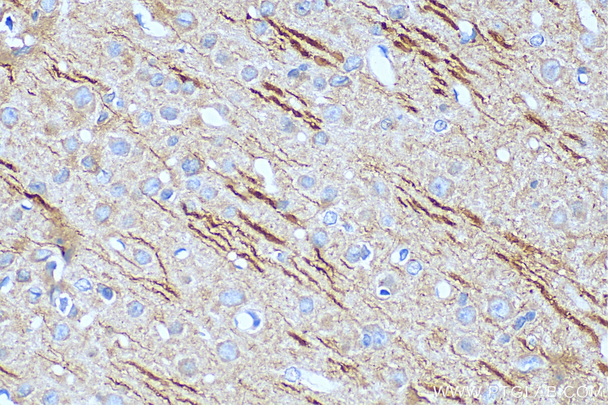
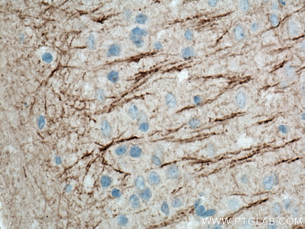
 staining of rat brain tissue using MAP2 Monoclonal antibody (67015-1-Ig)")
 staining of rat brain tissue using MAP2 Monoclonal antibody (67015-1-Ig)")
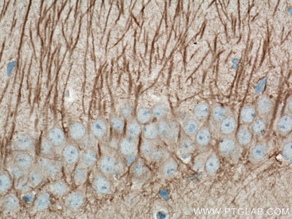
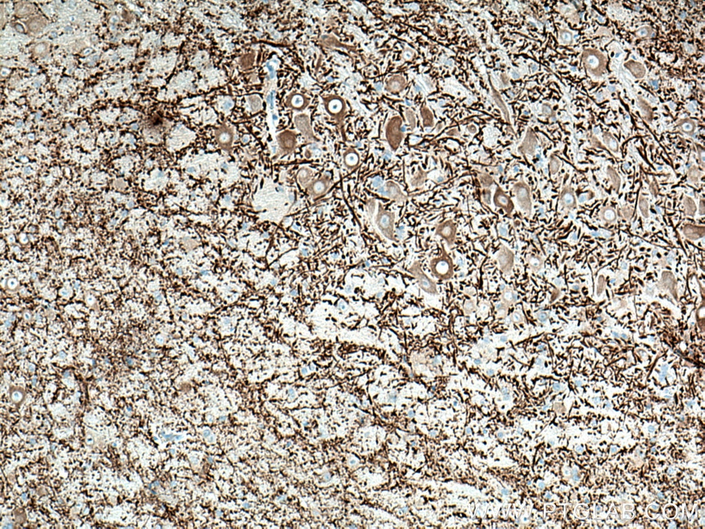
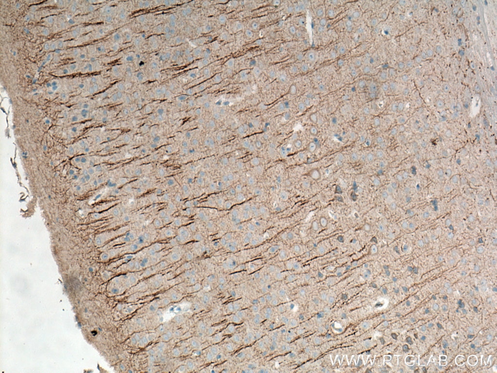
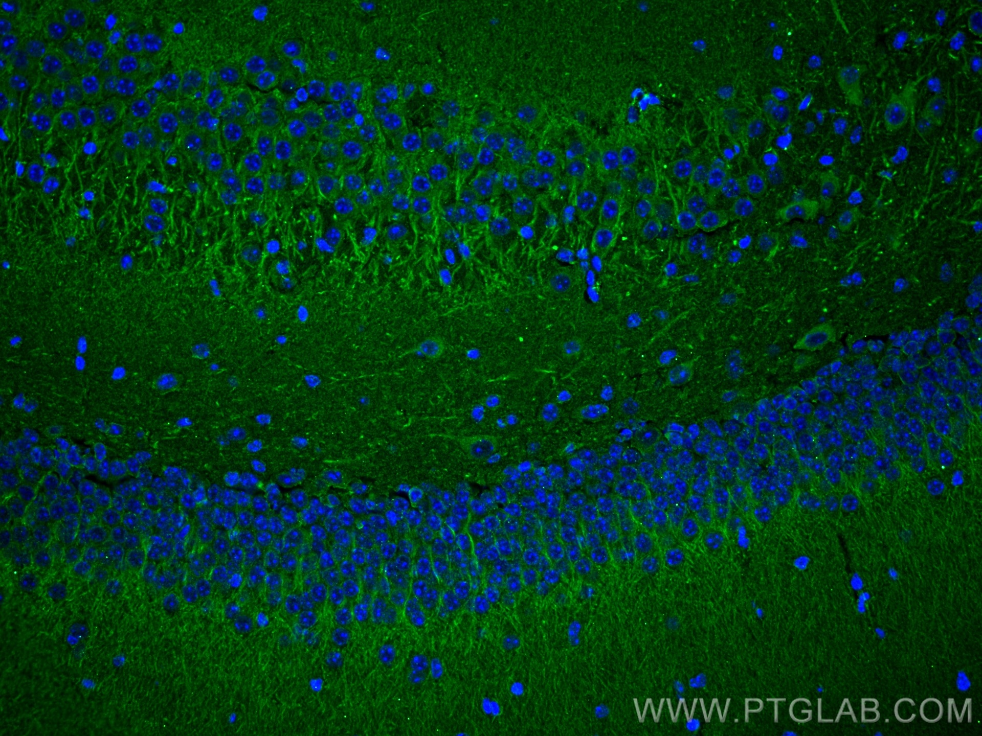
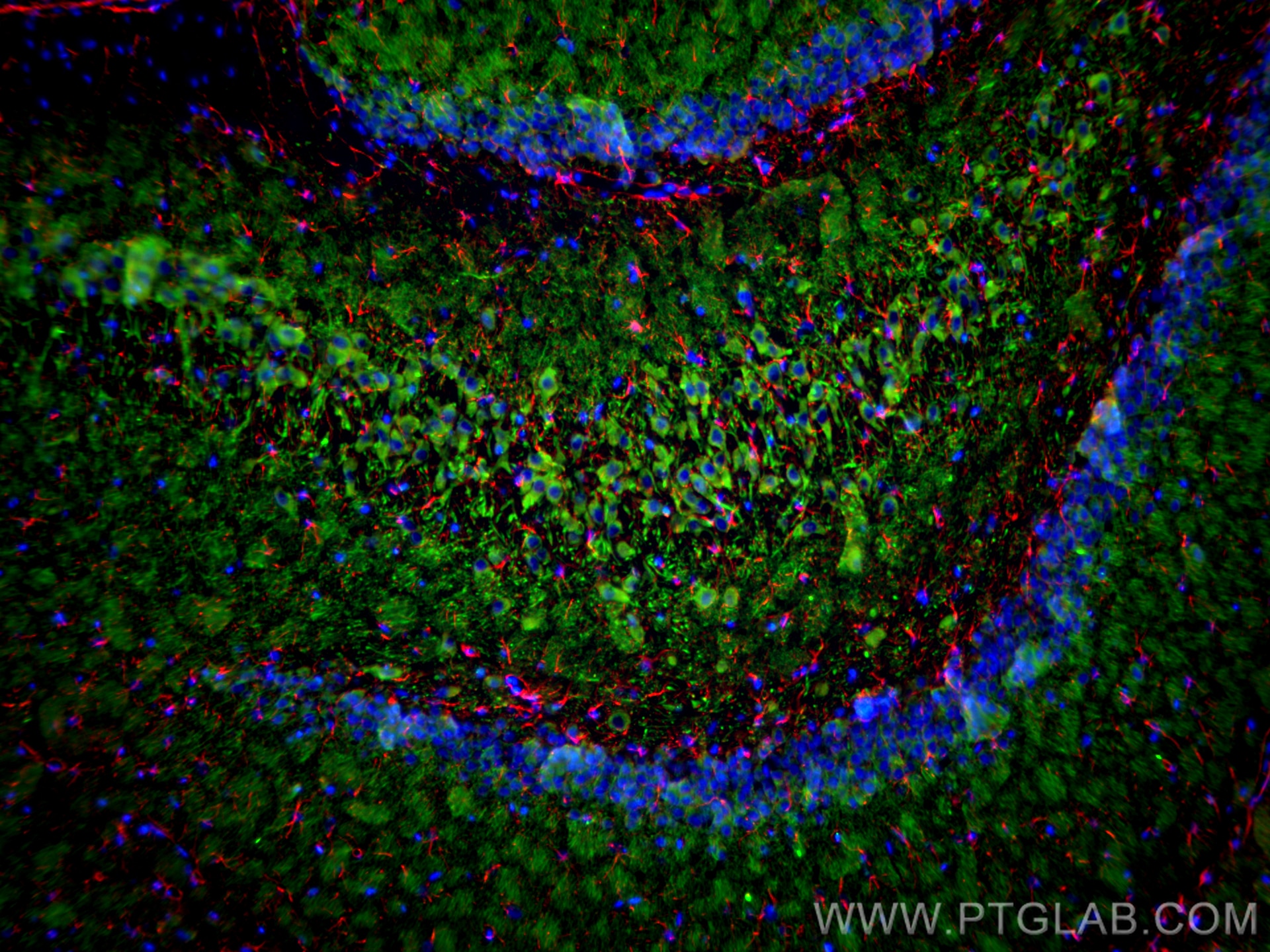
 staining of mouse cerebellum tissue using MAP2 Monoclonal antibody (67015-1-Ig)")
 staining of mouse cerebellum tissue using MAP2 Monoclonal antibody (67015-1-Ig)")
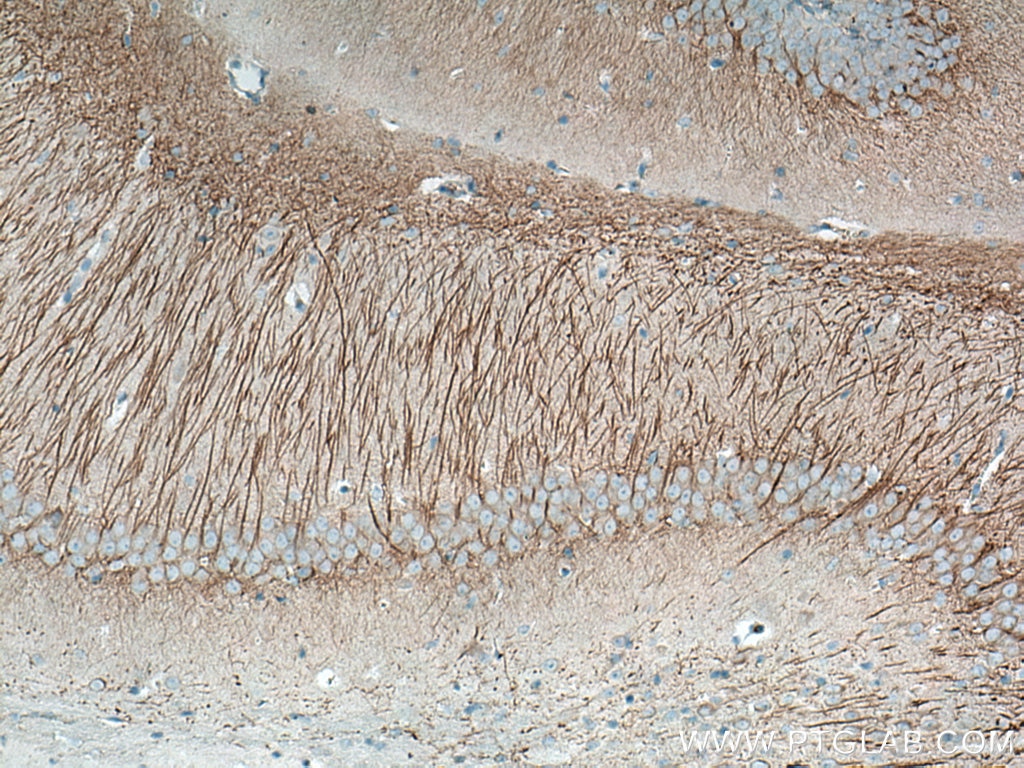
 staining of rat cerebellum tissue using MAP2 Monoclonal antibody (67015-1-Ig)")
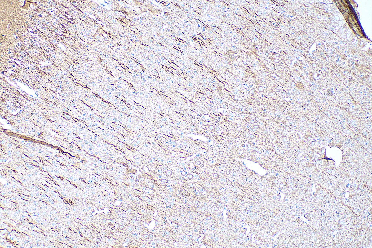
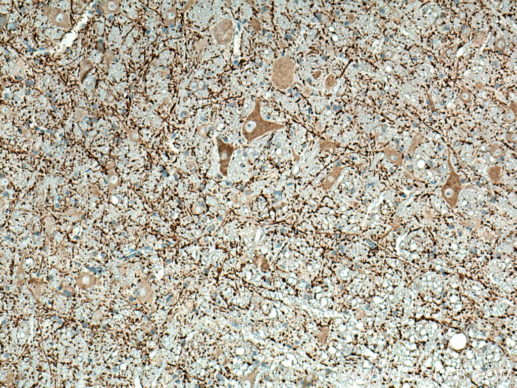
 staining of mouse brain tissue using MAP2 Monoclonal antibody (67015-1-Ig)")
 staining of mouse brain tissue using MAP2 Monoclonal antibody (67015-1-Ig)")
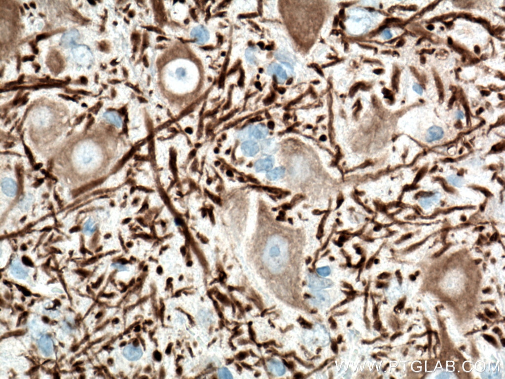
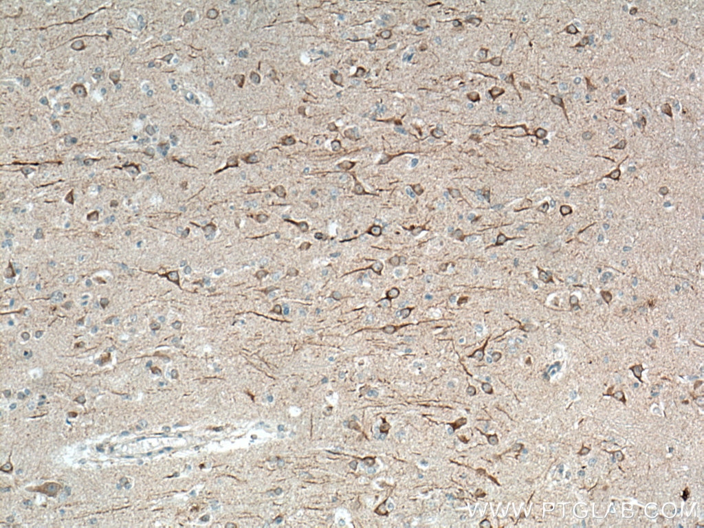
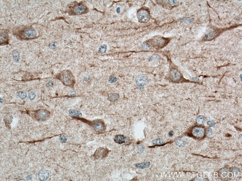
 staining of human brain tissue using MAP2 Monoclonal antibody (67015-1-Ig)")
 staining of human brain tissue using MAP2 Monoclonal antibody (67015-1-Ig)")
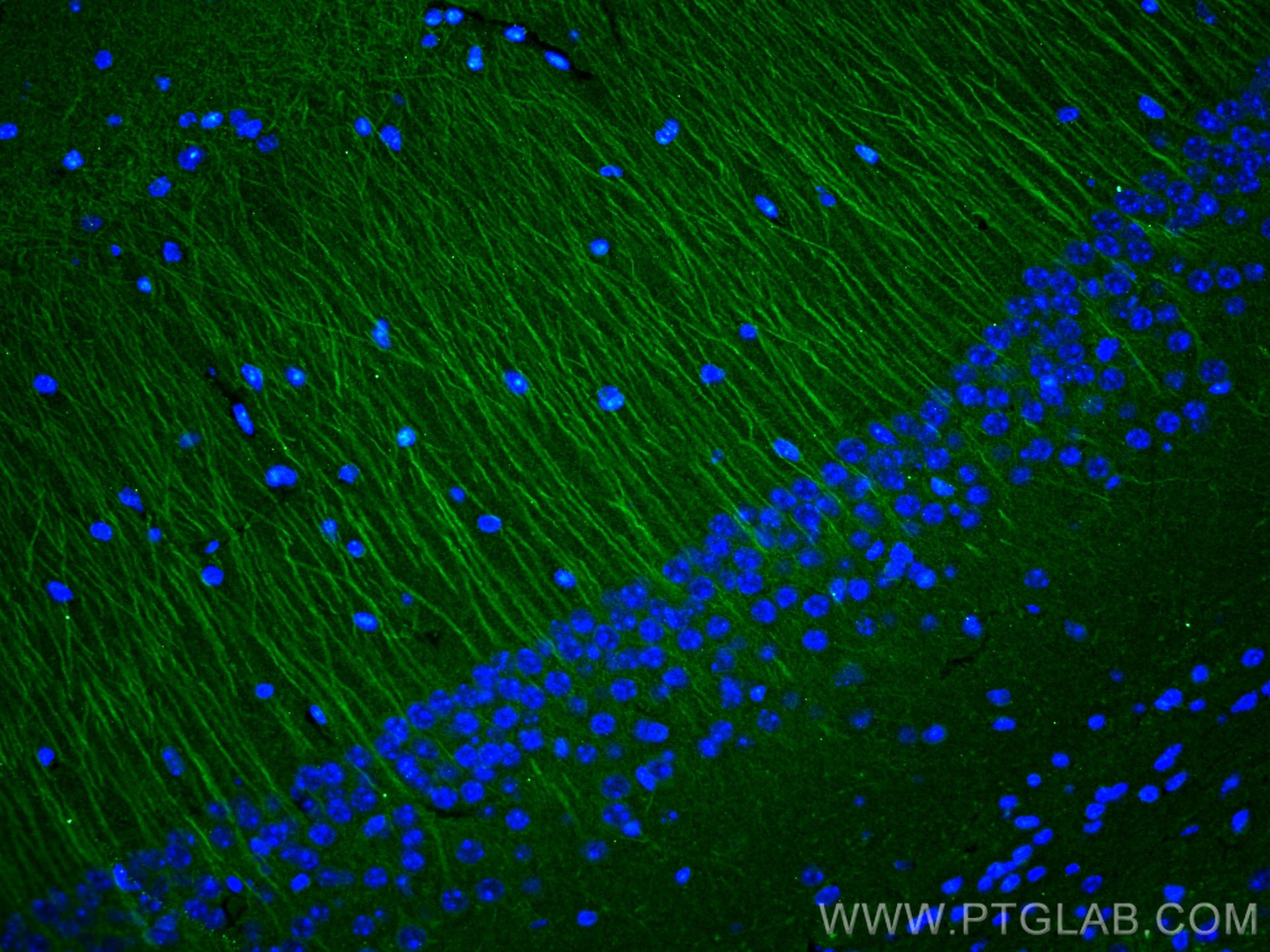
 / fluorescent staining of rat brain tissue using MAP2 Monoclonal antibody (67015-1-Ig)")
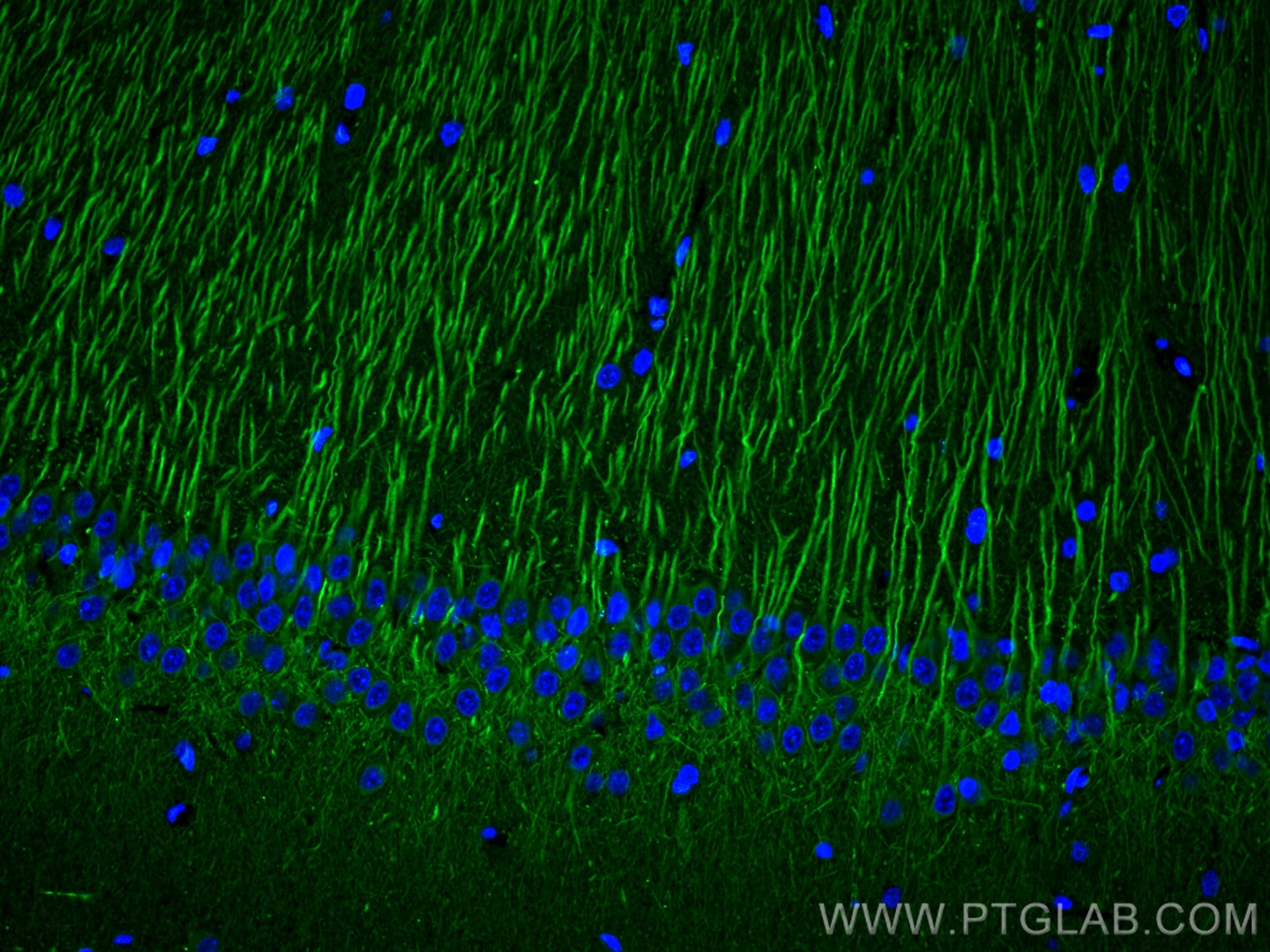
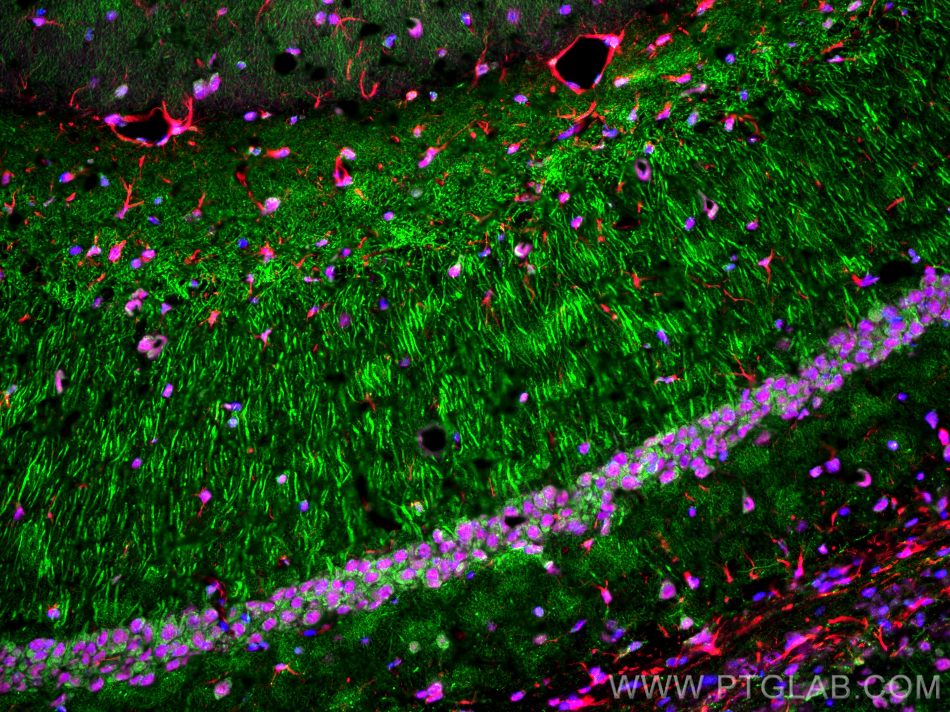
 / fluorescent staining of mouse brain tissue using MAP2 Monoclonal antibody (67015-1-Ig)")
 / fluorescent staining of mouse brain tissue using MAP2 Monoclonal antibody (67015-1-Ig)")
 / fluorescent staining of rat brain tissue using MAP2 Monoclonal antibody (67015-1-Ig)")
 / fluorescent staining of mouse brain tissue using MAP2 Monoclonal antibody (67015-1-Ig)")
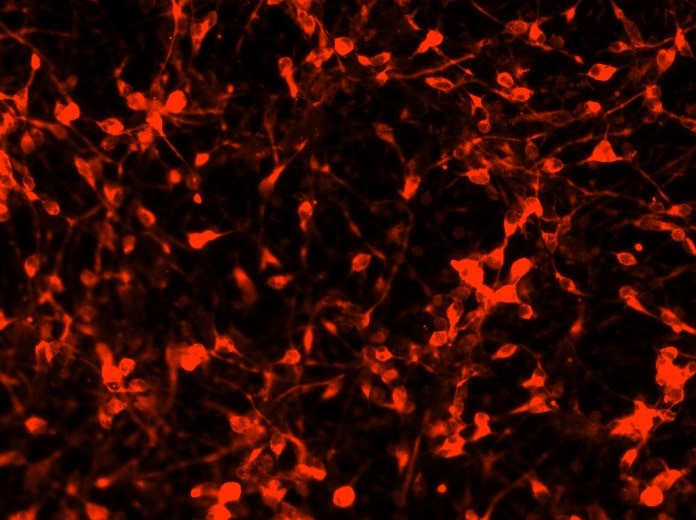
 experiment of Neuro-2a cells using MAP2 Monoclonal antibody (67015-1-Ig)")
Tested Applications
| Positive IHC detected in | mouse brain tissue, rat cerebellum tissue, rat brain tissue, human brain tissue, mouse cerebellum tissue Note: suggested antigen retrieval with TE buffer pH 9.0; (*) Alternatively, antigen retrieval may be performed with citrate buffer pH 6.0 |
| Positive IF-P detected in | rat brain tissue, mouse brain tissue |
| Positive IF-Fro detected in | rat brain tissue, mouse brain tissue |
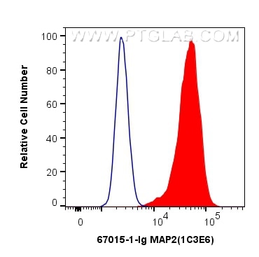
| Positive FC (Intra) detected in | Neuro-2a cells |
Recommended dilution
| Application | Dilution |
|---|---|
| Immunohistochemistry (IHC) | IHC : 1:1000-1:4000 |
| Immunofluorescence (IF)-P | IF-P : 1:200-1:800 |
| Immunofluorescence (IF)-FRO | IF-FRO : 1:1000-1:4000 |
| Flow Cytometry (FC) (INTRA) | FC (INTRA) : 0.40 ug per 10^6 cells in a 100 µl suspension |
| It is recommended that this reagent should be titrated in each testing system to obtain optimal results. | |
| Sample-dependent, Check data in validation data gallery. | |
Published Applications
| WB | See 4 publications below |
| IHC | See 1 publications below |
| IF | See 34 publications below |
| IP | See 1 publications below |
Product Information
67015-1-Ig targets MAP2 in WB, IHC, IF-P, IF-Fro, FC (Intra), IP, ELISA applications and shows reactivity with human, mouse, rat samples.
| Tested Reactivity | human, mouse, rat |
| Cited Reactivity | human, mouse, rat, monkey |
| Host / Isotype | Mouse / IgG2b |
| Class | Monoclonal |
| Type | Antibody |
| Immunogen |
CatNo: Ag11349 Product name: Recombinant human MAP2 protein Source: e coli.-derived, PET28a Tag: 6*His Domain: 213-559 aa of BC038857 Sequence: TSAGSTDRLPYSKSGNKDGVTKSPEKRSSLPRPSSILPPRRGVSGDRDENSFSLNSSISSSARRTTRSEPIRRAGKSGTSTPTTPGSTAITPGTPPSYSSRTPGTPGTPSYPRTPHTPGTPKSAILVPSEKKVAIIRTPPKSPATPKQLRLINQPLPDLKNVKSKIGSTDNIKYQPKGGQVRILNKKIDFSKVQSRCGSKDNIKHSAGGGNVQIVTKKIDLSHVTSKCGSLKNIRHRPGGGRVKIESVKLDFKEKAQAKVGSLDNAHHVPGGGNVKIDSQKLNFREHAKARVDHGAEIITQSPGRSSVASPRRLSNVSSSGSINLLESPQLATLAEDVTAALAKQGL Predict reactive species |
| Full Name | microtubule-associated protein 2 |
| Calculated Molecular Weight | 200 kDa |
| GenBank Accession Number | BC038857 |
| Gene Symbol | MAP2 |
| Gene ID (NCBI) | 4133 |
| RRID | AB_2882331 |
| Conjugate | Unconjugated |
| Form | Liquid |
| Purification Method | Protein A purification |
| UNIPROT ID | P11137 |
| Storage Buffer | PBS with 0.02% sodium azide and 50% glycerol, pH 7.3. |
| Storage Conditions | Store at -20°C. Stable for one year after shipment. Aliquoting is unnecessary for -20oC storage. 20ul sizes contain 0.1% BSA. |
Background Information
MAP2 (microtubule-associated protein 2) is a cytoskeleton protein abundant in brain and has important role in neuronal morphogenesis. Multiple high molecular weight (MW) and low molecular weight (MW) MAP2 isoforms are expressed within axons, dendrites, and cell bodies. The expression of MAP2 is regulated in both a tissue- and developmentally specific manner. MAP2 antibodies have been widely used to mark the neuron or dendrite formation.
Protocols
| Product Specific Protocols | |
|---|---|
| FC protocol for MAP2 antibody 67015-1-Ig | Download protocol |
| IF protocol for MAP2 antibody 67015-1-Ig | Download protocol |
| IHC protocol for MAP2 antibody 67015-1-Ig | Download protocol |
| Standard Protocols | |
|---|---|
| Click here to view our Standard Protocols |
Publications
| Species | Application | Title |
|---|---|---|
Cell Stem Cell In vivo reprogramming of NG2 glia enables adult neurogenesis and functional recovery following spinal cord injury. | ||
Nat Chem Biol Tracking endogenous proteins based on RNA editing-mediated genetic code expansion | ||
Small Stemness Maintenance and Massproduction of Neural Stem Cells on Poly L-Lactic Acid Nanofibrous Membrane Based on Piezoelectriceffect. | ||
Cell Death Dis ChemR23 activation attenuates cognitive impairment in chronic cerebral hypoperfusion by inhibiting NLRP3 inflammasome-induced neuronal pyroptosis | ||
Cell Death Dis Pyruvate dehydrogenase kinase 1 protects against neuronal injury and memory loss in mouse models of diabetes |
Reviews
The reviews below have been submitted by verified Proteintech customers who received an incentive for providing their feedback.
FH Marion (Verified Customer) (03-11-2025) | Very good product
 |
FH Marianne (Verified Customer) (11-24-2024) | good
|
FH Alessandro (Verified Customer) (12-09-2023) | good IF antibody. no unspecific staining
|
FH Azita (Verified Customer) (05-31-2021) | Human primary cortical cells went trough ICC and the MAP2 antibody at the 1/1000 dilution was used overnight at 4°C.
 |