

Tested Applications
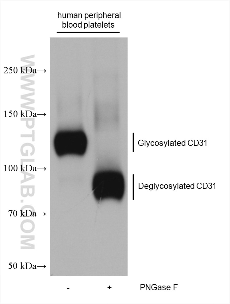
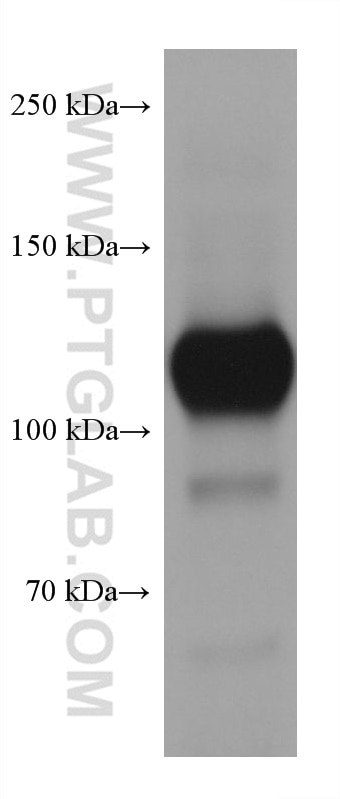
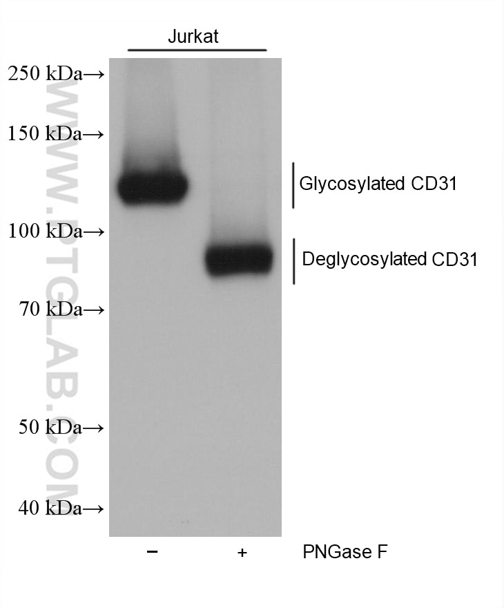
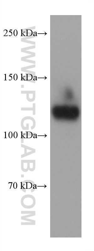
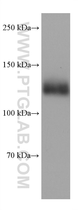
| Positive WB detected in | human peripheral blood platelets, Jurkat cells, THP-1 cells |
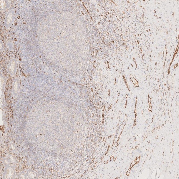
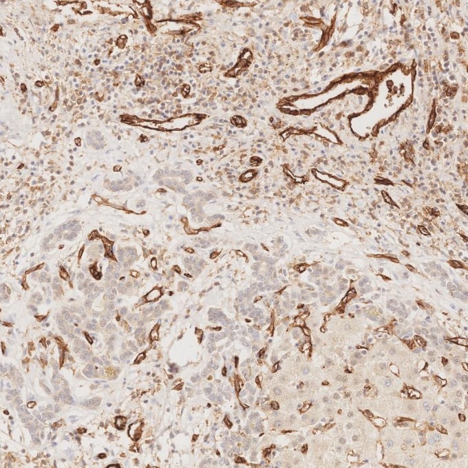
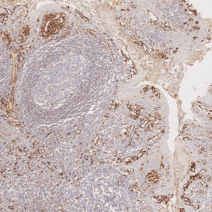
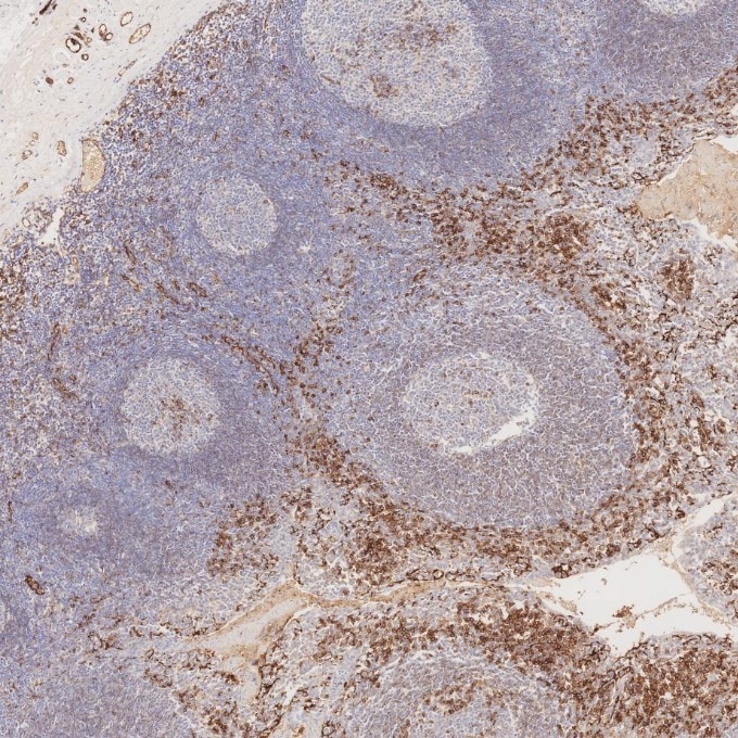
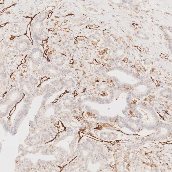
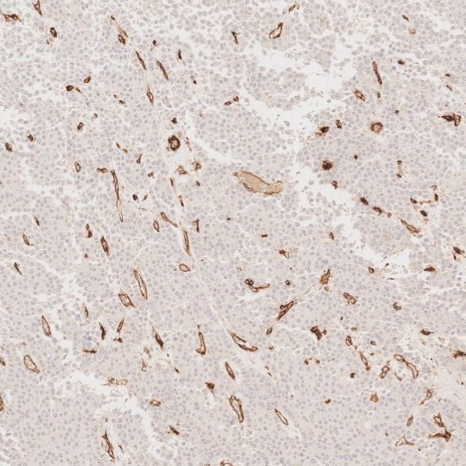
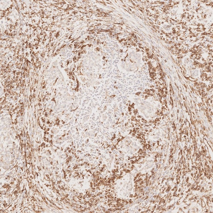
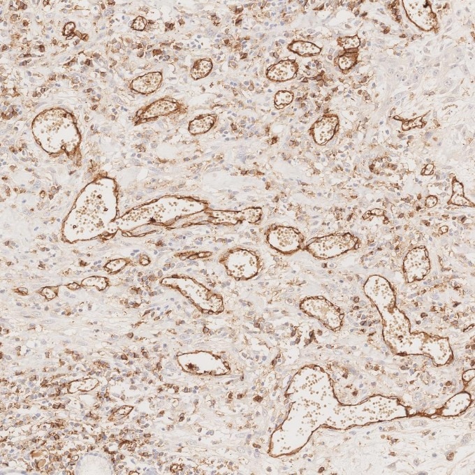
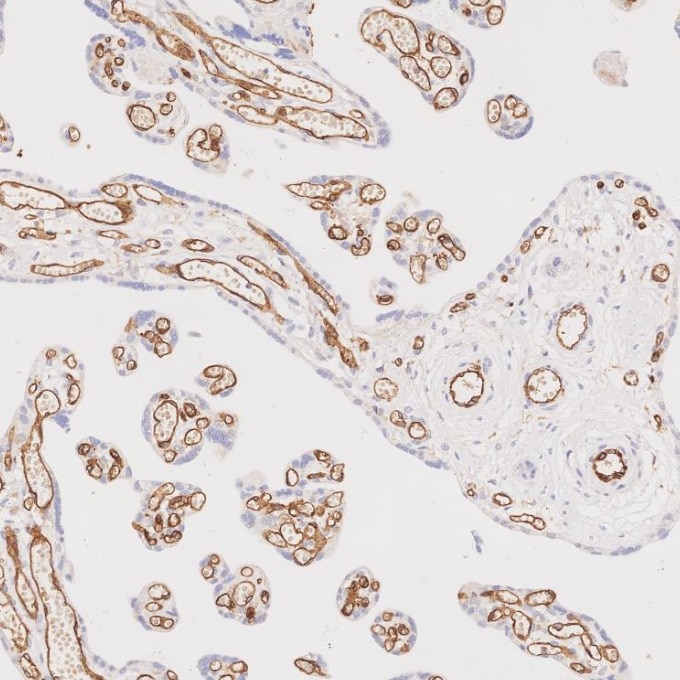
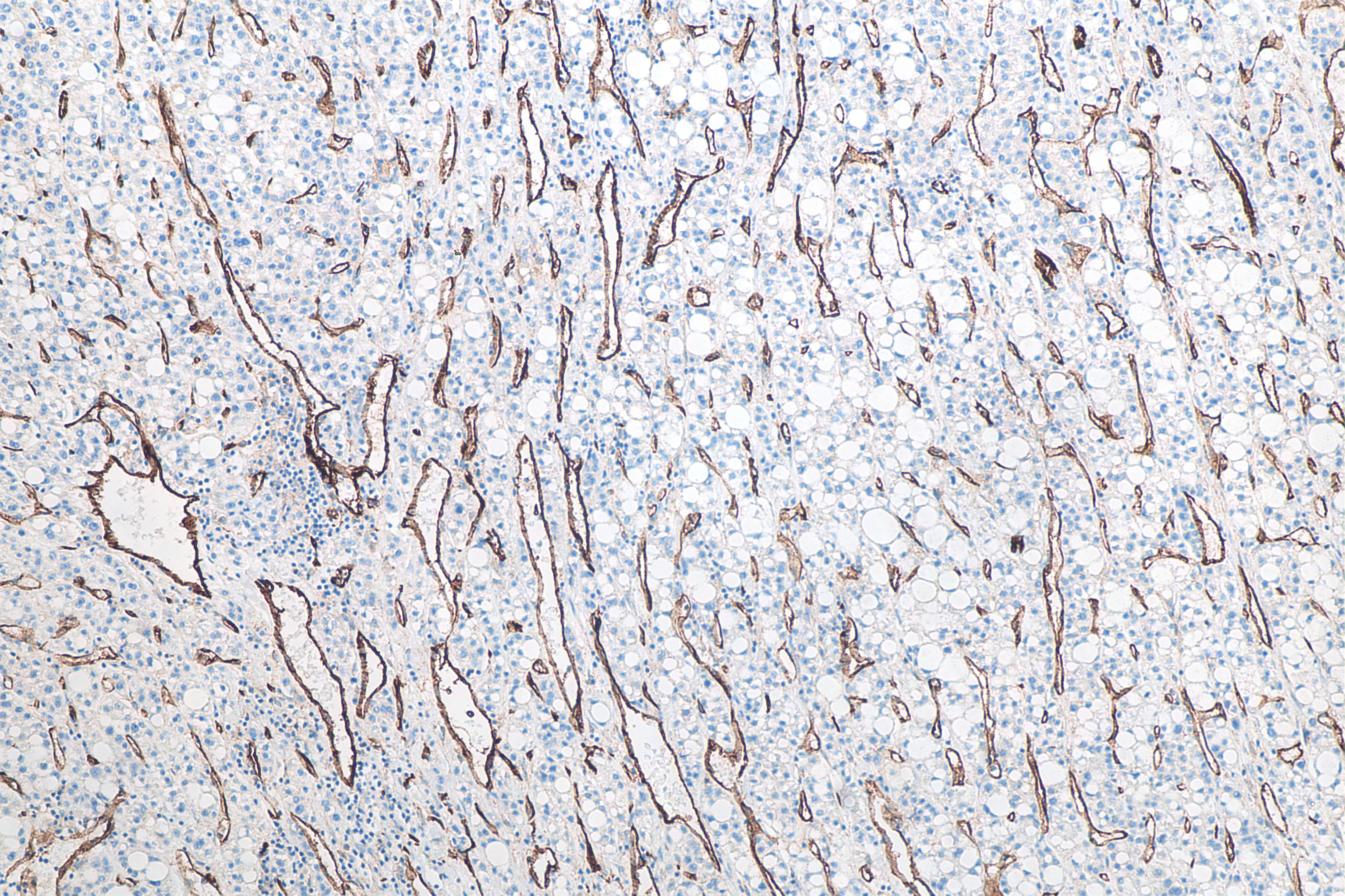
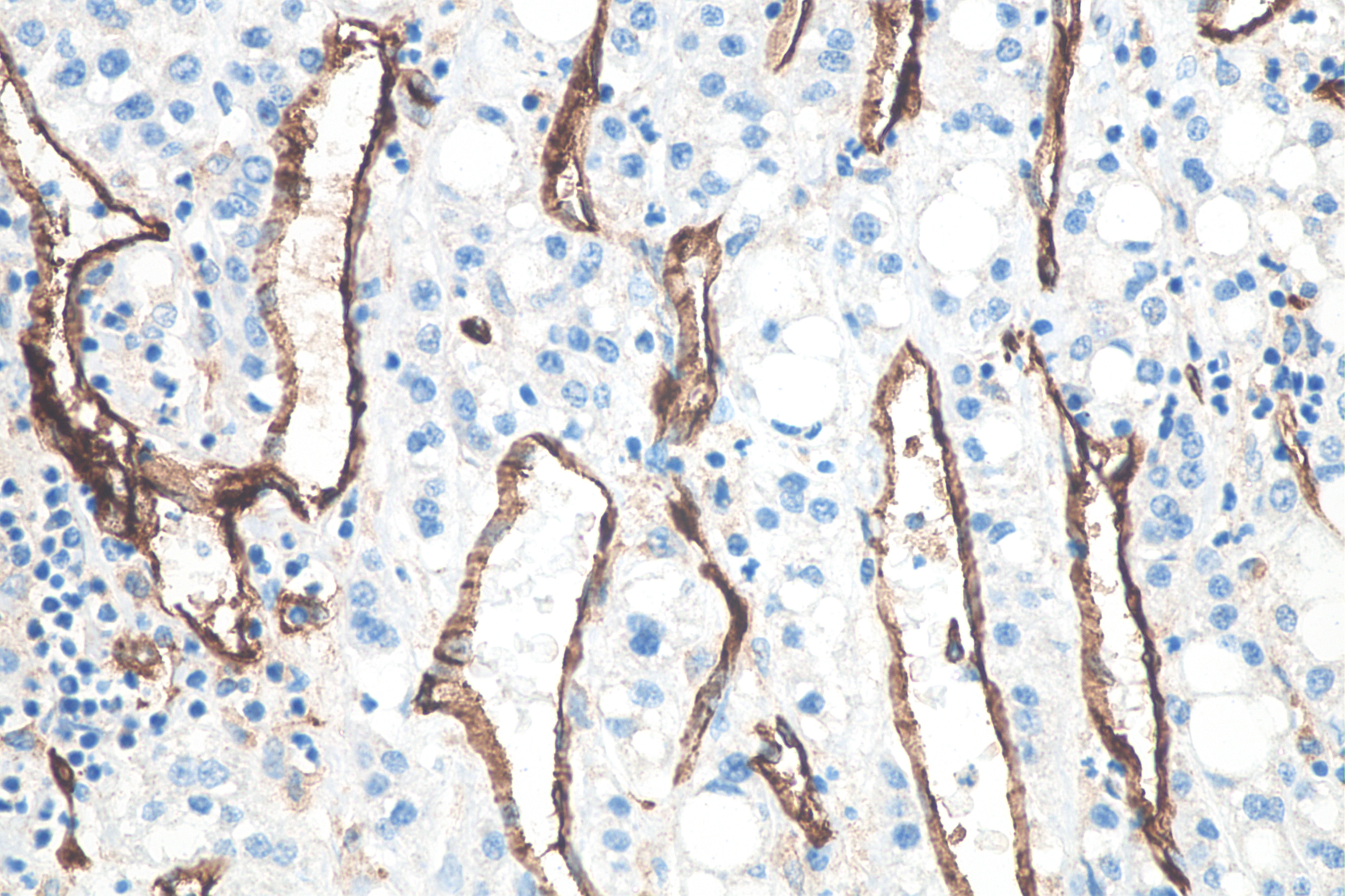
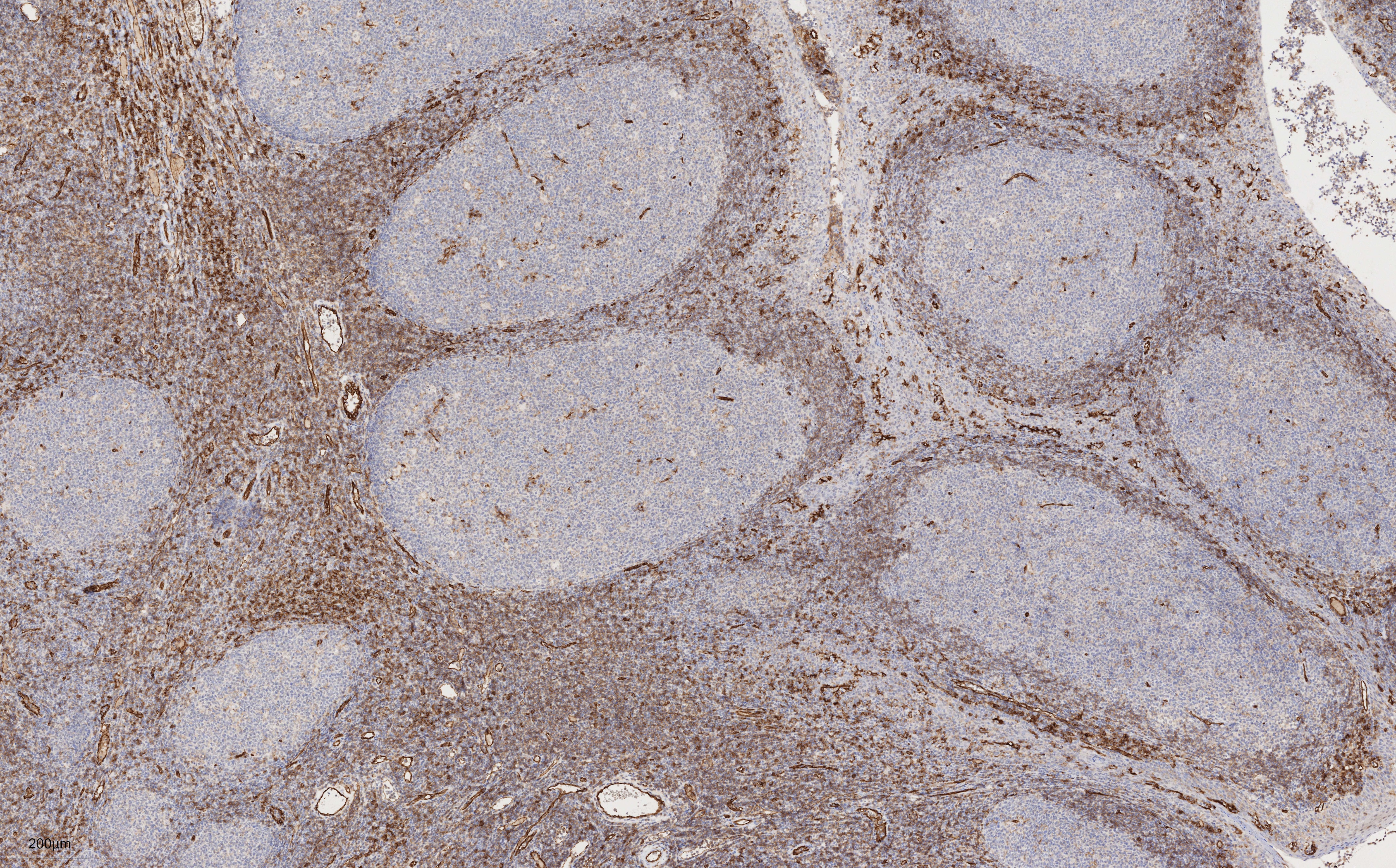
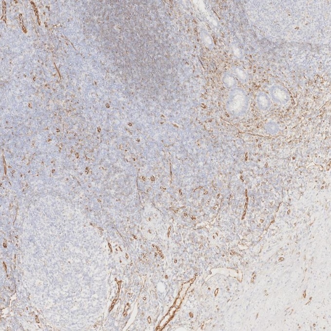
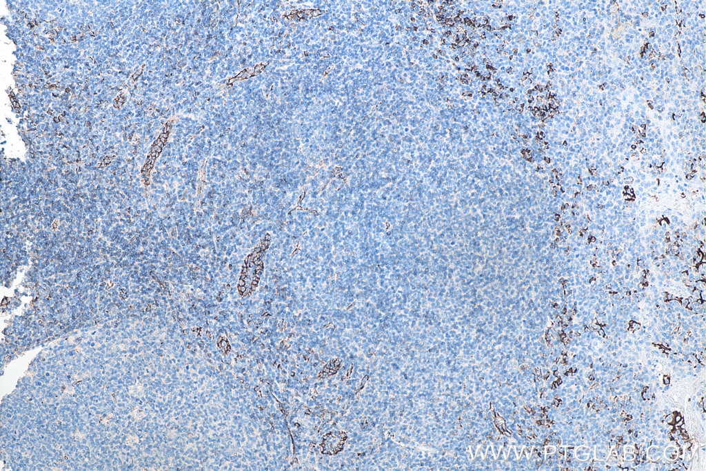
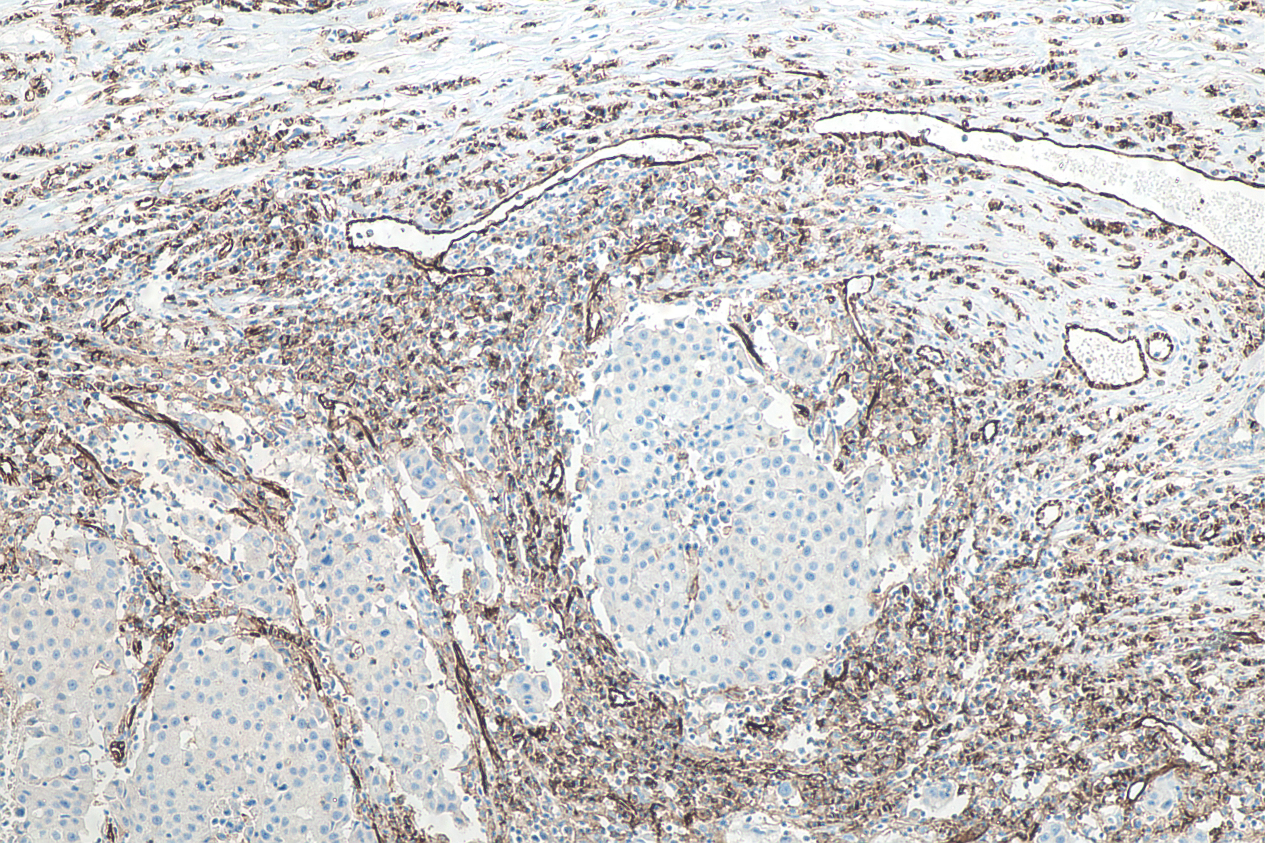
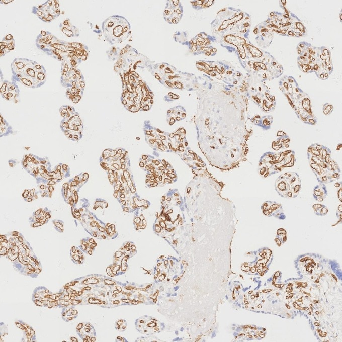
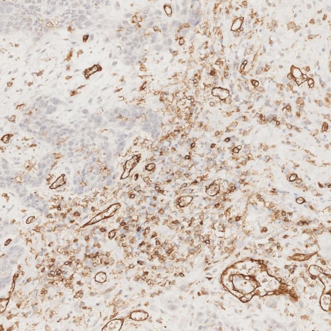
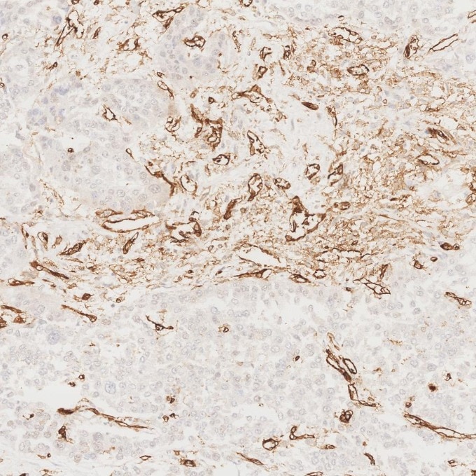
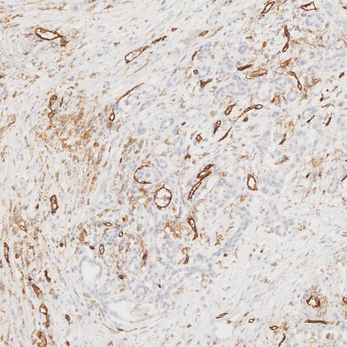
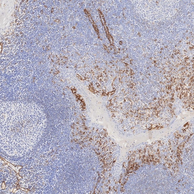
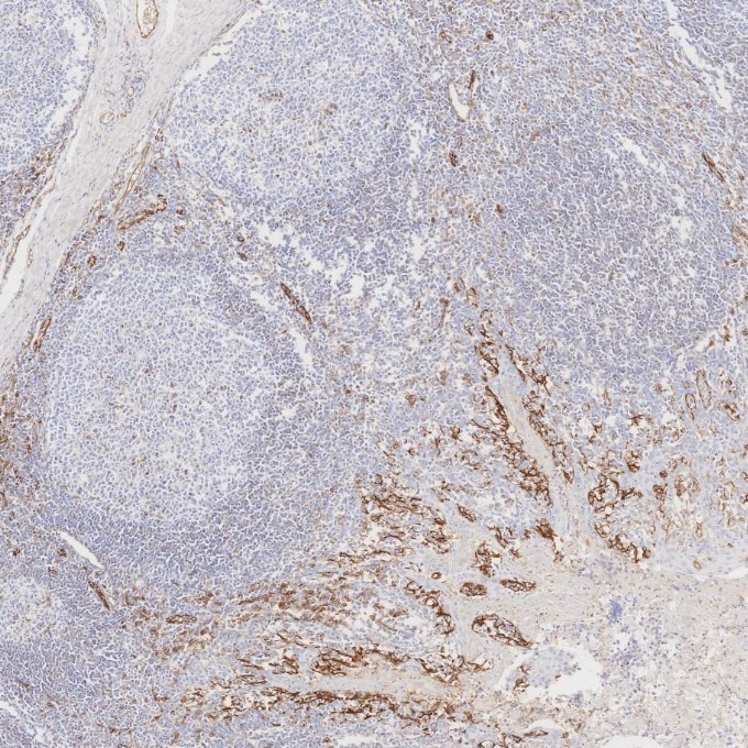
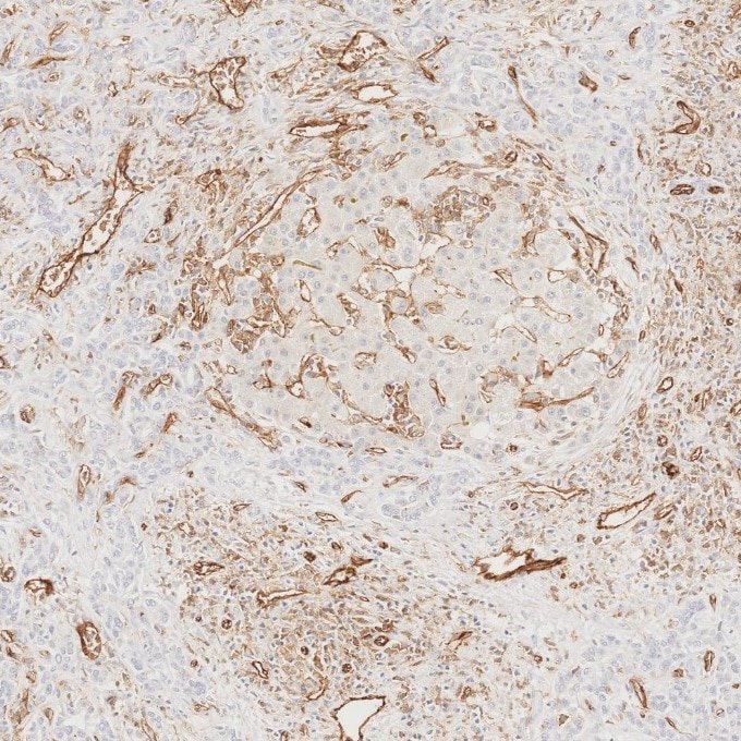
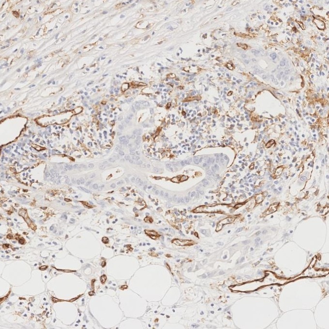
| Positive IHC-Autostainer detected in | human liver cancer tissue, human placenta tissue, human oesophagus cancer tissue, human stomach cancer tissue, human pancreas cancer tissue, human tonsillitis tissue, human liver tissue, human pancreas tissue, human appendicitis tissue |
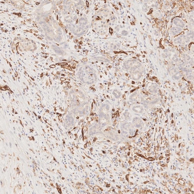
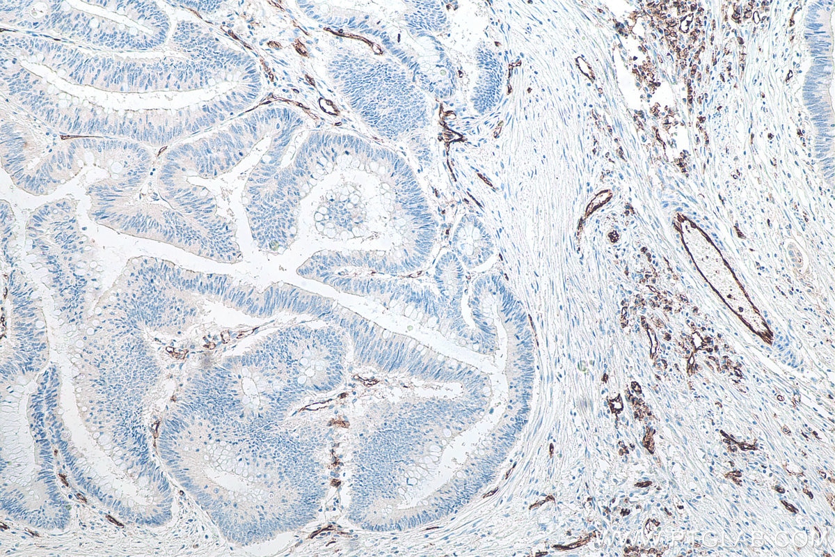
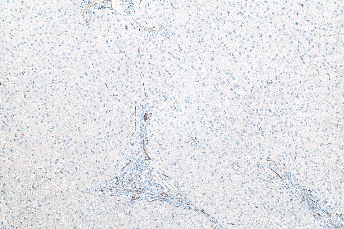
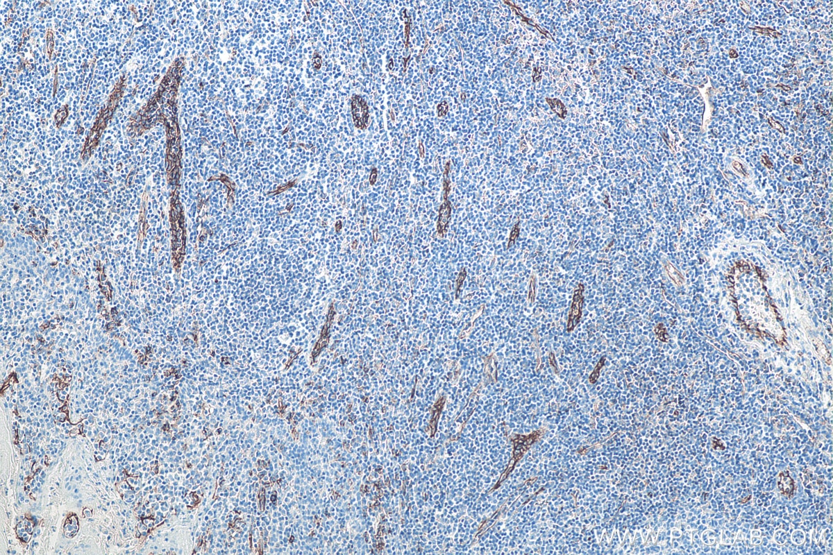
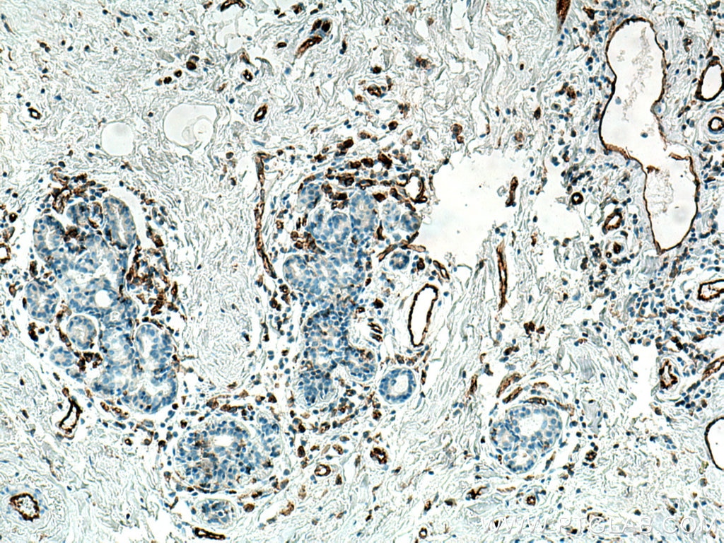
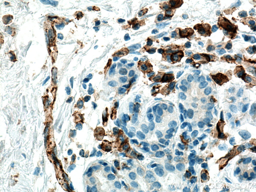
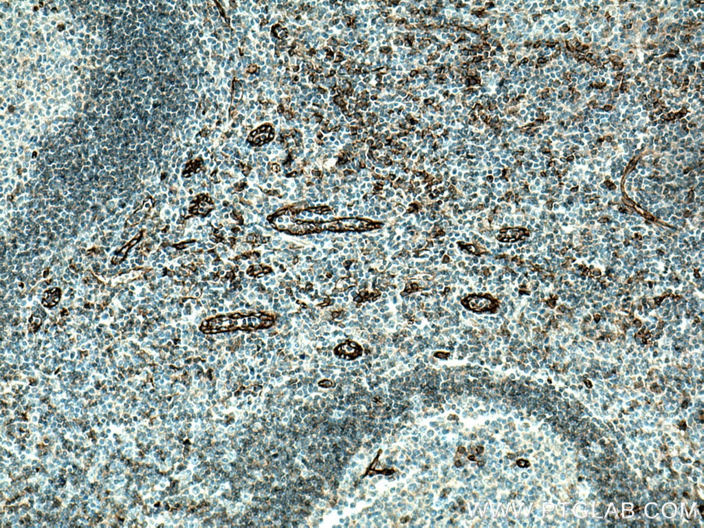
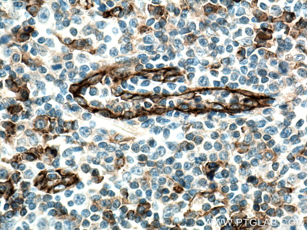
| Positive IHC detected in | human liver cancer tissue, human oesophagus cancer tissue, human liver tissue, human tonsillitis tissue, human pancreas tissue, human placenta tissue, human pancreas cancer tissue, human colon cancer tissue, human liver cancer tissue, human appendicitis tissue, human breast cancer tissue Note: suggested antigen retrieval with TE buffer pH 9.0; (*) Alternatively, antigen retrieval may be performed with citrate buffer pH 6.0 |
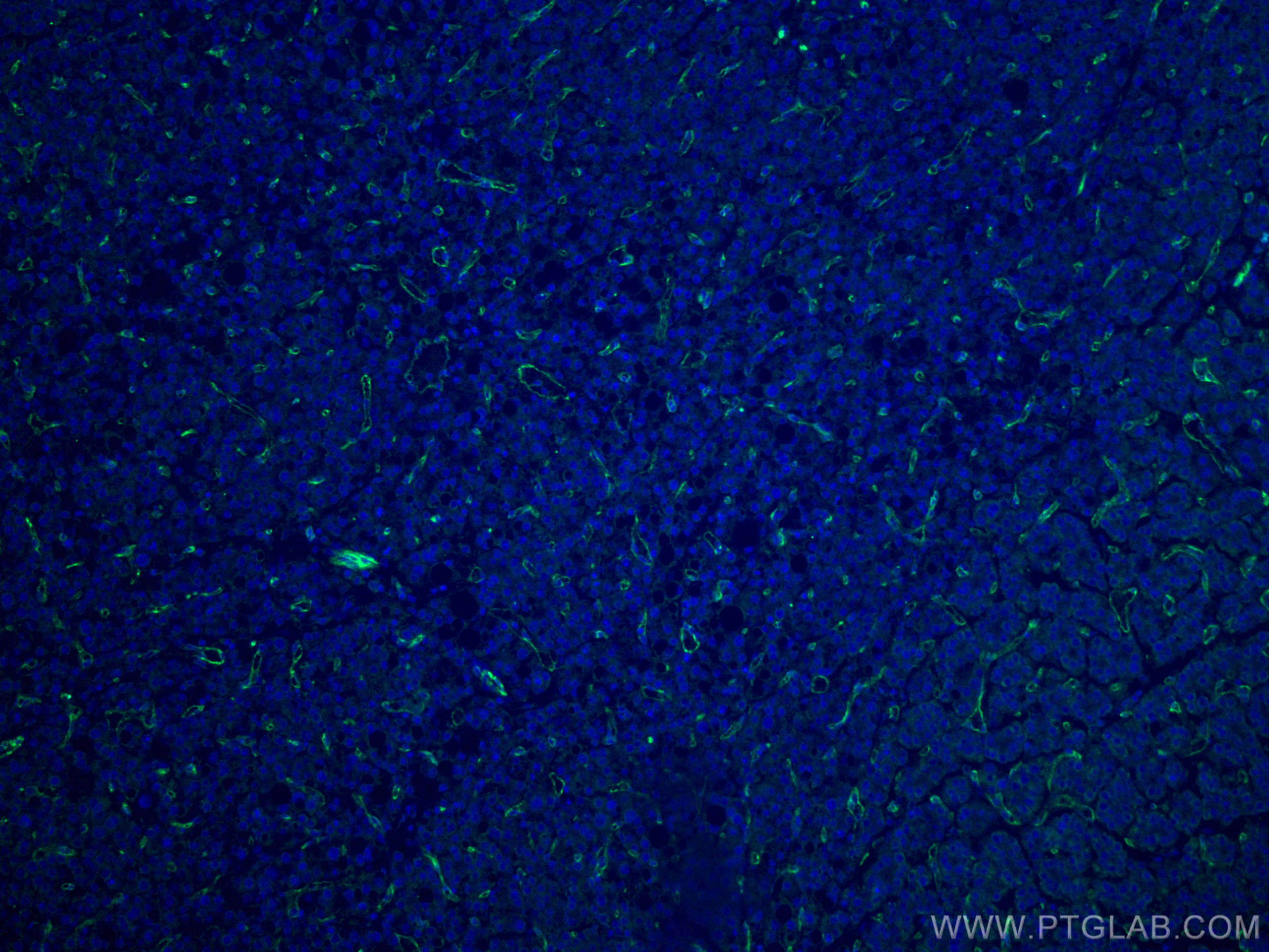
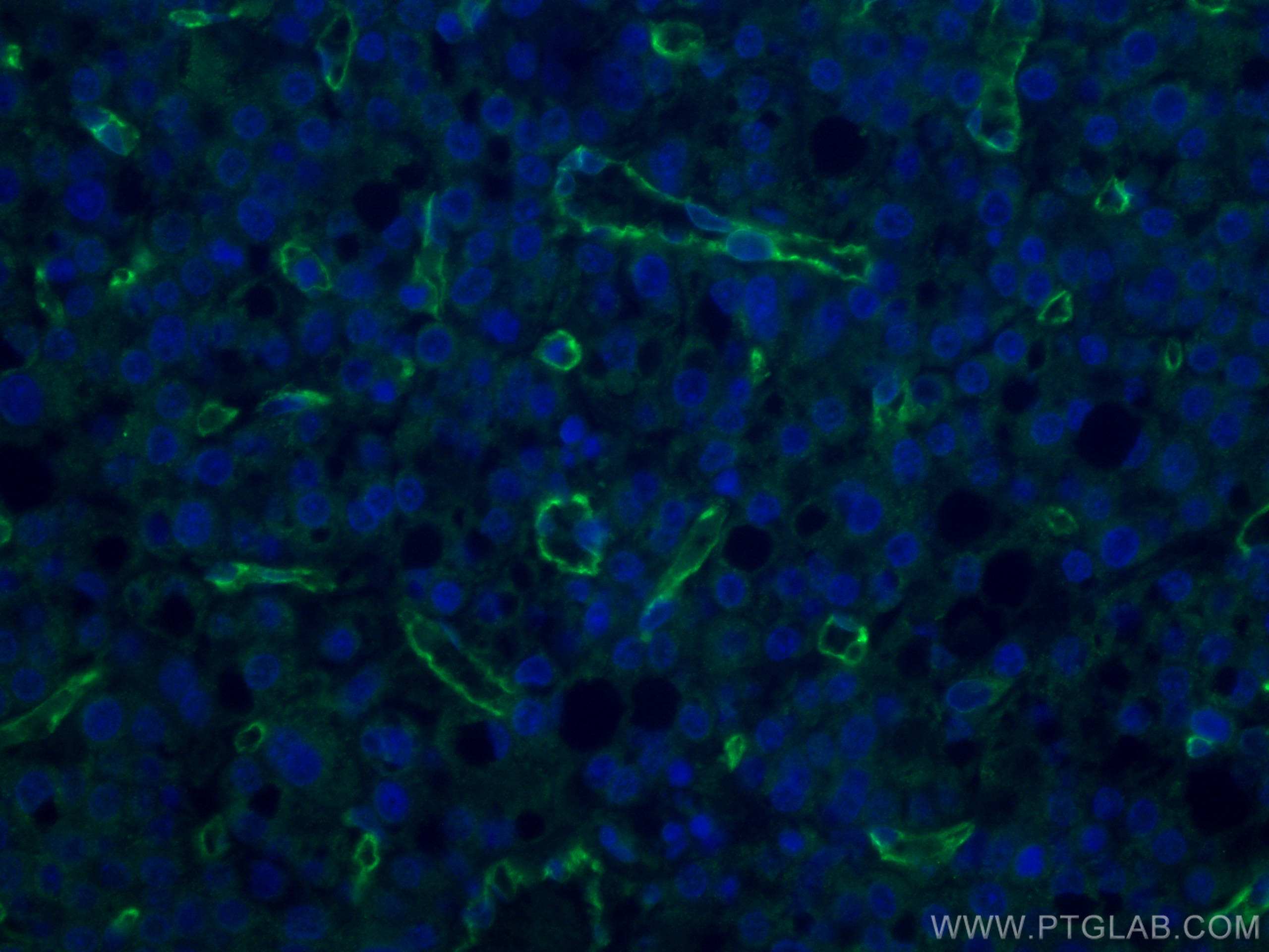
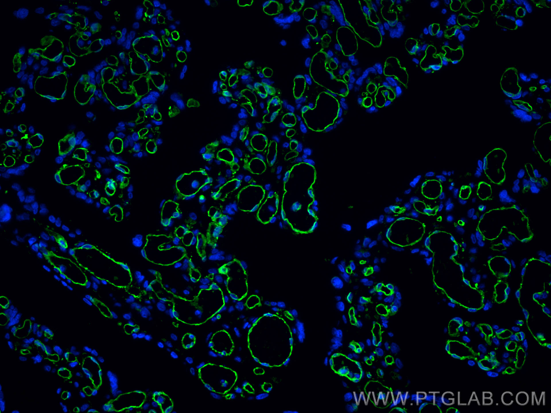
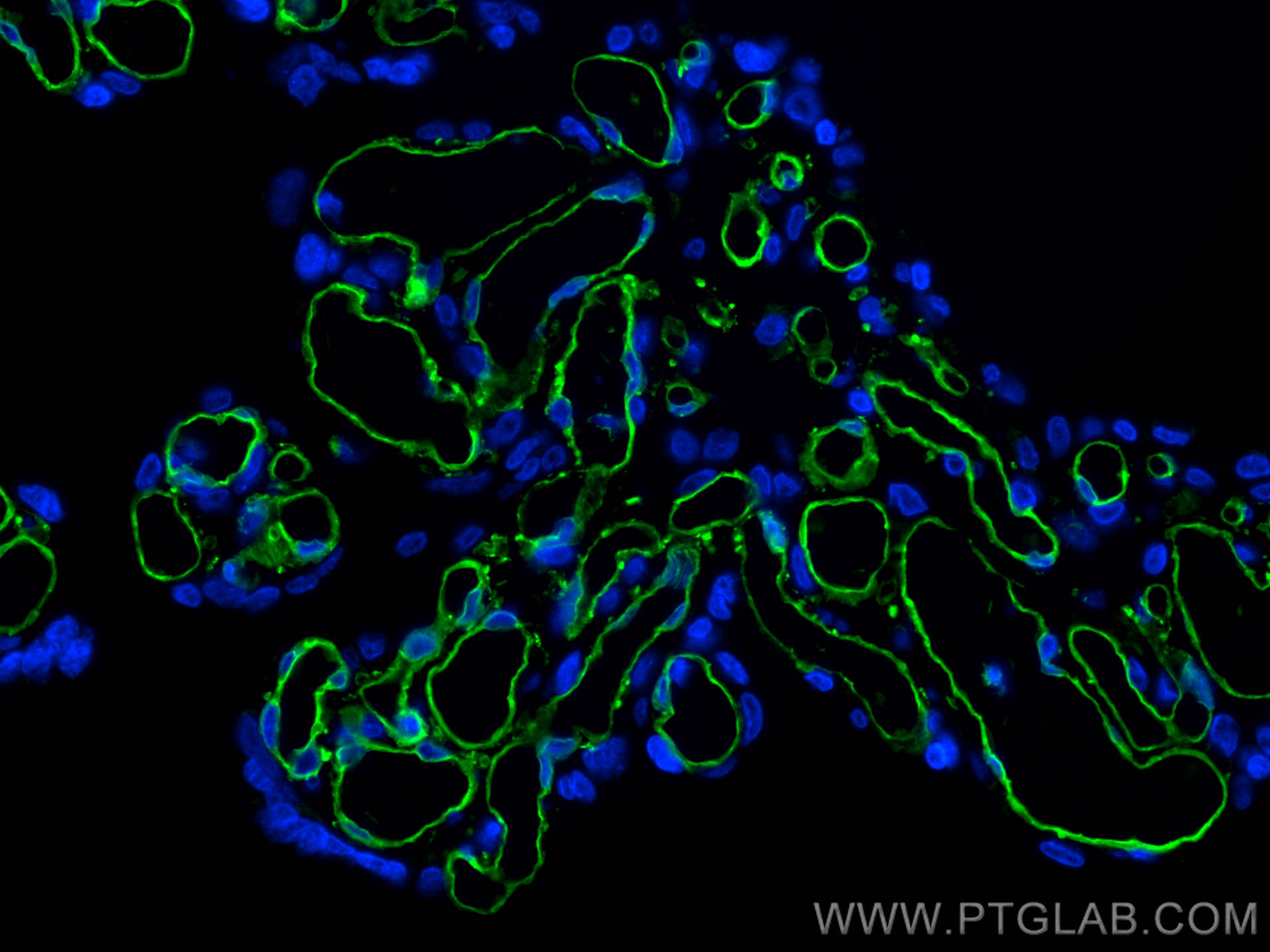
| Positive IF-P detected in | human placenta tissue, human liver cancer tissue |
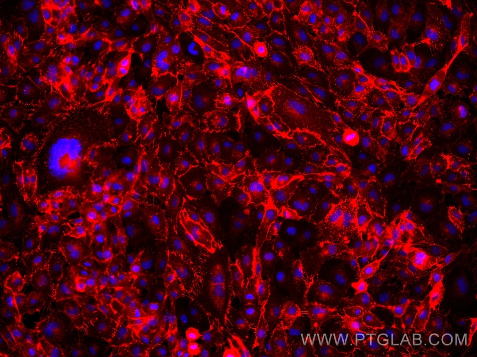
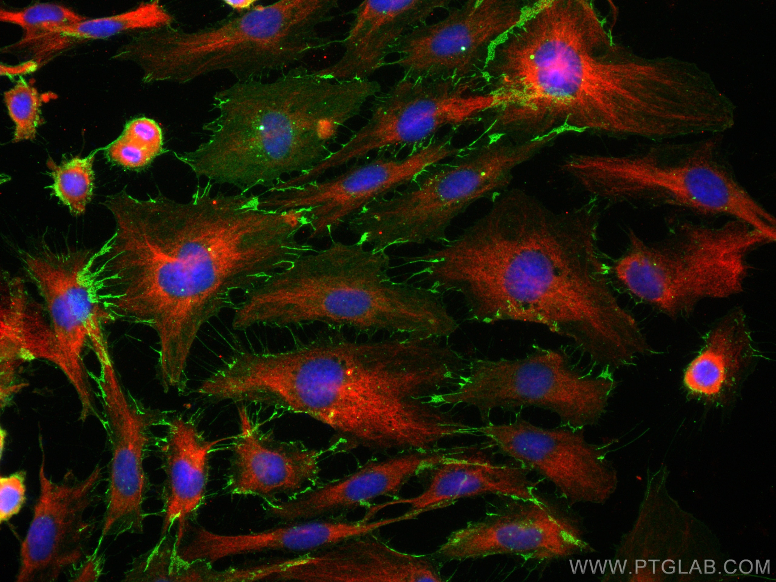
| Positive IF/ICC detected in | HUVEC cells |
Recommended dilution
| Application | Dilution |
|---|---|
| Western Blot (WB) | WB : 1:5000-1:50000 |
| Immunohistochemistry (IHC)-AUTOSTAINER | IHC-AUTOSTAINER : 1:2500-1:10000 |
| Immunohistochemistry (IHC) | IHC : 1:5000-1:20000 |
| Immunofluorescence (IF)-P | IF-P : 1:400-1:1600 |
| Immunofluorescence (IF)/ICC | IF/ICC : 1:1000-1:4000 |
| It is recommended that this reagent should be titrated in each testing system to obtain optimal results. | |
| Sample-dependent, Check data in validation data gallery. | |
Product Information
66065-2-Ig targets CD31 in WB, IHC, IHC-Autostainer, IF/ICC, IF-P, ELISA applications and shows reactivity with human samples.
| Tested Reactivity | human |
| Cited Reactivity | human, pig, rabbit |
| Host / Isotype | Mouse / IgG1 |
| Class | Monoclonal |
| Type | Antibody |
| Immunogen |
CatNo: Ag19730 Product name: Recombinant human CD31 protein Source: e coli.-derived, PET28a Tag: 6*His Domain: 428-738 aa of BC022512 Sequence: EVRCESISGTLPISYQLLKTSKVLENSTKNSNDPAVFKDNPTEDVEYQCVADNCHSHAKMLSEVLRVKVIAPVDEVQISILSSKVVESGEDIVLQCAVNEGSGPITYKFYREKEGKPFYQMTSNATQAFWTKQKANKEQEGEYYCTAFNRANHASSVPRSKILTVRVILAPWKKGLIAVVIIGVIIALLIIAAKCYFLRKAKAKQMPVEMSRPAVPLLNSNNEKMSDPNMEANSHYGHNDDVGNHAMKPINDNKEPLNSDVQYTEVQVSSAESHKDLGKKDTETVYSEVRKAVPDAVESRYSRTEGSLDGT Predict reactive species |
| Full Name | platelet/endothelial cell adhesion molecule |
| Calculated Molecular Weight | 83 kDa |
| Observed Molecular Weight | 120-130 kDa |
| GenBank Accession Number | BC022512 |
| Gene Symbol | CD31 |
| Gene ID (NCBI) | 5175 |
| ENSEMBL Gene ID | ENSG00000261371 |
| RRID | AB_2918476 |
| Conjugate | Unconjugated |
| Form | Liquid |
| Purification Method | Protein G purification |
| UNIPROT ID | P16284 |
| Storage Buffer | PBS with 0.02% sodium azide and 50% glycerol, pH 7.3. |
| Storage Conditions | Store at -20°C. Stable for one year after shipment. Aliquoting is unnecessary for -20oC storage. 20ul sizes contain 0.1% BSA. |
Background Information
Platelet endothelial cell adhesion molecule-1 (PECAM-1, CD31) is a member of the immunoglobulin gene superfamily of cell adhesion molecules. CD31 is a transmembrane glycoprotein that is highly expressed on the surface of the endothelium, making up a large portion of its intracellular junctions. PECAM-1 is also present on the surface of hematopoietic cells and immune cells including platelets, monocytes, neutrophils, natural killer cells, megakaryocytes and some types of T-cell (PMID: 9011572). As well as its role in cell-cell adhesion, PECAM-1 functions as a signaling receptor, and is involved in important physiological events such as nitric oxide production, regulation of T-cell immunity and tolerance, leukocyte transendothelial migration and inflammation and angiogenesis (PMID: 21183735; 20978210; 17872453; 20634489).
Protocols
| Product Specific Protocols | |
|---|---|
| IF protocol for CD31 antibody 66065-2-Ig | Download protocol |
| IHC protocol for CD31 antibody 66065-2-Ig | Download protocol |
| WB protocol for CD31 antibody 66065-2-Ig | Download protocol |
| Standard Protocols | |
|---|---|
| Click here to view our Standard Protocols |
Publications
What published studies show
The CD31 Monoclonal antibody (Cat# 66065-2-Ig) has accumulated 142 citations across high-impact journals including Nature Communications, Cell, Cancer Cell, Nature Nanotechnology, Advanced Science, Acta Biomaterialia, Cell Death & Differentiation, and Journal of Nanobiotechnology. Research fields covered include angiogenesis, vascular biology, oncology, cardiovascular disease, tissue engineering, diabetic complications, neurology, immunology, and wound healing. The antibody is predominantly applied in immunofluorescence (IF), immunohistochemistry (IHC), and Western blot (WB).
| Species | Application | Title |
|---|---|---|
Cancer Cell Conserved spatial subtypes and cellular neighborhoods of cancer-associated fibroblasts revealed by single-cell spatial multi-omics | ||
Nat Nanotechnol Prevention of acute thrombosis with vascular endothelium antioxidative nanoscavenger. | ||
Cancer Commun (Lond) Targeting SPHK1 in macrophages remodels the tumor microenvironment and enhances anti-PD-1 immunotherapy efficacy in colorectal cancer liver metastasis. | ||
Bioact Mater Throw out an oligopeptide to catch a protein: Deep learning and natural language processing-screened tripeptide PSP promotes Osteolectin-mediated vascularized bone regeneration | ||
Nat Commun Lymph nodes molecular subtypes unravel lymph nodes heterogeneity and clinical implications in colorectal cancer |
Reviews
What customers say
Both reviewers rated this antibody 5 stars. One user reported excellent performance in immunofluorescence staining of human iPSC-derived vascular organoids, using a 1:500 dilution with overnight incubation on 5% PFA-fixed, Triton-X-permeabilized tissue. The other user described it as a good IF antibody with no unspecific staining. Overall, customers find it reliable for immunofluorescence applications with clean, specific signal.
The reviews below have been submitted by verified Proteintech customers who received an incentive for providing their feedback.
Refik (Verified Customer) (12-07-2025) | Works well with overnight incubation of human iPSC-derived vascular specific organoids using concentration 1:500 for ICC of 5% PFA fixed and 0.1/1% triton-X permeabilized cells and tissues.
|
Alessandro (Verified Customer) (12-09-2023) | good IF antibody. no unspecific staining
|











































