



Tested Applications
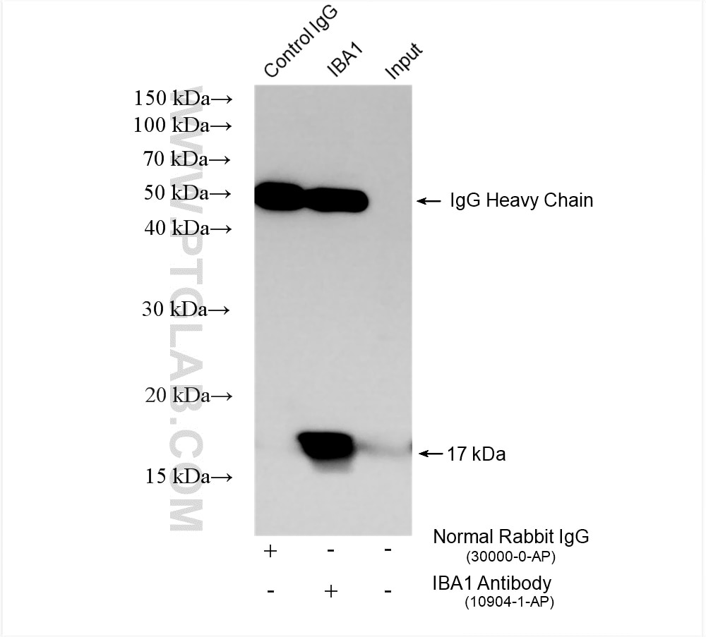
| Positive IP detected in | THP-1 cells |
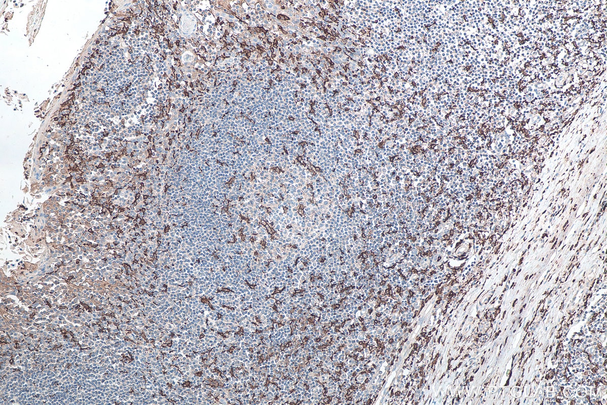
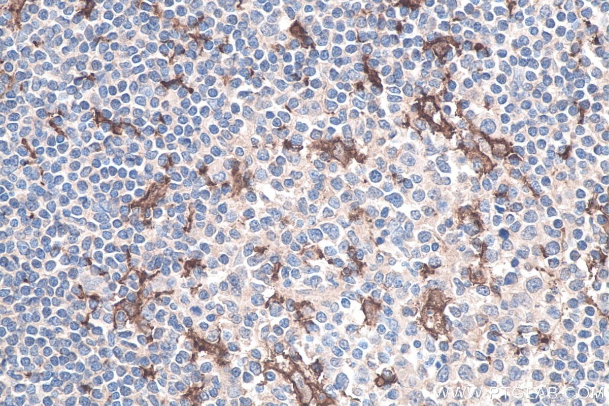
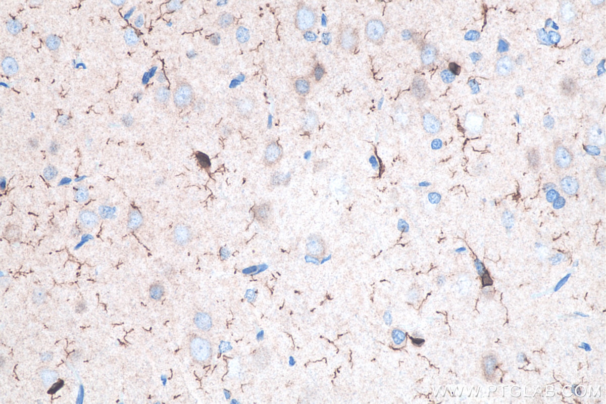
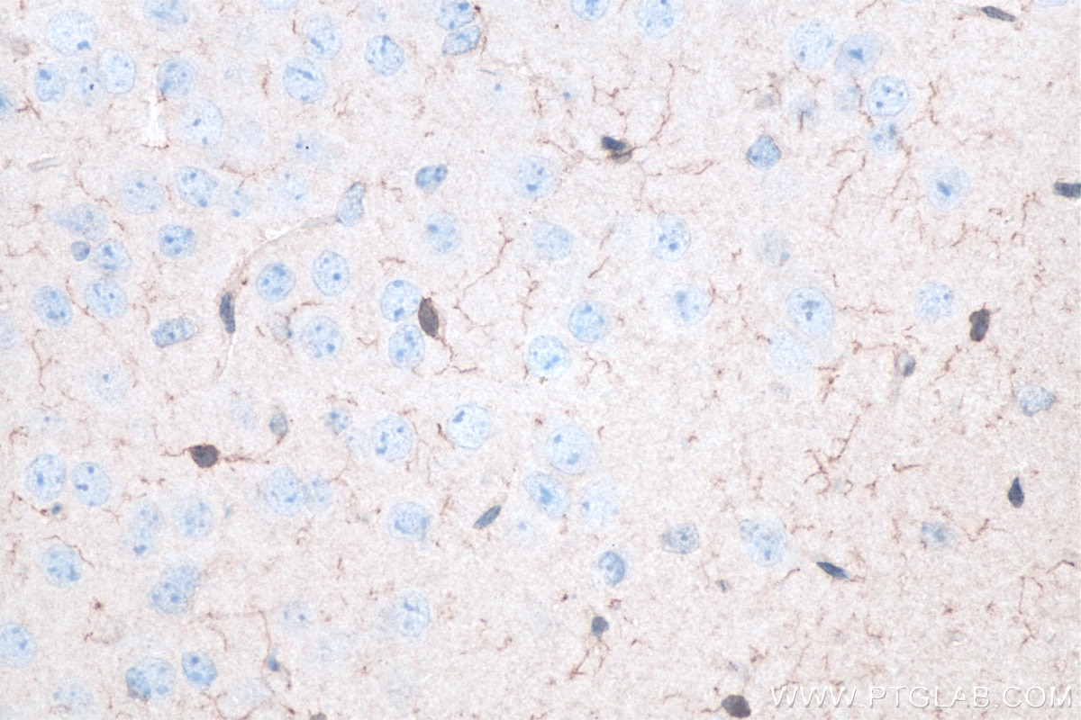
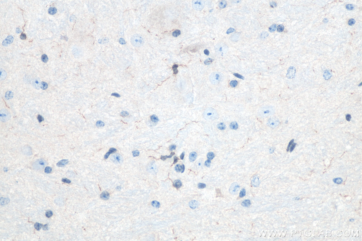
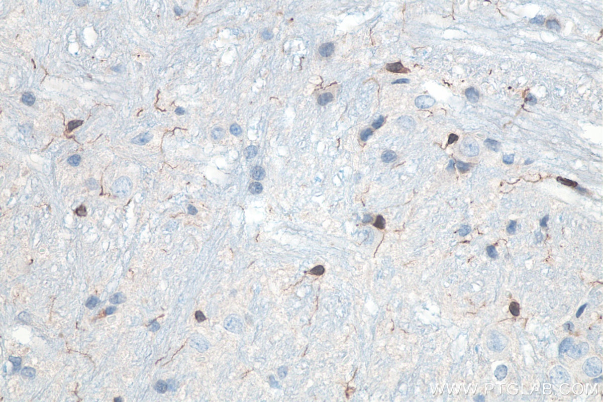
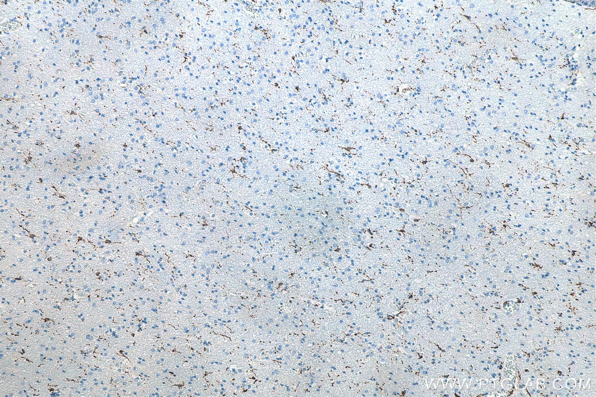
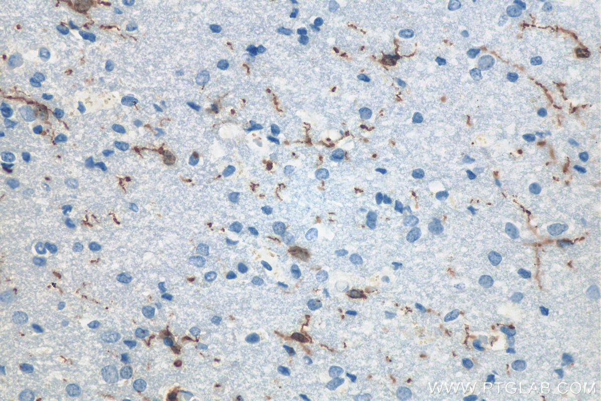
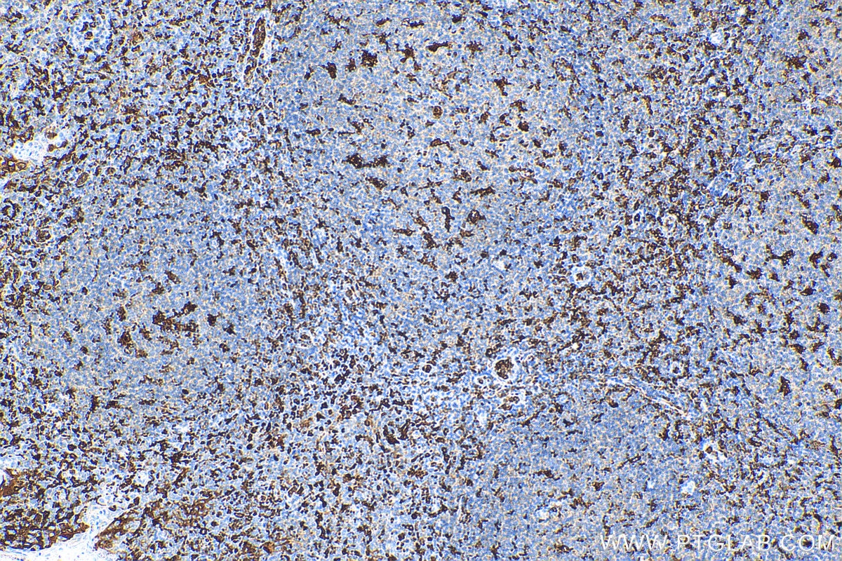
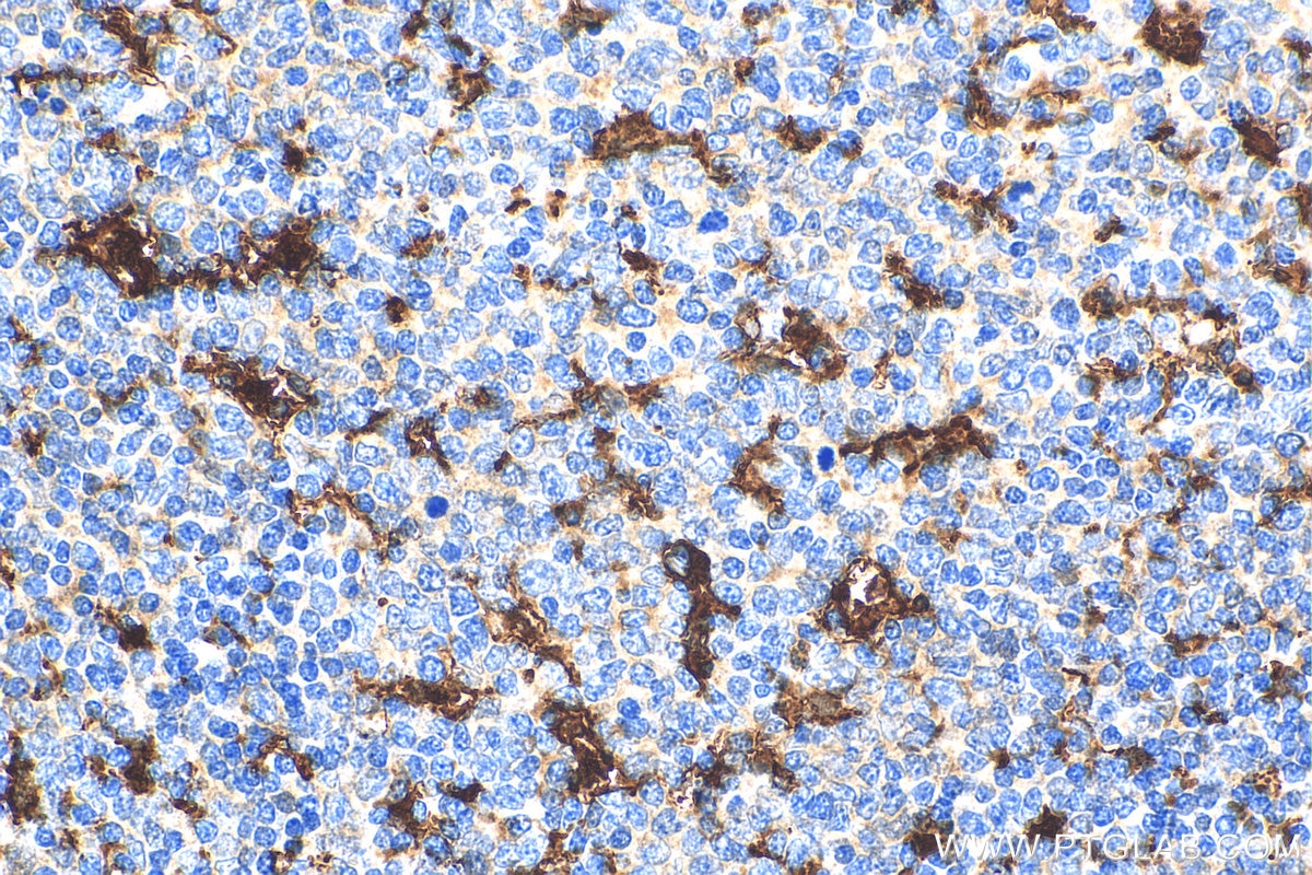
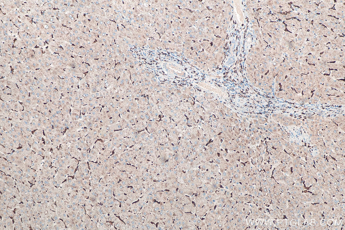
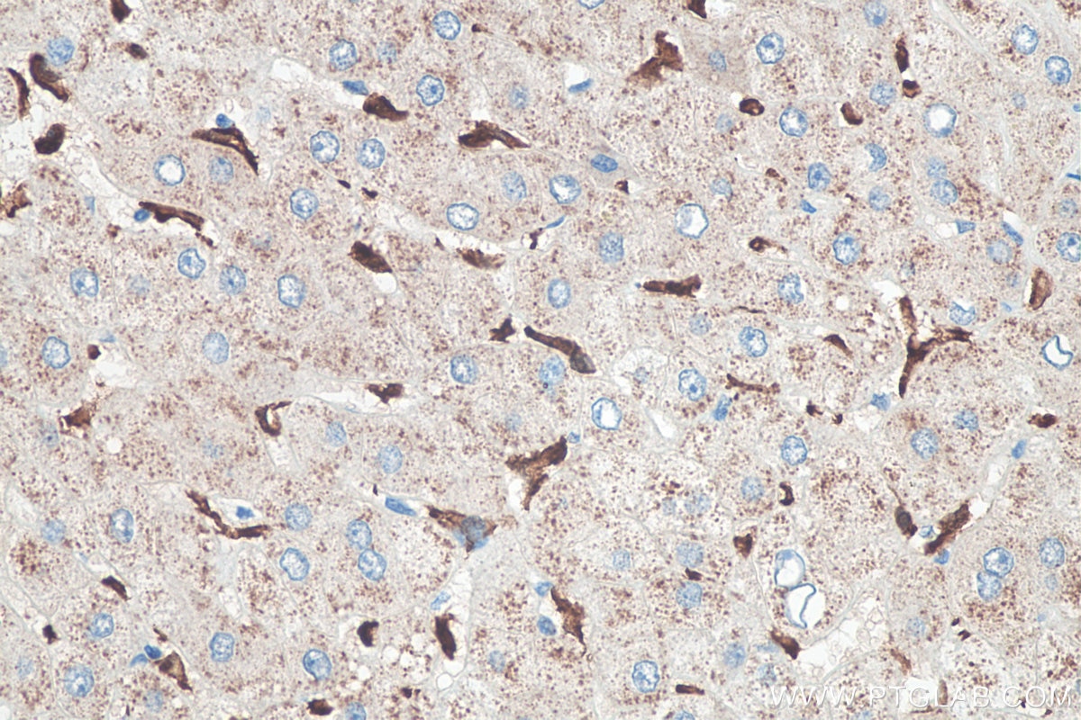
| Positive IHC detected in | rat brain tissue, rat cerebellum tissue, human tonsillitis tissue, human liver tissue, mouse brain tissue, human brain tissue, rat brain tissue, mouse cerebellum tissue Note: suggested antigen retrieval with TE buffer pH 9.0; (*) Alternatively, antigen retrieval may be performed with citrate buffer pH 6.0 |
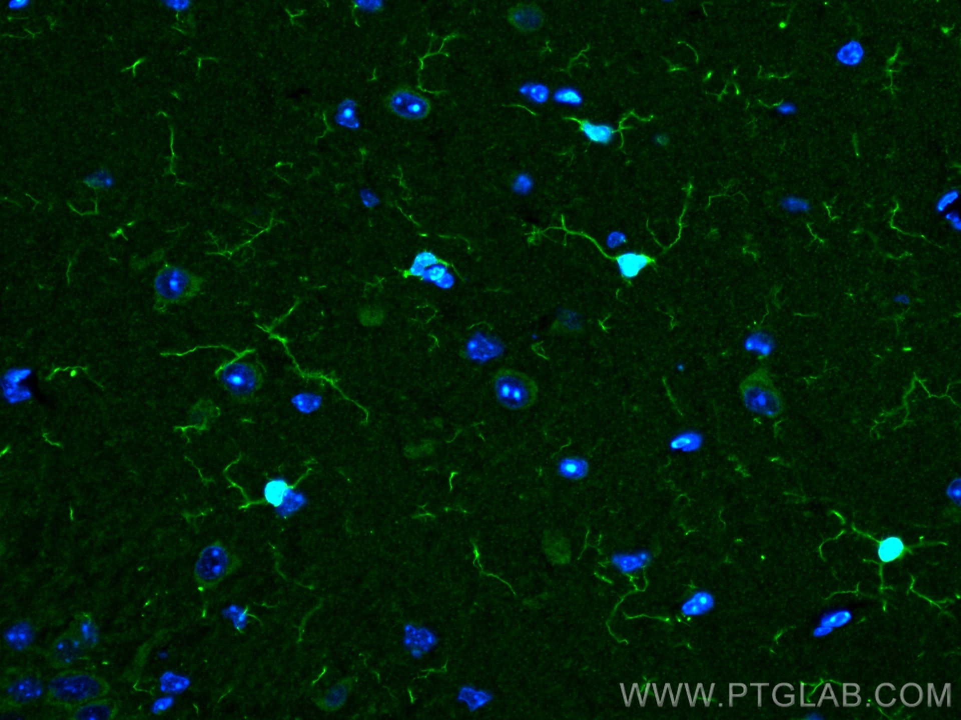
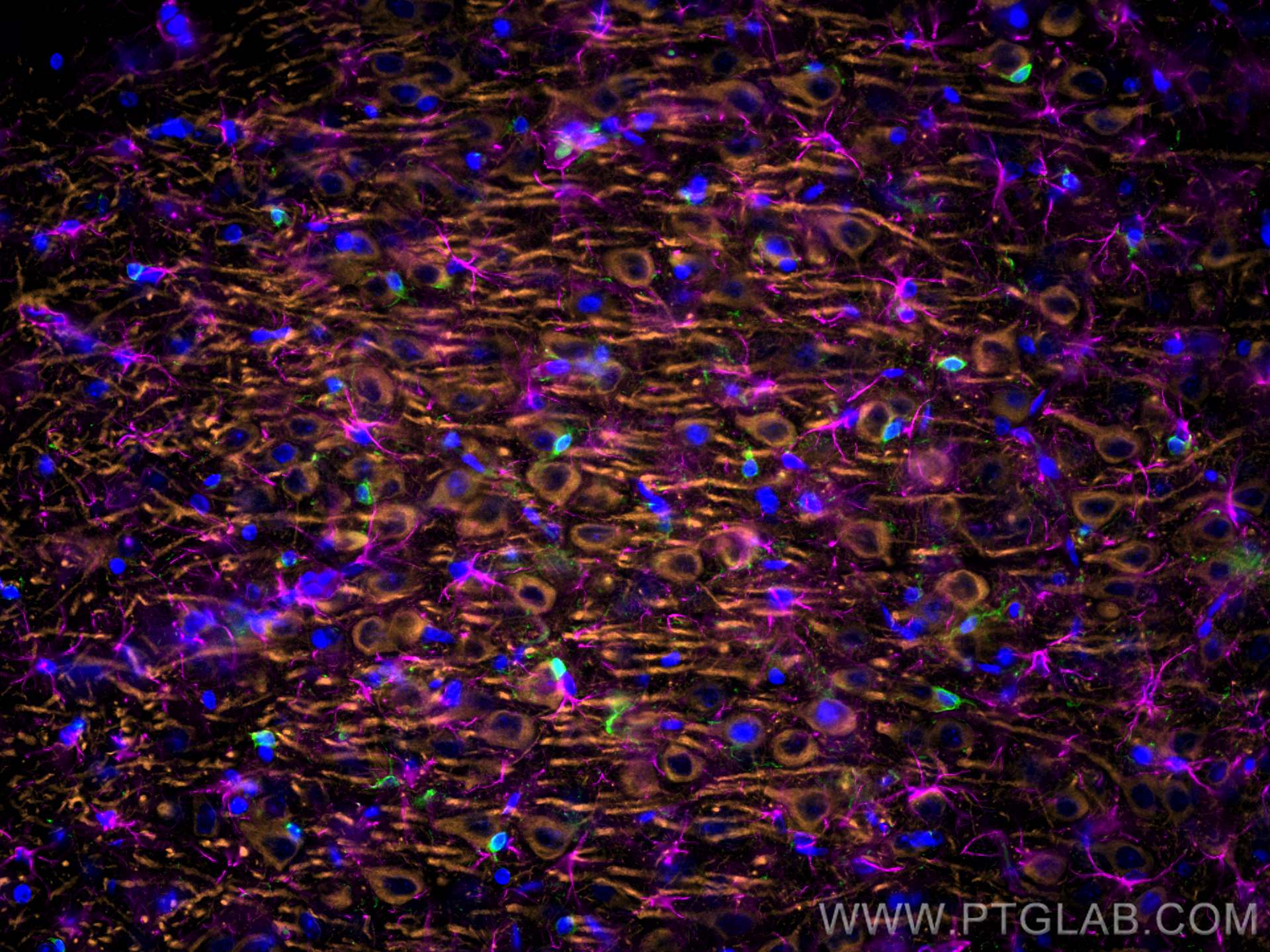
| Positive IF-P detected in | mouse brain tissue, rat brain tissue |
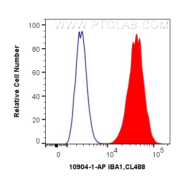
| Positive FC (Intra) detected in | THP-1 cells |
Recommended dilution
| Application | Dilution |
|---|---|
| Immunoprecipitation (IP) | IP : 0.5-4.0 ug for 1.0-3.0 mg of total protein lysate |
| Immunohistochemistry (IHC) | IHC : 1:2000-1:8000 |
| Immunofluorescence (IF)-P | IF-P : 1:50-1:500 |
| Flow Cytometry (FC) (INTRA) | FC (INTRA) : 0.40 ug per 10^6 cells in a 100 µl suspension |
| It is recommended that this reagent should be titrated in each testing system to obtain optimal results. | |
| Sample-dependent, Check data in validation data gallery. | |
Product Information
10904-1-AP targets IBA1 in IHC, IF-P, FC (Intra), IP, ELISA applications and shows reactivity with human, mouse, rat samples.
| Tested Reactivity | human, mouse, rat |
| Cited Reactivity | human, mouse, rat, pig, monkey, zebrafish, hamster, sheep |
| Host / Isotype | Rabbit / IgG |
| Class | Polyclonal |
| Type | Antibody |
| Immunogen |
CatNo: Ag1363 Product name: Recombinant human IBA1 protein Source: e coli.-derived, PGEX-4T Tag: GST Domain: 1-147 aa of BC009474 Sequence: MSQTRDLQGGKAFGLLKAQQEERLDEINKQFLDDPKYSSDEDLPSKLEGFKEKYMEFDLNGNGDIDIMSLKRMLEKLGVPKTHLELKKLIGEVSSGSGETFSYPDFLRMMLGKRSAILKMILMYEEKAREKEKPTGPPAKKAISELP Predict reactive species |
| Full Name | allograft inflammatory factor 1 |
| Calculated Molecular Weight | 17 kDa |
| GenBank Accession Number | BC009474 |
| Gene Symbol | IBA1 |
| Gene ID (NCBI) | 199 |
| ENSEMBL Gene ID | ENSG00000204472 |
| RRID | AB_2224377 |
| Conjugate | Unconjugated |
| Form | Liquid |
| Purification Method | Antigen affinity purification |
| UNIPROT ID | P55008 |
| Storage Buffer | PBS with 0.02% sodium azide and 50% glycerol, pH 7.3. |
| Storage Conditions | Store at -20°C. Stable for one year after shipment. Aliquoting is unnecessary for -20oC storage. 20ul sizes contain 0.1% BSA. |
Background Information
What is the molecular weight of IBA1?
The molecular weight of IBA1 is 16.7 kD.
What is IBA1?
Ionized calcium-binding adaptor molecule 1 (IBA1), also known as Allograft inflammatory factor-1 (AIF-1), is an inflammation-responsive scaffold protein expressed and secreted from macrophages. Microglia response factor (MRF-1) and daintain are also similar, and likely identical, proteins (PMIDs: 29749461, 9630473, 23792284).
What is the function of IBA1?
IBA1 is necessary for macrophage survival, and it is also a key molecule in proinflammatory activity (PMID: 29749461).
Where is IBA1 localized?
IBA1 is a cytoplasmic protein, often expressed in immunocytes, macrophages, and microglia. It can be used as a marker for normal (not 'dark') microglia in brain tissue, as IBA1 is expressed by all microglial cell subpopulations (PMIDs: 29749461, 11943136, 26847266, 9630473).
Is IBA1 upregulated in active immunophages?
Yes; its expression is associated with inflammatory activity (PMID: 29749461).
How do IBA1-positive microglia differ from microglia that express less or no IBA1?
'Dark' microglia is a recently described phenotype associated with Alzheimer's disease pathology and chronic stress; these dark microglia express decreased IBA1 (often punctiform), and show distinct ultrastructural differences from 'normal' microglia as well as condensed and electron-dense cytoplasm and nucleoplasm. Normal microglia generally display strong, diffuse expression of IBA1. IBA1-positive microglial processes in normal conditions also have a strong tendency to exclusively contact synaptic elements, such as axon terminals, dendritic spines, and synaptic clefts (PMIDs: 29992181, 26847266).
How is IBA1 related to Alzheimer's disease and pain?
Exacerbated immunoactivity of IBA1, particularly in proximity to amyloid plaques, is prominently featured in AD pathology. IBA1, along with other microglial markers, are also associated with pain, and robust microglial reactions often follow spinal cord injury (PMIDs: 29992181, 23792284).
Protocols
| Product Specific Protocols | |
|---|---|
| FC protocol for IBA1 antibody 10904-1-AP | Download protocol |
| IF protocol for IBA1 antibody 10904-1-AP | Download protocol |
| IHC protocol for IBA1 antibody 10904-1-AP | Download protocol |
| IP protocol for IBA1 antibody 10904-1-AP | Download protocol |
| Standard Protocols | |
|---|---|
| Click here to view our Standard Protocols |
Publications
What published studies show
The IBA1 Polyclonal antibody (Cat# 10904-1-AP) has accumulated 666 citations across high-impact journals including Nature Medicine, Nature Communications, Nature Neuroscience, Cell Stem Cell, ACS Nano, Theranostics, Advanced Science, Biomaterials, Cell Death & Differentiation, Journal of Neuroinflammation, Brain, Alzheimer's & Dementia, and Phytomedicine. Research fields span neuroinflammation, microglial biology, Alzheimer's and Parkinson's disease, stroke, spinal cord injury, traumatic brain injury, glioma, neuropathic pain, ferroptosis, neurogenesis, depression, and nanomedicine-based therapeutic strategies.
| Species | Application | Title |
|---|---|---|
Nat Med Aged blood impairs hippocampal neural precursor activity and activates microglia via brain endothelial cell VCAM1. | ||
Nat Aging Aged bone marrow macrophages drive systemic aging and age-related dysfunction via extracellular vesicle-mediated induction of paracrine senescence | ||
Bioact Mater M2 macrophage-derived exosomes delivering haptoglobin and interleukin-10 plasmids for synergistic therapy of intracerebral hemorrhage. | ||
Cell Stem Cell Auditory activity sustains adult neurogenesis and cognition through the locus coeruleus-norepinephrine system. | ||
Nat Neurosci Mitochondrial respiratory complex IV deficiency recapitulates amyotrophic lateral sclerosis | ||
Mol Neurodegener Islet amyloid polypeptide cross-seeds tau and drives the neurofibrillary pathology in Alzheimer's disease. |
Reviews
What customers say
All three reviews rated the antibody 5 stars. Users reported clear, strong signal in immunofluorescence on human brain cortex and mouse ventricle tissue, with one noting crisp microglia labeling. A Western blot user detected endogenous Iba1 in mouse brain lysate at 1:500 dilution and described the result as clean. Overall, reviewers expressed high satisfaction with specificity and signal quality across both IF and WB applications.
The reviews below have been submitted by verified Proteintech customers who received an incentive for providing their feedback.
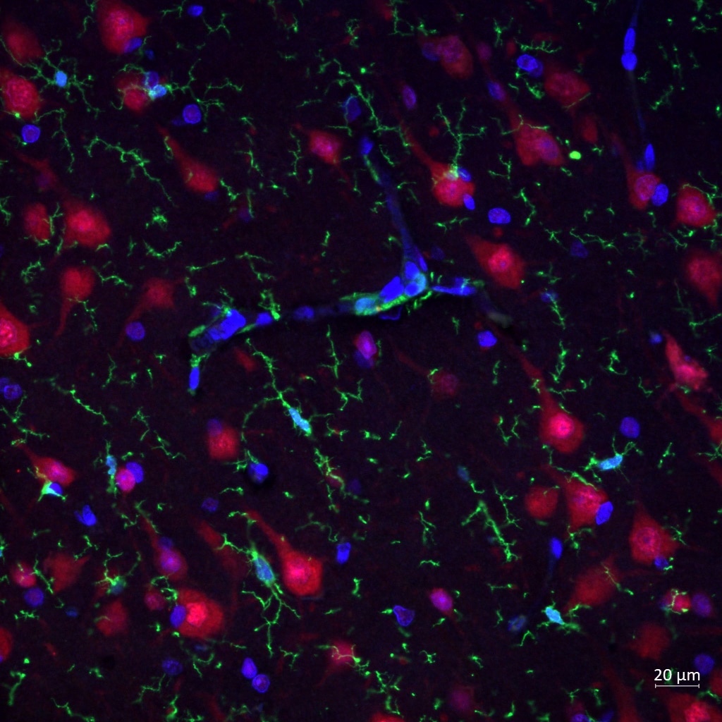
Reyes (Verified Customer) (04-05-2024) | Iba1 (in green) marks clearly my microglia in human brain FFPE cortex.
 |
Alexandru (Verified Customer) (11-13-2023) | Very pleased with the antibody! I had a claer signal in all my stains.
|
NX (Verified Customer) (02-27-2023) | A clean antibody to detect endogenous Iba1 from mouse brain lysate.
|



















