

Tested Applications
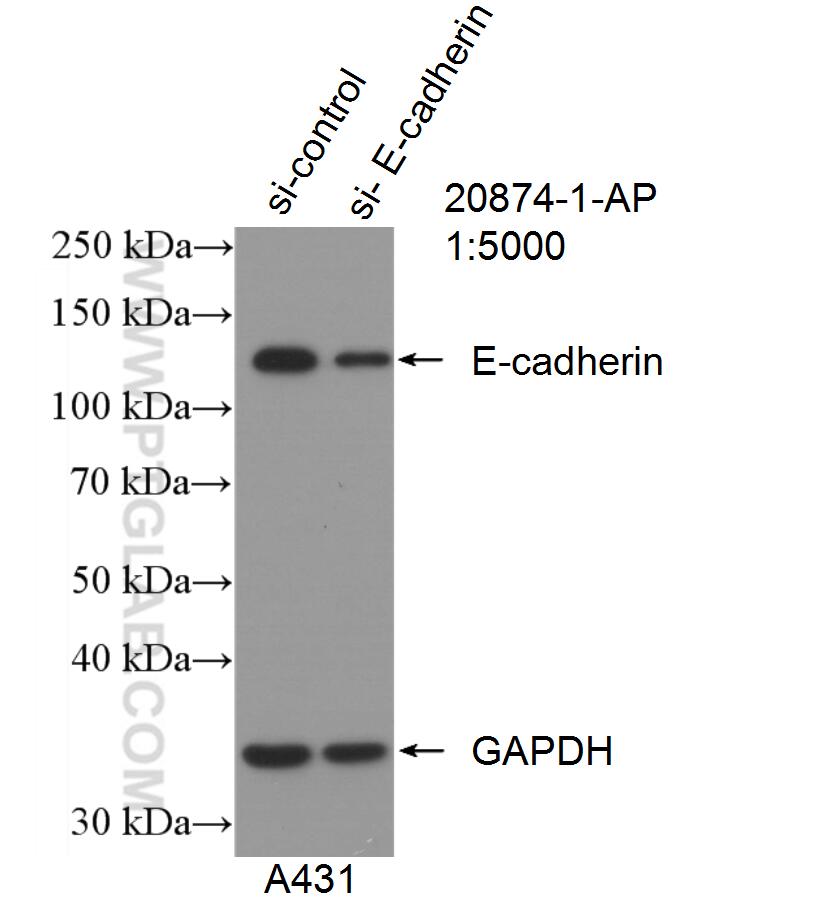
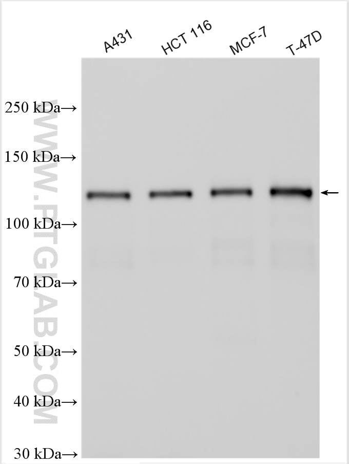
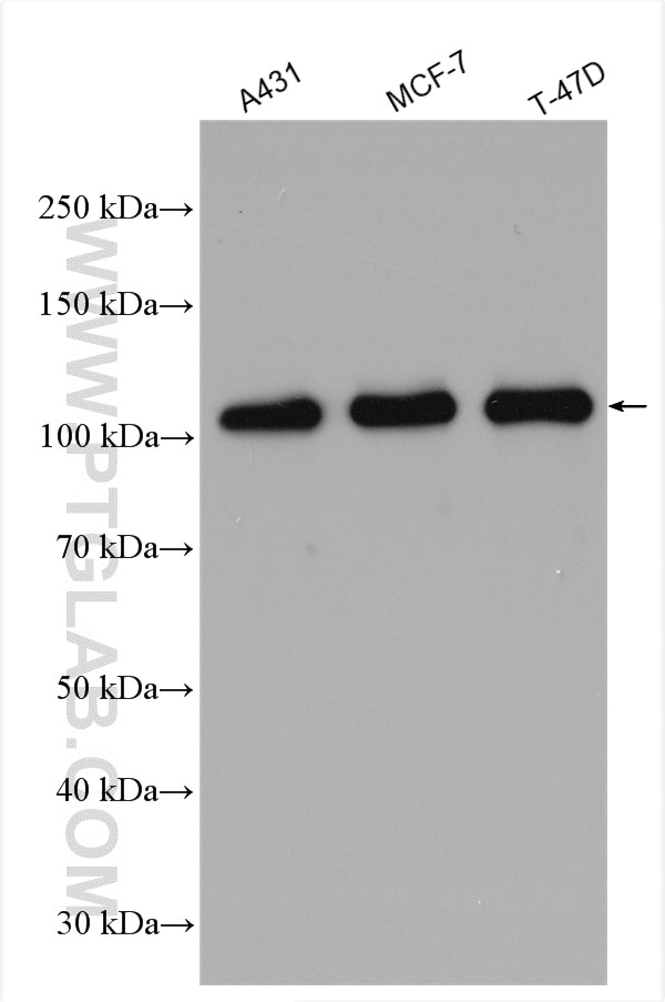
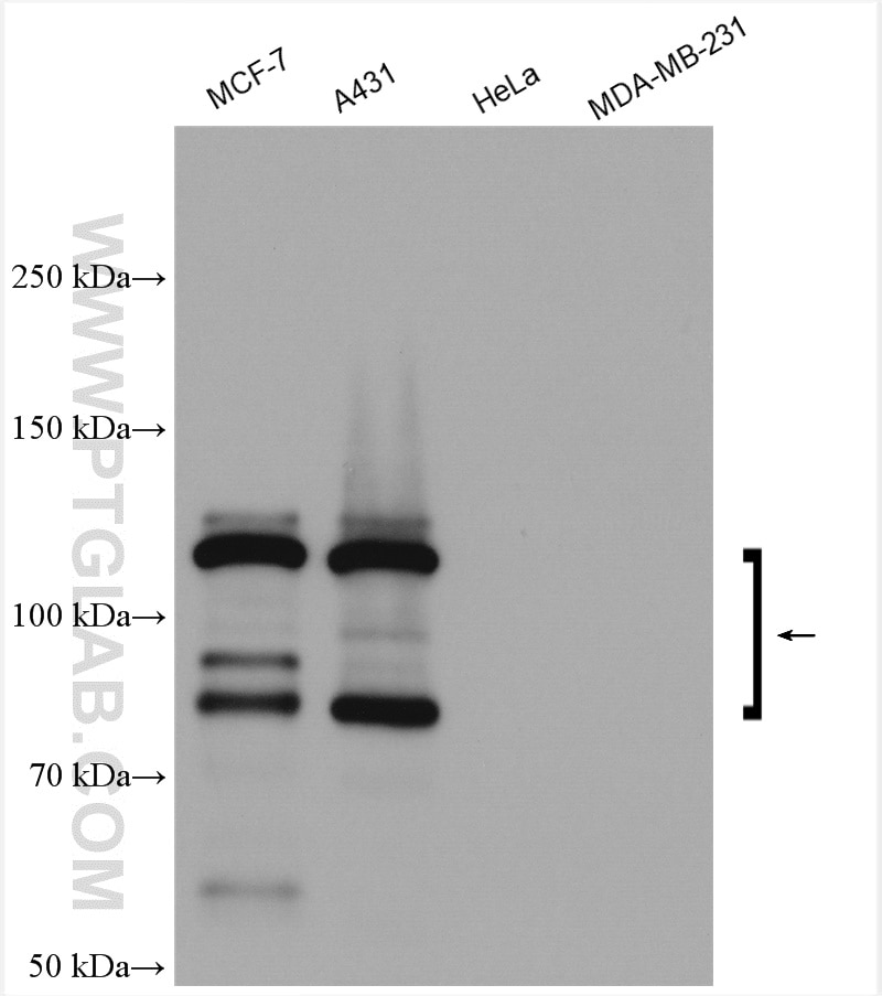
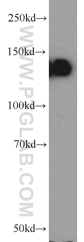
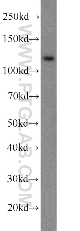
| Positive WB detected in | A431 cells, DU 145 cells, mouse testis tissue, HCT 116 cells, MCF-7 cells, T-47D cells |
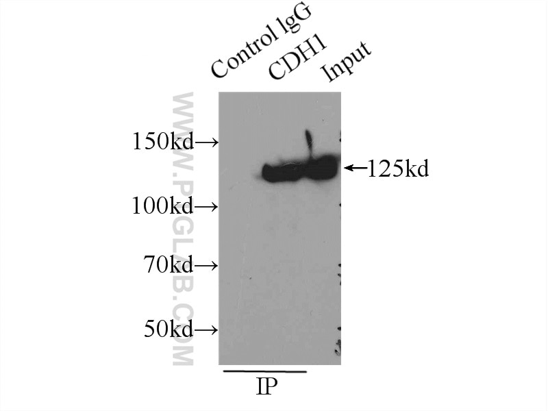
| Positive IP detected in | A431 cells |
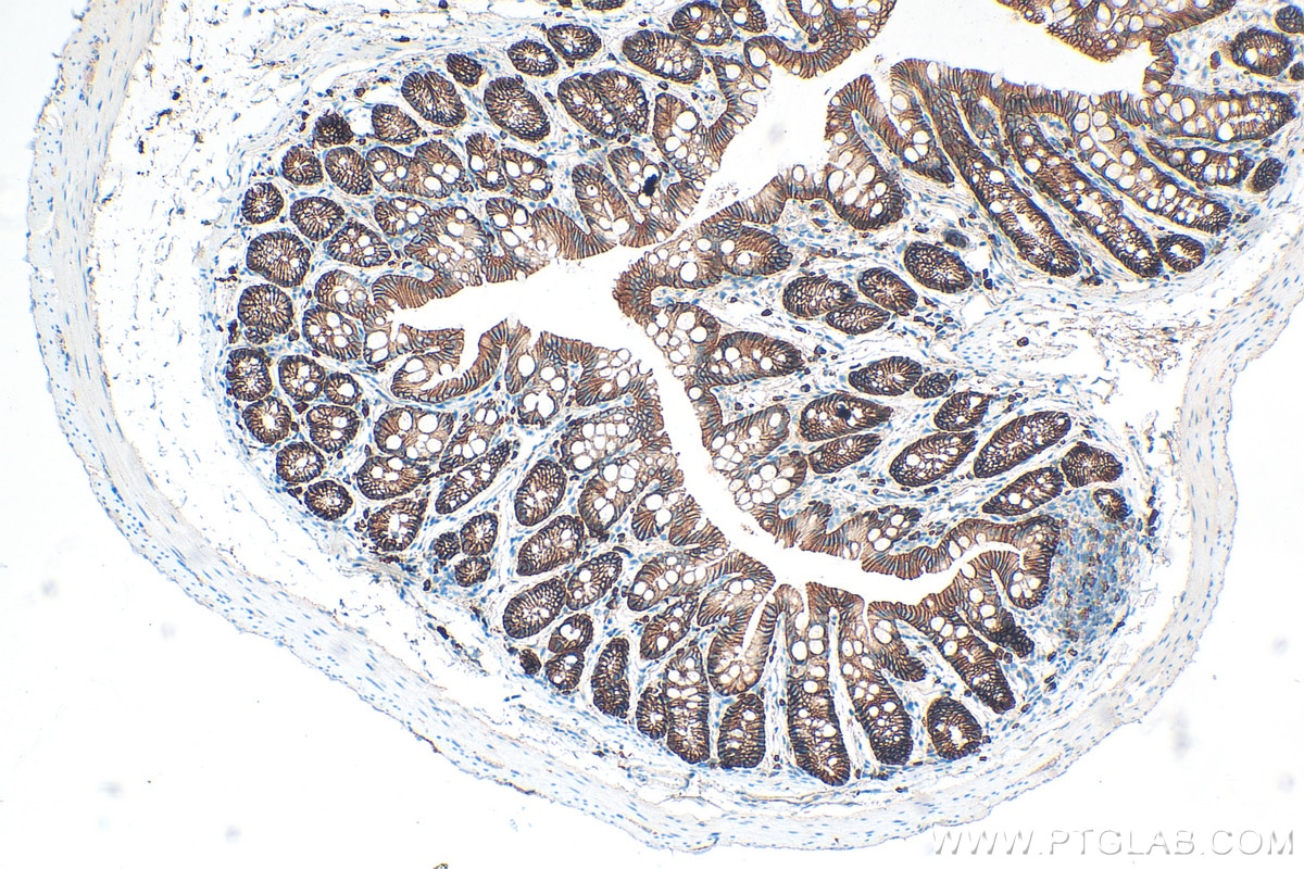
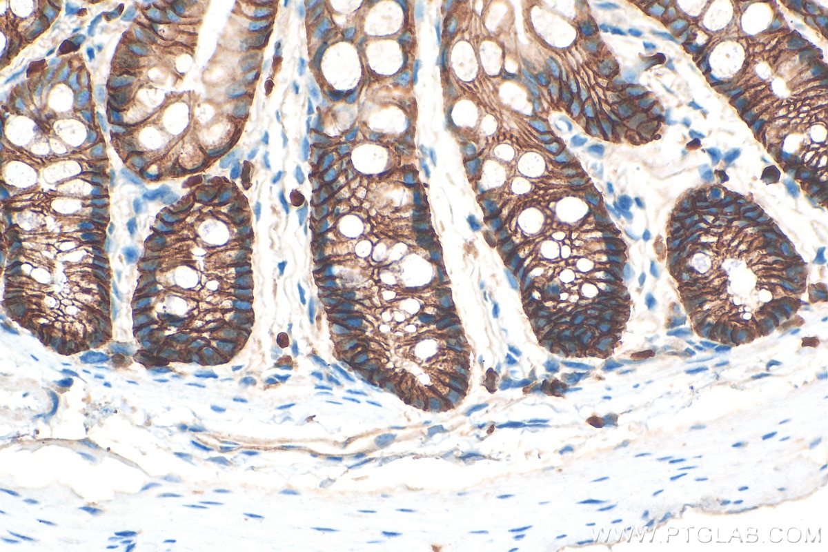
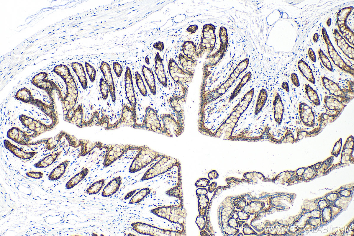
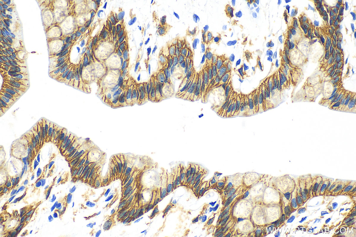
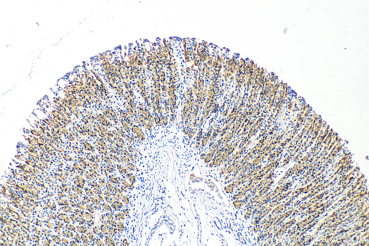
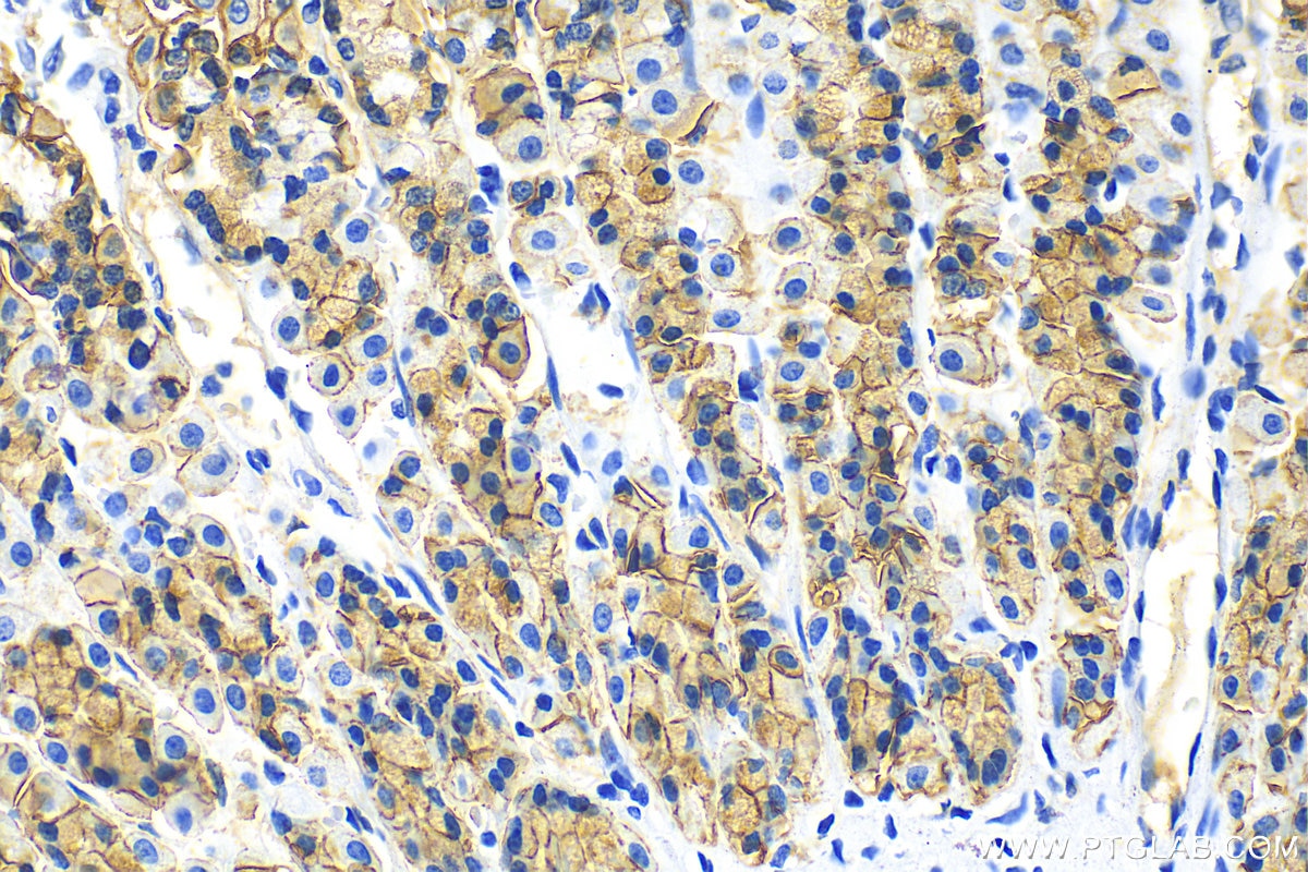
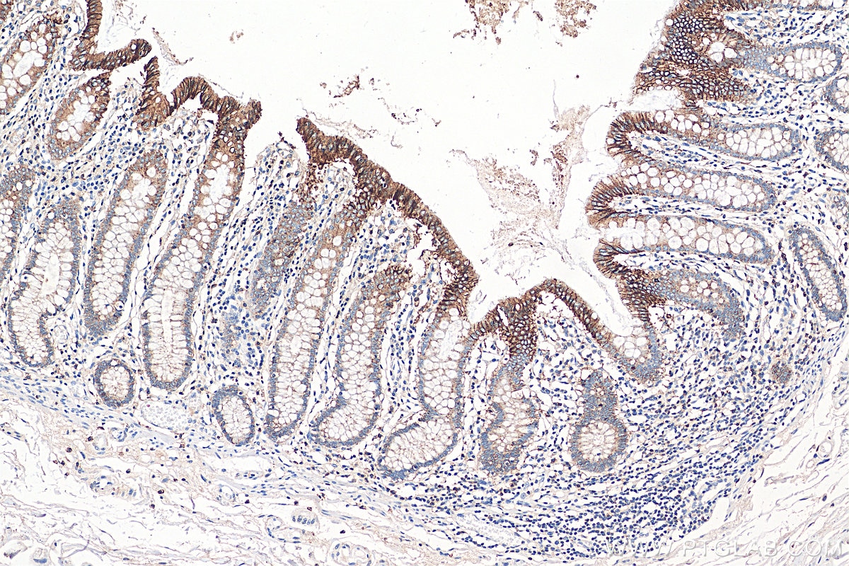
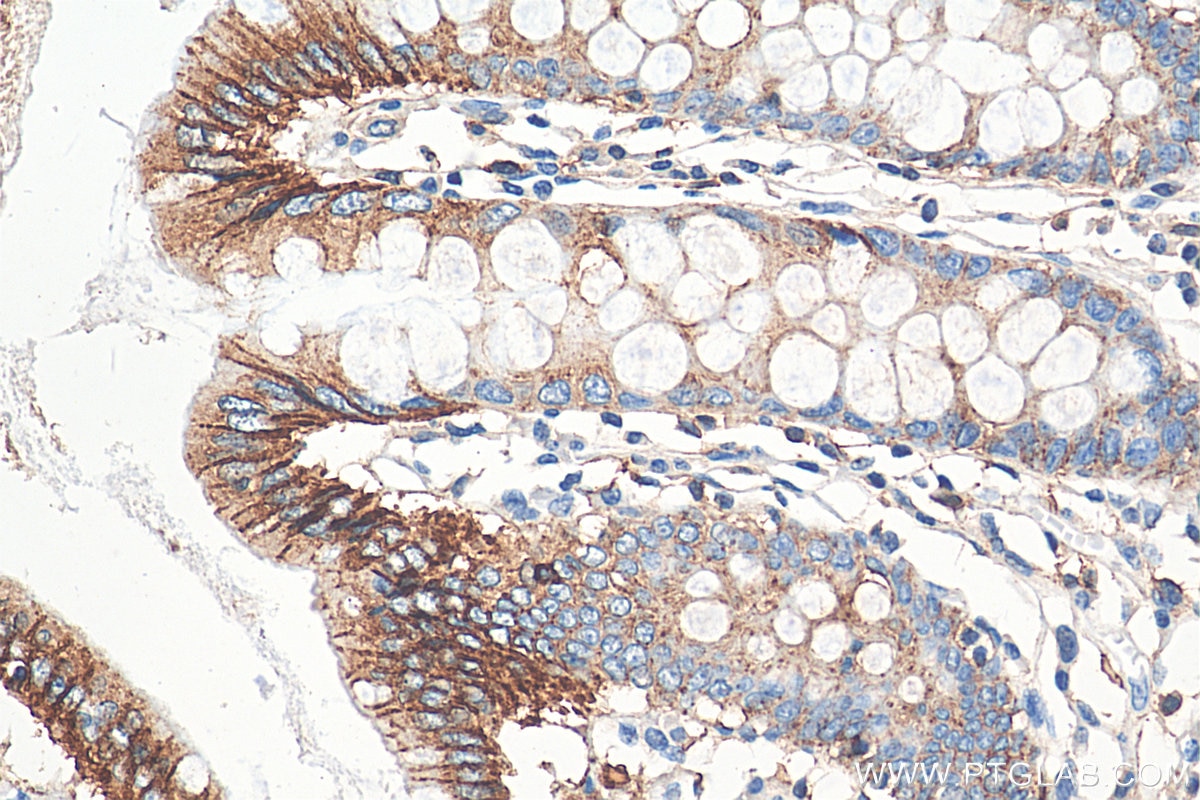
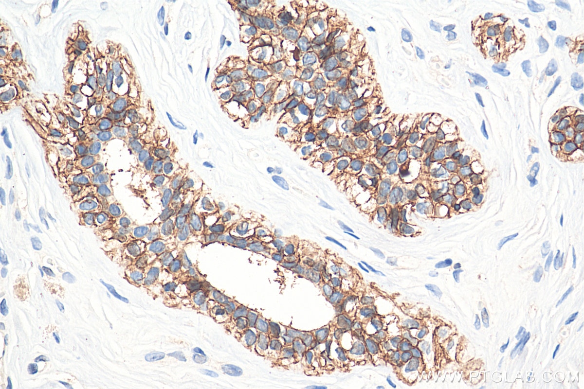
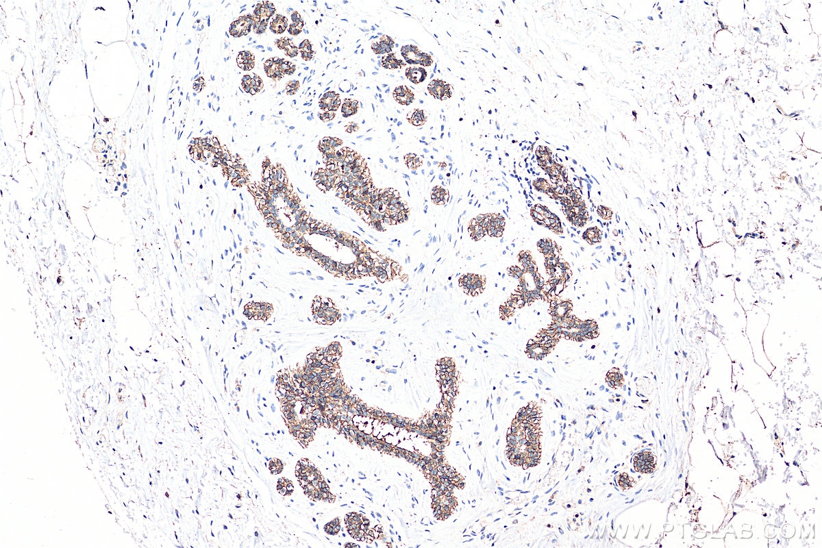
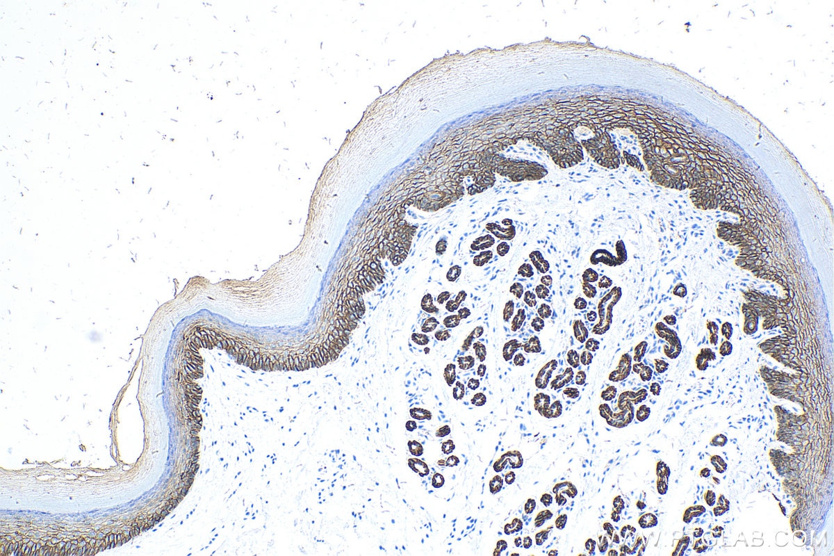
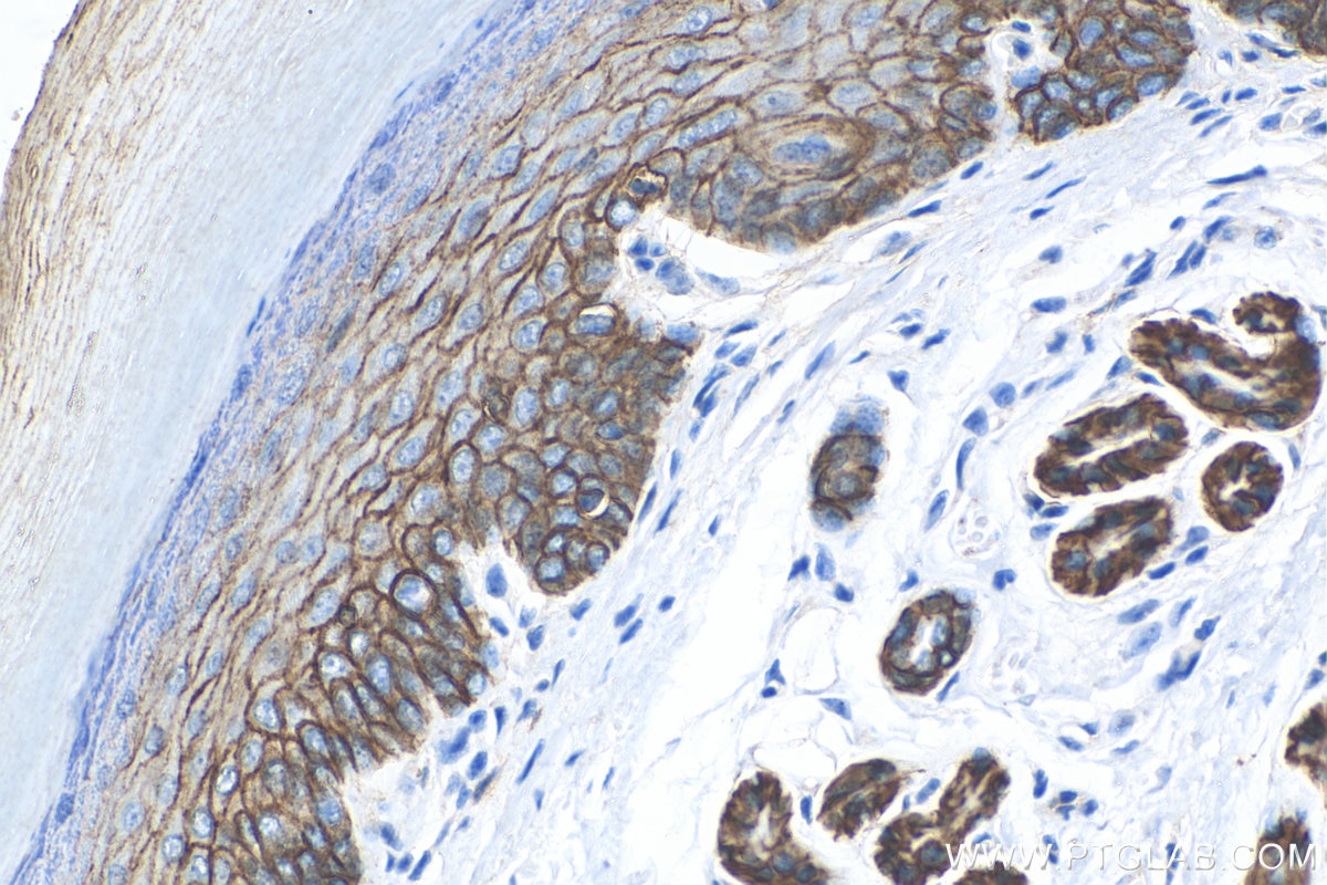
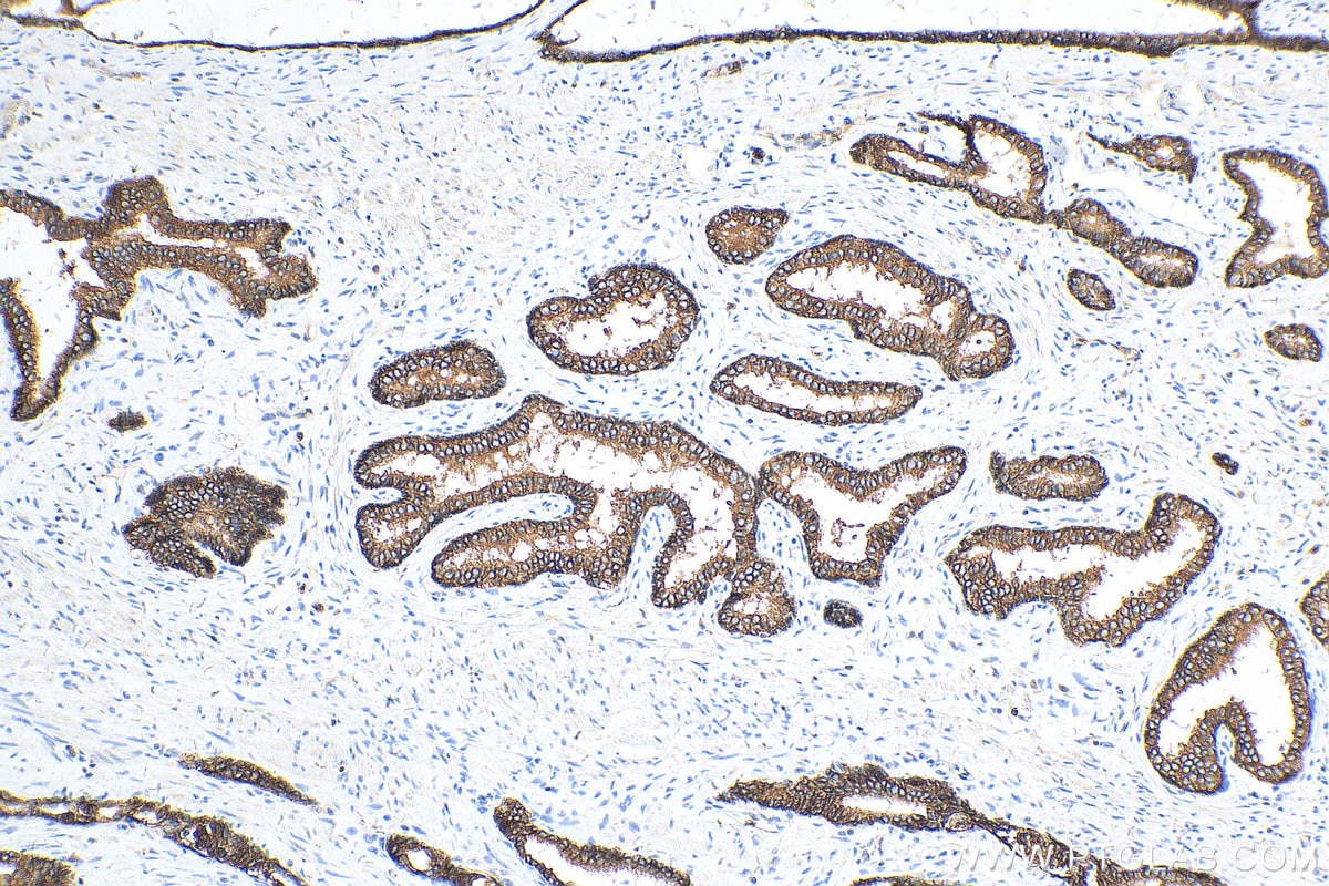
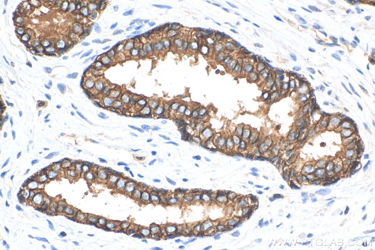
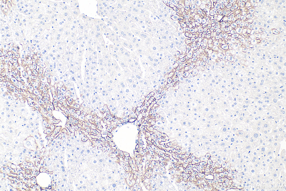
| Positive IHC detected in | mouse colon tissue, human breast cancer tissue, human colon tissue, human prostate cancer tissue, mouse liver tissue, mouse skin tissue, rat colon tissue, rat stomach tissue Note: suggested antigen retrieval with TE buffer pH 9.0; (*) Alternatively, antigen retrieval may be performed with citrate buffer pH 6.0 |
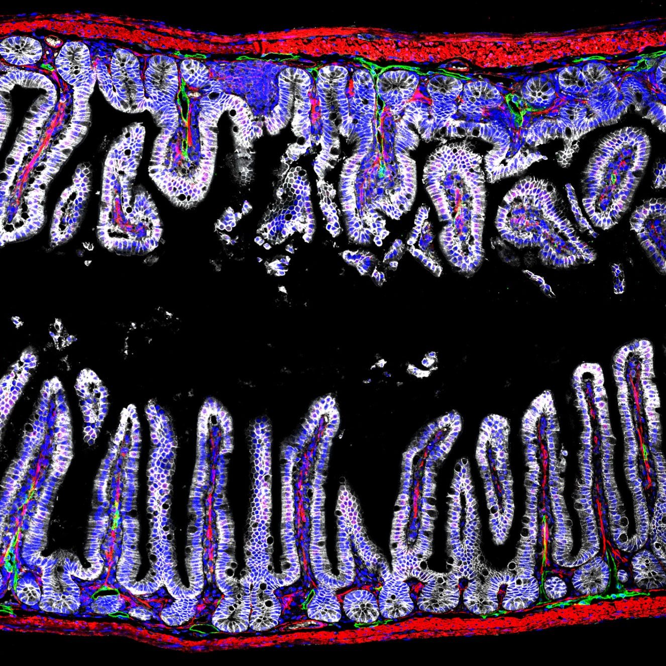
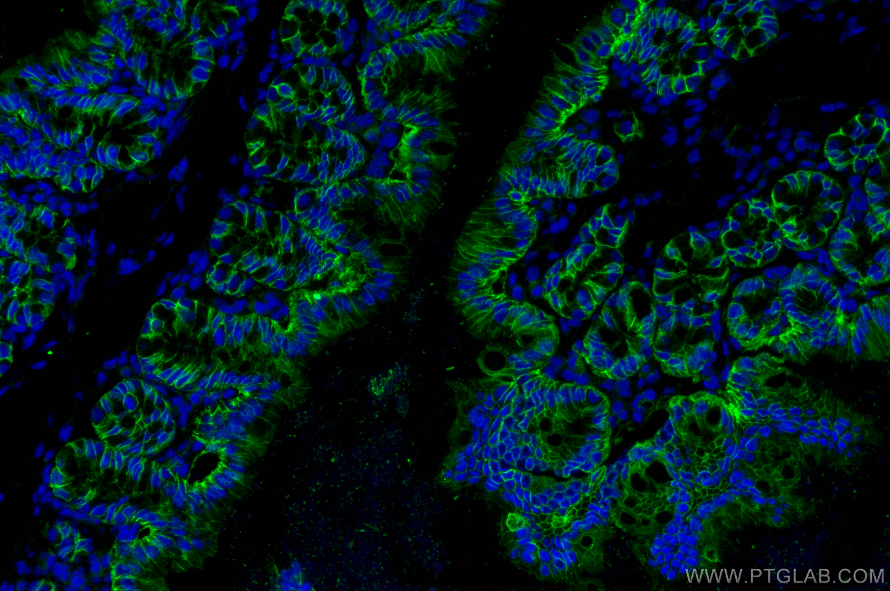
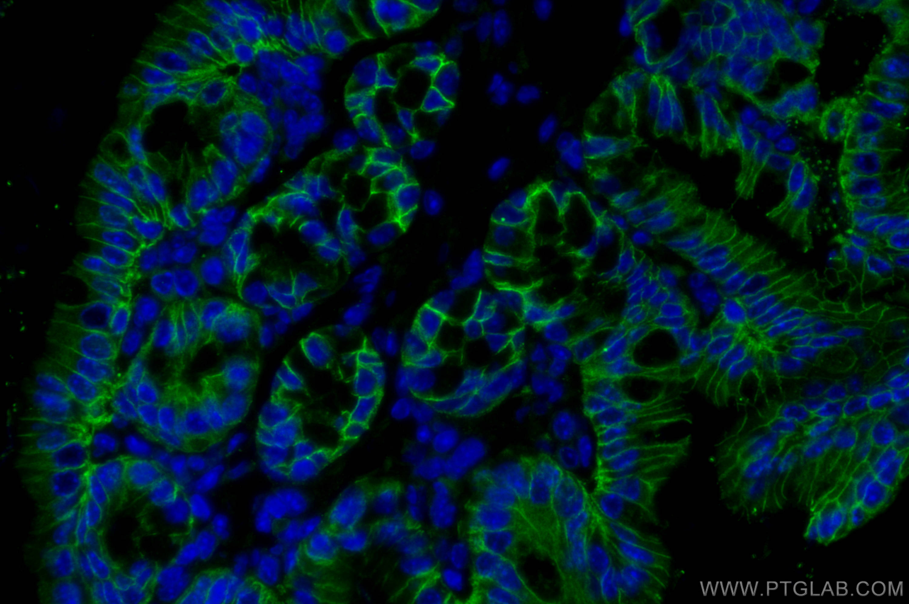
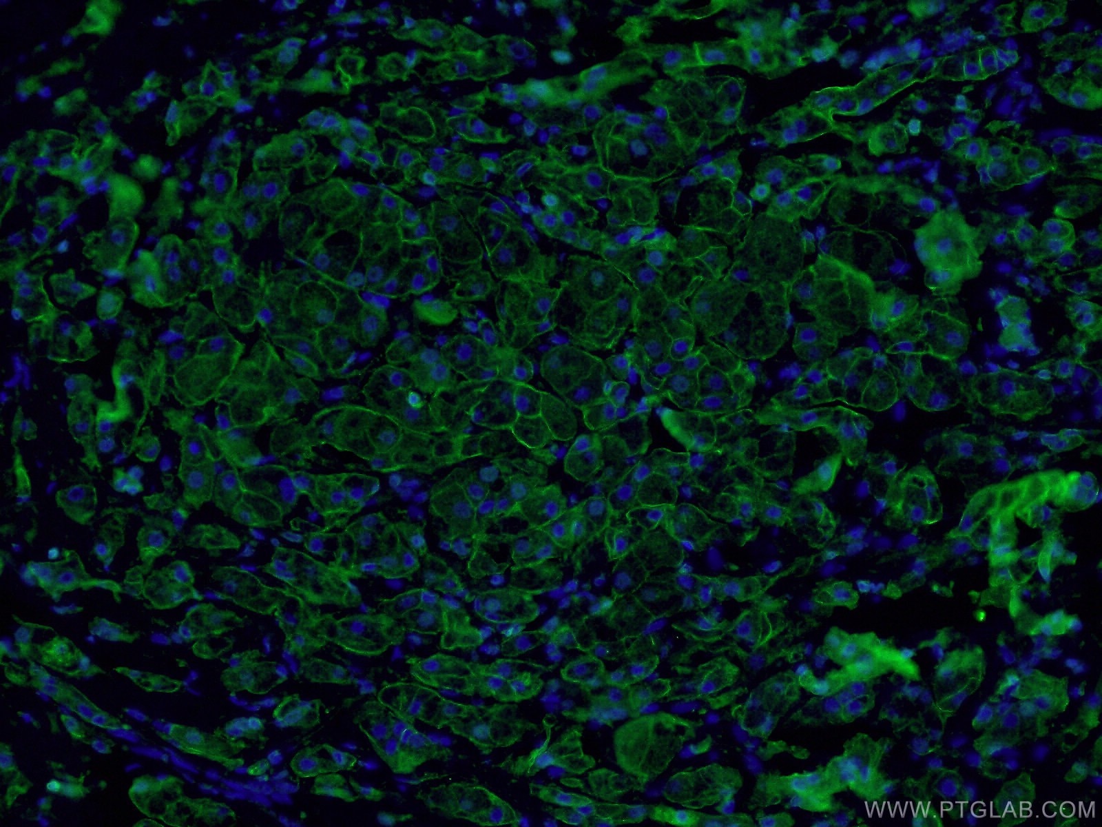
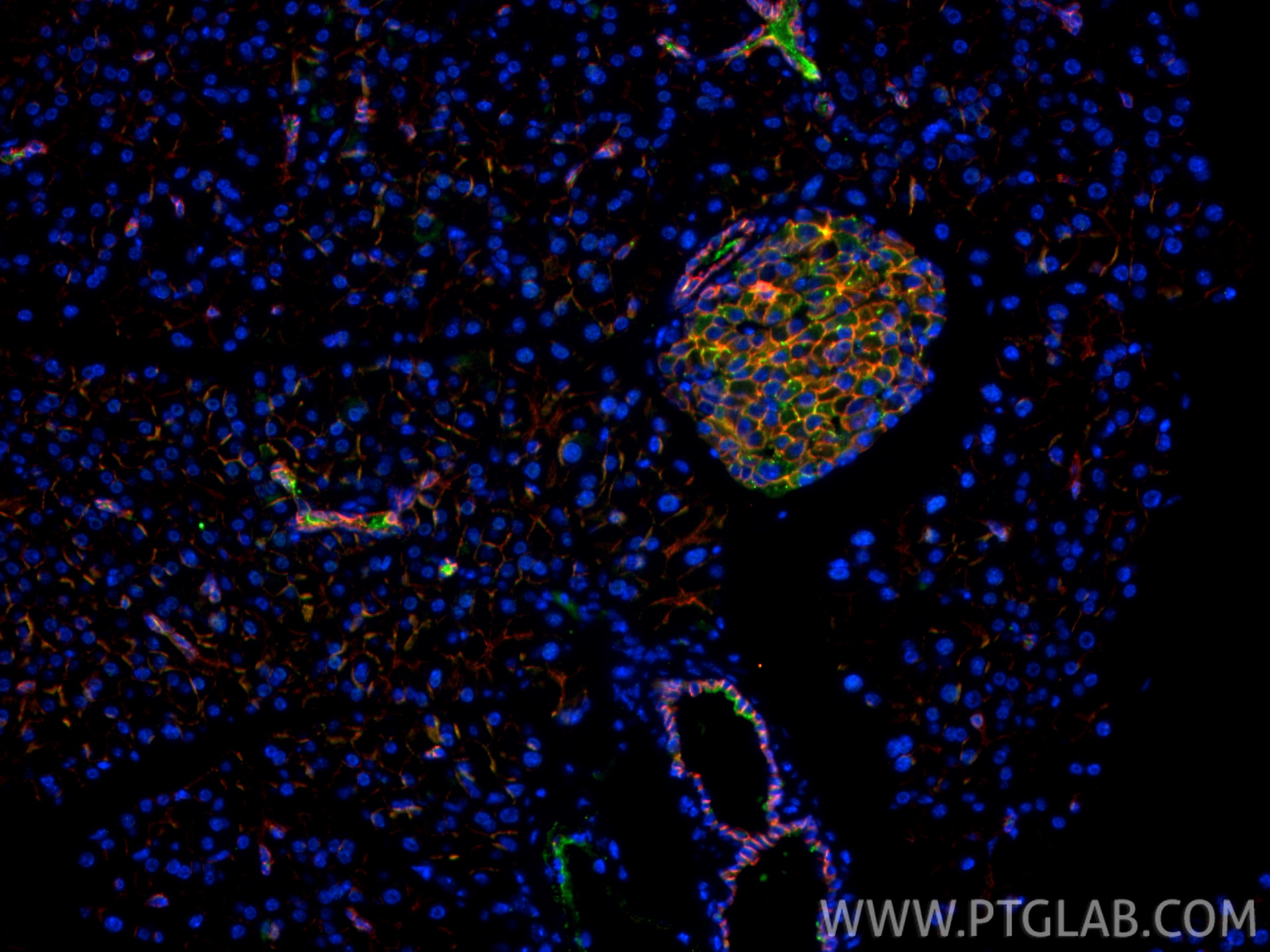
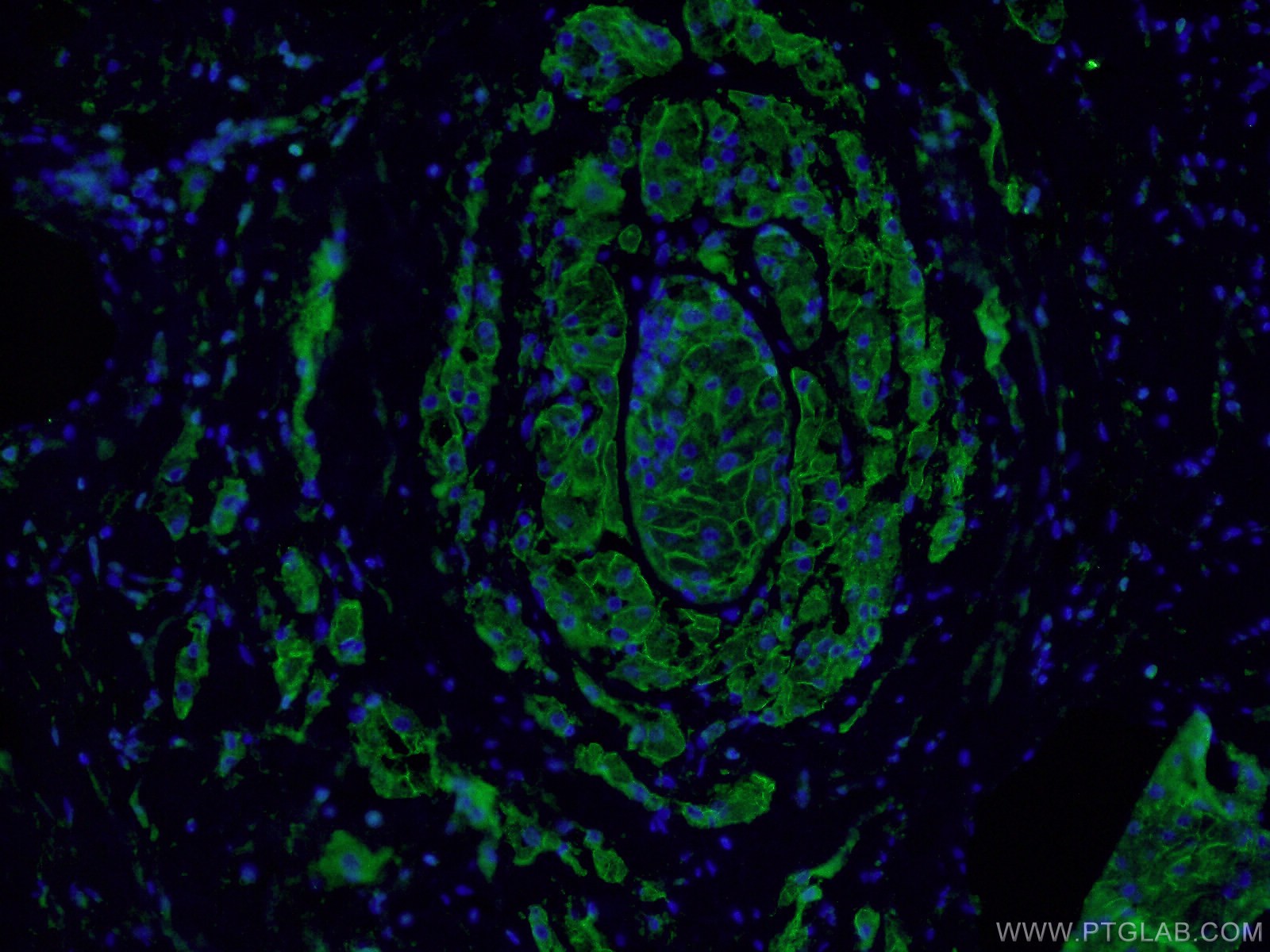
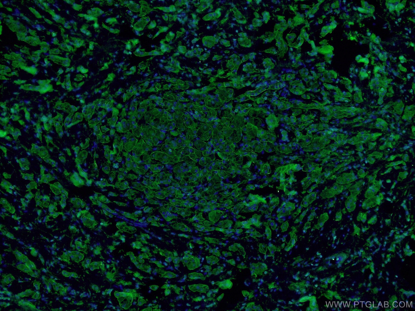
| Positive IF-P detected in | mouse colon tissue, human breast cancer tissue, mouse small intestine tissue, mouse pancreas tissue |
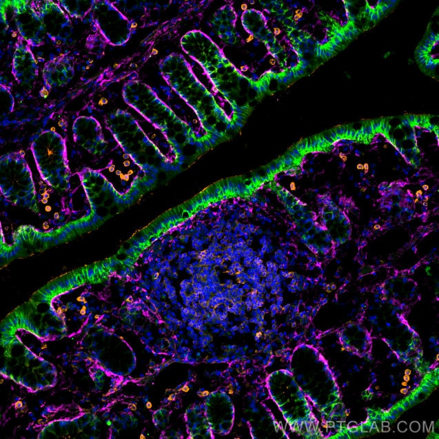
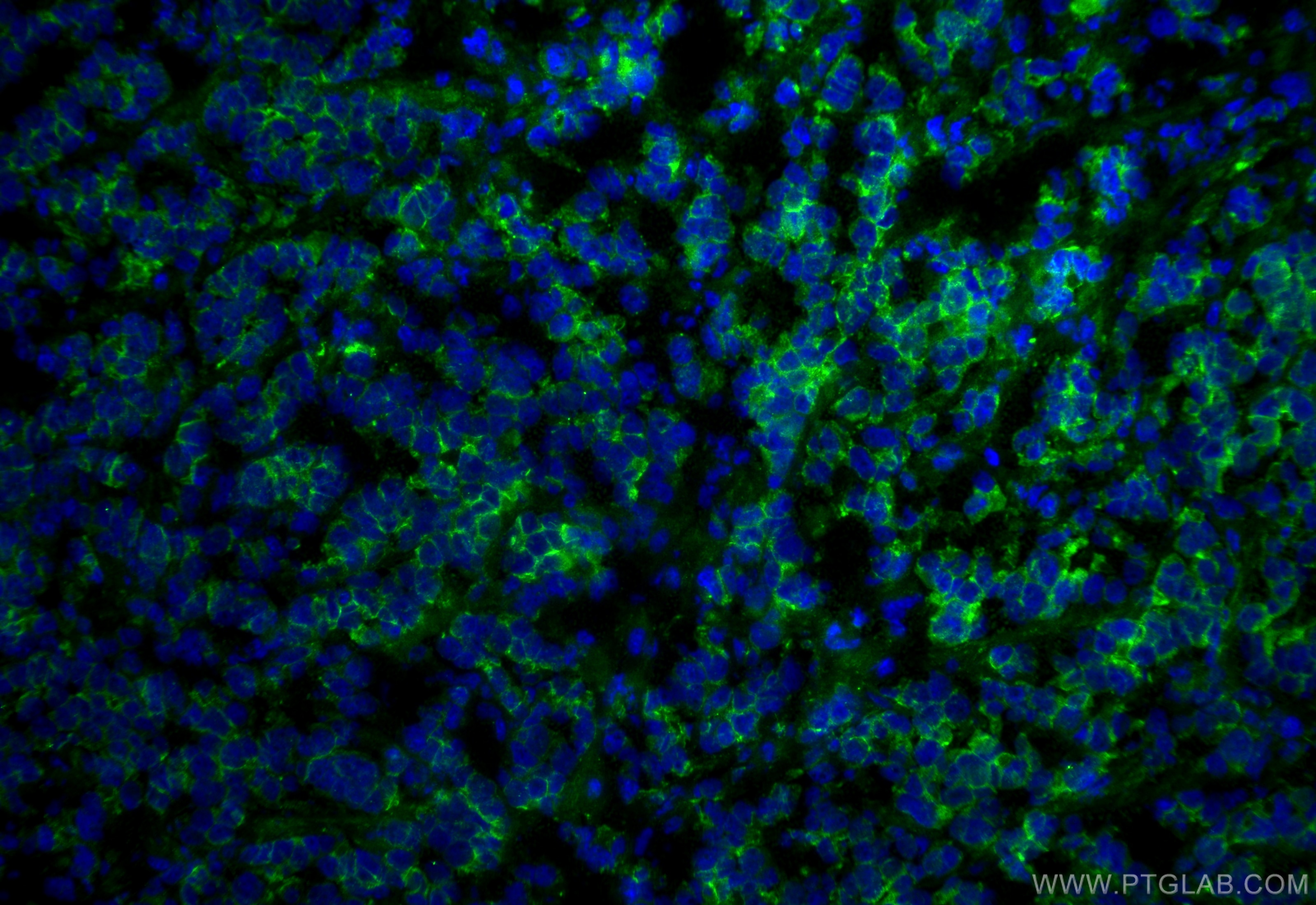
| Positive IF-Fro detected in | mouse colon tissue, mouse breast cancer |
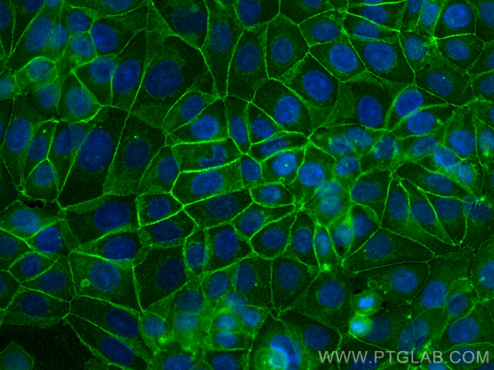
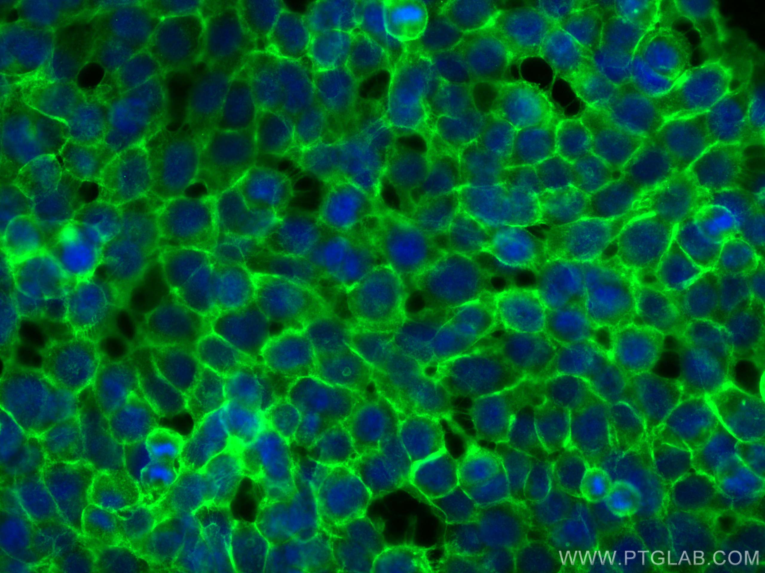
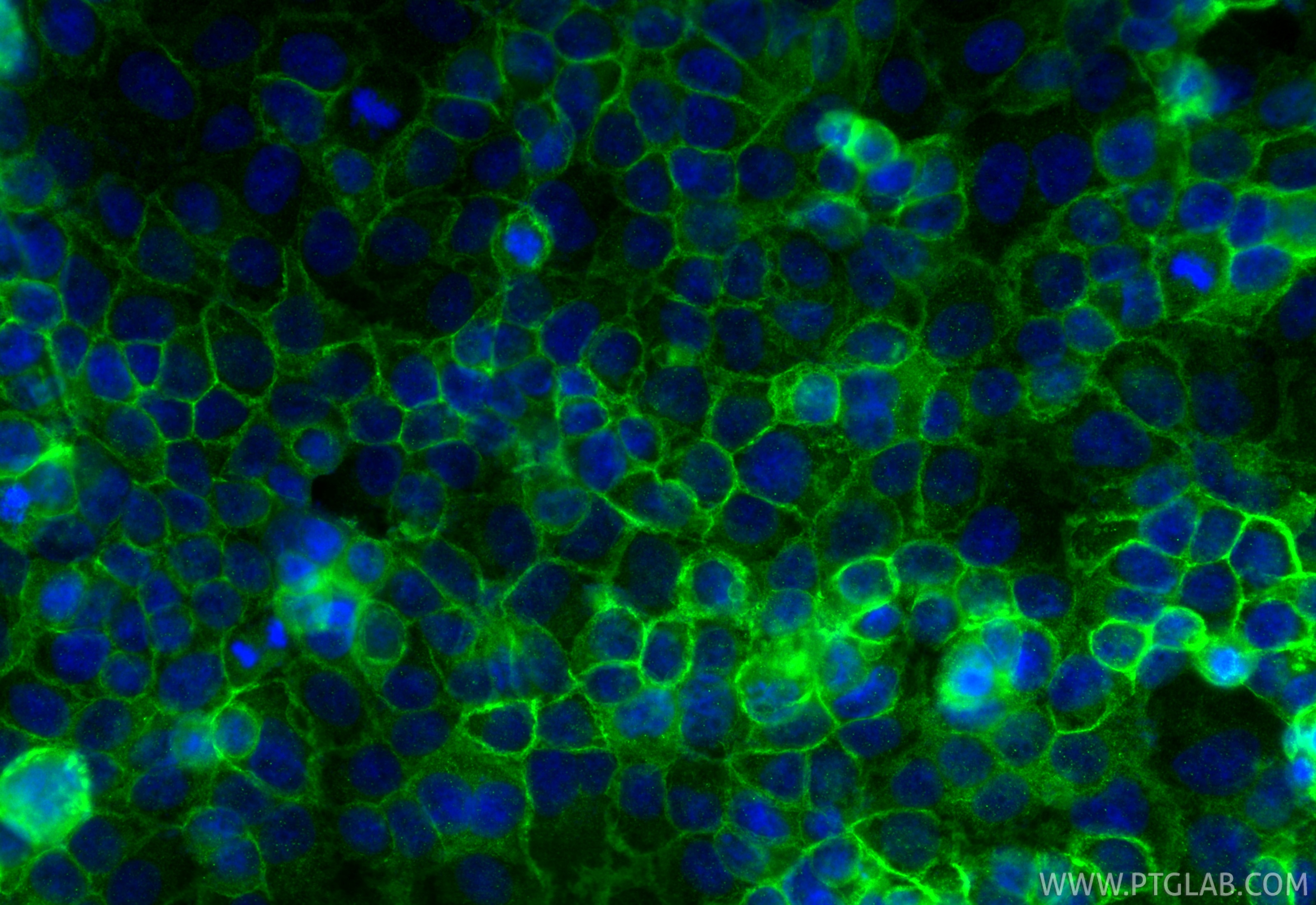
| Positive IF/ICC detected in | MCF-7 cells, A431 cells, OVCAR-3 cells |
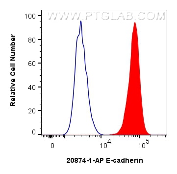
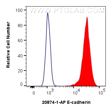
| Positive FC (Intra) detected in | MCF-7 cells |
| Positive FC detected in | MCF-7 cells |
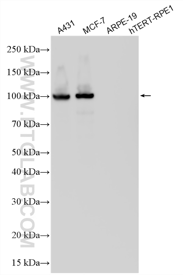
Some bands between 80 and 120 kDa may be observed due to proteolytic cleavage.
Recommended dilution
| Application | Dilution |
|---|---|
| Western Blot (WB) | WB : 1:20000-1:200000 |
| Immunoprecipitation (IP) | IP : 0.5-4.0 ug for 1.0-3.0 mg of total protein lysate |
| Immunohistochemistry (IHC) | IHC : 1:5000-1:20000 |
| Immunofluorescence (IF)-P | IF-P : 1:200-1:800 |
| Immunofluorescence (IF)-FRO | IF-FRO : 1:50-1:500 |
| Immunofluorescence (IF)/ICC | IF/ICC : 1:200-1:800 |
| Flow Cytometry (FC) (INTRA) | FC (INTRA) : 0.25 ug per 10^6 cells in a 100 µl suspension |
| Flow Cytometry (FC) | FC : 0.25 ug per 10^6 cells in a 100 µl suspension |
| It is recommended that this reagent should be titrated in each testing system to obtain optimal results. | |
| Sample-dependent, Check data in validation data gallery. | |
Product Information
20874-1-AP targets E-cadherin in WB, IHC, IF/ICC, IF-P, IF-Fro, FC (Intra), IP, CoIP, ELISA applications and shows reactivity with human, mouse, rat samples.
| Tested Reactivity | human, mouse, rat |
| Cited Reactivity | human, mouse, rat, pig, canine, chicken, zebrafish, bovine |
| Host / Isotype | Rabbit / IgG |
| Class | Polyclonal |
| Type | Antibody |
| Immunogen |
CatNo: Ag14973 Product name: Recombinant human E-cadherin protein Source: e coli.-derived, PGEX-4T Tag: GST Domain: 373-622 aa of BC141838 Sequence: PIFNPTTYKGQVPENEANVVITTLKVTDADAPNTPAWEAVYTILNDDGGQFVVTTNPVNNDGILKTAKGLDFEAKQQYILHVAVTNVVPFEVSLTTSTATVTVDVLDVNEAPIFVPPEKRVEVSEDFGVGQEITSYTAQEPDTFMEQKITYRIWRDTANWLEINPDTGAISTRAELDREDFEHVKNSTYTALIIATDNGSPVATGTGTLLLILSDVNDNAPIPEPRTIFFCERNPKPQVINIIDADLPPI Predict reactive species |
| Full Name | cadherin 1, type 1, E-cadherin (epithelial) |
| Calculated Molecular Weight | 882 aa, 97 kDa |
| Observed Molecular Weight | 120-125 kDa |
| GenBank Accession Number | BC141838 |
| Gene Symbol | E-cadherin |
| Gene ID (NCBI) | 999 |
| RRID | AB_10697811 |
| Conjugate | Unconjugated |
| Form | Liquid |
| Purification Method | Antigen affinity purification |
| UNIPROT ID | P12830 |
| Storage Buffer | PBS with 0.02% sodium azide and 50% glycerol, pH 7.3. |
| Storage Conditions | Store at -20°C. Stable for one year after shipment. Aliquoting is unnecessary for -20oC storage. 20ul sizes contain 0.1% BSA. |
Background Information
Cadherins are a family of transmembrane glycoproteins that mediate calcium-dependent cell-cell adhesion and play an important role in the maintenance of normal tissue architecture. E-cadherin (epithelial cadherin), also known as CDH1 (cadherin 1) or CAM 120/80, is a classical member of the cadherin superfamily which also includes N-, P-, R-, and B-cadherins. E-cadherin is expressed on the cell surface in most epithelial tissues. The extracellular region of E-cadherin establishes calcium-dependent homophilic trans binding, providing specific interaction with adjacent cells, while the cytoplasmic domain is connected to the actin cytoskeleton through the interaction with p120-, α-, β-, and γ-catenin (plakoglobin). E-cadherin is important in the maintenance of epithelial integrity and is involved in mechanisms regulating proliferation, differentiation, and survival of epithelial cells. E-cadherin may also play a role in tumorigenesis. It is considered to be an invasion suppressor protein, and its loss is an indicator of high tumor aggressiveness. E-cadherin is sensitive to trypsin digestion in the absence of Ca2+. This polyclonal antibody recognizes 120-125 kDa intact E-cadherin and its cleaved fragments of 80-120 kDa.
Protocols
| Product Specific Protocols | |
|---|---|
| FC protocol for E-cadherin antibody 20874-1-AP | Download protocol |
| IF protocol for E-cadherin antibody 20874-1-AP | Download protocol |
| IHC protocol for E-cadherin antibody 20874-1-AP | Download protocol |
| IP protocol for E-cadherin antibody 20874-1-AP | Download protocol |
| WB protocol for E-cadherin antibody 20874-1-AP | Download protocol |
| Standard Protocols | |
|---|---|
| Click here to view our Standard Protocols |
Publications
What published studies show
The E-cadherin Polyclonal antibody (Cat# 20874-1-AP) has accumulated 3,297 citations. Publications appear in high-impact journals including Nature, Science, Nature Communications, Molecular Cancer, Cancer Cell, Gastroenterology, Advanced Science, Theranostics, and Journal of Experimental & Clinical Cancer Research. Research fields span cancer biology (EMT, metastasis, chemoresistance), autophagy, inflammation, tissue regeneration, stem cell biology, nanomedicine, gut microbiome, and cardiovascular disease, with applications primarily in WB, IHC, and IF.
| Species | Application | Title |
|---|---|---|
Signal Transduct Target Ther Prostaglandin E2-driven dedifferentiation of Schwann cells leads to perineural invasion in pancreatic ductal adenocarcinoma | ||
Signal Transduct Target Ther Hypoxia-inducible factor upregulation by roxadustat attenuates drug reward by altering brain iron homoeostasis | ||
Cancer Cell A conserved re-epithelialization program underlies malignancy in pancreatic ductal adenocarcinoma. | ||
J Hematol Oncol The LINC00623/NAT10 signaling axis promotes pancreatic cancer progression by remodeling ac4C modification of mRNA | ||
J Hematol Oncol LincK contributes to breast tumorigenesis by promoting proliferation and epithelial-to-mesenchymal transition. |
Reviews
What customers say
All 14 reviews are highly positive (12 five-star, 2 four-star). Users report excellent performance in Western Blot, Immunofluorescence, IHC, and Immunoprecipitation across human, mouse, and rat samples. Commonly tested cell types include Caco2, ARPE-19, Müller glia, corneal cells, and lung cancer cells. Recommended dilutions range from 1:100 to 1:5000 depending on the application. Reviewers consistently highlight strong signal, specific bands, and reliable staining, making it a dependable choice for E-cadherin detection in adhesion-related studies.
The reviews below have been submitted by verified Proteintech customers who received an incentive for providing their feedback.
P (Verified Customer) (03-22-2026) | Works for cells and mouse tissue
|
Sai Sindhura (Verified Customer) (12-22-2025) | E-cadherin is working well.
|
ray (Verified Customer) (12-11-2025) | Reliable polyclonal anti-E-cadherin antibody with strong performance in Western blot, IF/ICC, IHC, and more, delivering clear, specific detection across human, mouse, and rat samples. Widely cited in publications and dependable for adhesion-related studies.
|
Dhanwini (Verified Customer) (09-24-2025) | GOOD
|
Robert (Verified Customer) (09-24-2025) | Works as intended!
|
Sneha (Verified Customer) (09-24-2025) | GOOD
|
Zeeshan (Verified Customer) (09-18-2025) | Work great
|
Michael (Verified Customer) (09-18-2025) | Works perfectly for western blots
|
Henry (Verified Customer) (09-12-2025) | Excellent product Perfect bands
|
Jimmy (Verified Customer) (05-20-2025) | Great results for western blot application
|
Davide (Verified Customer) (11-22-2022) | The antibody works optimally at 1:100. By the strenght of the signal it could probably also work at 1:50. Optimal product. E-CAD in red in the picture (abundant background signal due to non optimal blocking)
 |
Sophie (Verified Customer) (02-07-2022) | Highly efficient staining when used at 1/1000 overnight for IF on paraffin embedded tissues
|
Kishor (Verified Customer) (01-30-2019) | This antibody working very good for human cancer cells in western blotting (1:1000)
|
Martin (Verified Customer) (12-13-2017) |
|






































