

Tested Applications
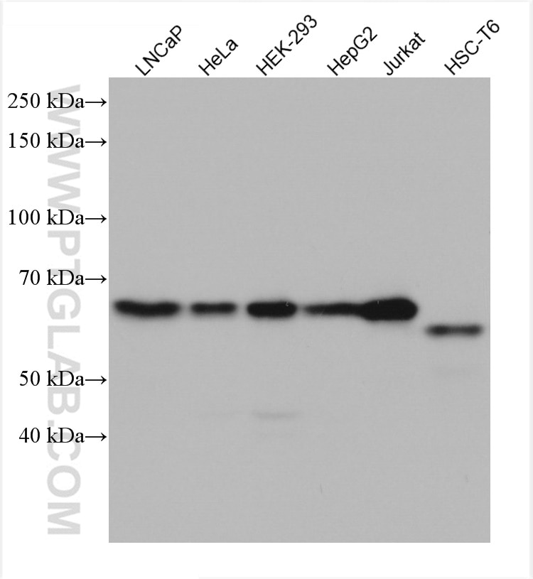
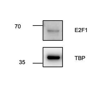
| Positive WB detected in | LNCaP cells, HepG2 cells, HeLa cells, HEK-293 cells, Jurkat cells, HSC-T6 cells |
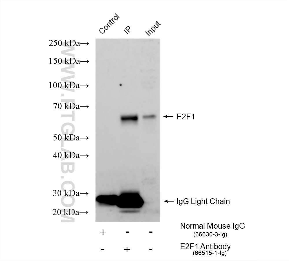
| Positive IP detected in | HEK-293T cells |
Recommended dilution
| Application | Dilution |
|---|---|
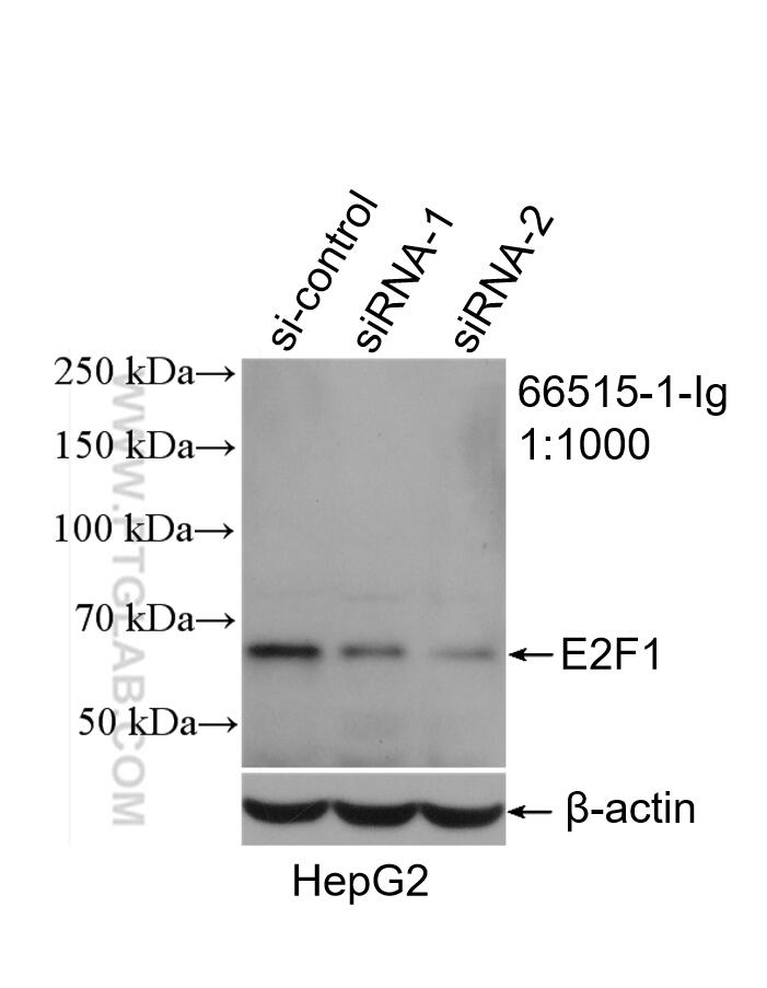
| Western Blot (WB) | WB : 1:1000-1:6000 |
| Immunoprecipitation (IP) | IP : 0.5-4.0 ug for 1.0-3.0 mg of total protein lysate |
| It is recommended that this reagent should be titrated in each testing system to obtain optimal results. | |
| Sample-dependent, Check data in validation data gallery. | |
Product Information
66515-1-Ig targets E2F1 in WB, IHC, IF, IP, CoIP, chIP, RIP, ELISA applications and shows reactivity with human, rat samples.
| Tested Reactivity | human, rat |
| Cited Reactivity | human, mouse, rat |
| Host / Isotype | Mouse / IgG2b |
| Class | Monoclonal |
| Type | Antibody |
| Immunogen |
CatNo: Ag17363 Product name: Recombinant human E2F1 protein Source: e coli.-derived, PET28a Tag: 6*His Domain: 88-438 aa of BC050369 Sequence: VKRRLDLETDHQYLAESSGPARGRGRHPGKGVKSPGEKSRYETSLNLTTKRFLELLSHSADGVVDLNWAAEVLKVQKRRIYDITNVLEGIQLIAKKSKNHIQWLGSHTTVGVGGRLEGLTQDLRQLQESEQQLDHLMNICTTQLRLLSEDTDSQRLAYVTCQDLRSIADPAEQMVMVIKAPPETQLQAVDSSENFQISLKSKQGPIDVFLCPEETVGGISPGKTPSQEVTSEEENRATDSATIVSPPPSSPPSSLTTDPSQSLLSLEQEPLLSRMGSLRAPVDEDRLSPLVAADSLLEHVREDFSGLLPEEFISLSPPHEALDYHFGLEEGEGIRDLFDCDFGDLTPLDF Predict reactive species |
| Full Name | E2F transcription factor 1 |
| Calculated Molecular Weight | 437 aa, 47 kDa |
| Observed Molecular Weight | 55-60 kDa |
| GenBank Accession Number | BC050369 |
| Gene Symbol | E2F1 |
| Gene ID (NCBI) | 1869 |
| RRID | AB_2881878 |
| Conjugate | Unconjugated |
| Form | Liquid |
| Purification Method | Protein A purification |
| UNIPROT ID | Q01094 |
| Storage Buffer | PBS with 0.02% sodium azide and 50% glycerol, pH 7.3. |
| Storage Conditions | Store at -20°C. Stable for one year after shipment. Aliquoting is unnecessary for -20oC storage. 20ul sizes contain 0.1% BSA. |
Background Information
Transcription factor E2F1 (E2F1), also known as RBBP3, is a transcription activator that binds DNA cooperatively with dp proteins through the E2 recognition site, 5'-TTTC[CG]CGC-3' found in the promoter region of a number of genes whose products are involved in cell cycle regulation or in DNA replication. The DRTF1/E2F complex functions in the control of cell-cycle progression from G1 to S phase. E2F-1 binds preferentially RB1 protein, in a cell-cycle dependent manner. It can mediate both cell proliferation and p53-dependent apoptosis. The calculated molecular weight of E2F1 is 47 kDa, but the sumoylated E2F1 is bout 55-60 kDa.
Protocols
| Product Specific Protocols | |
|---|---|
| IP protocol for E2F1 antibody 66515-1-Ig | Download protocol |
| WB protocol for E2F1 antibody 66515-1-Ig | Download protocol |
| Standard Protocols | |
|---|---|
| Click here to view our Standard Protocols |
Publications
What published studies show
E2F1 antibody (Cat# 66515-1-Ig) has accumulated 102 citations across journals including Nature Communications, Cancer Research, Cell Death & Disease, Molecular Cancer, Signal Transduction and Targeted Therapy, Cell Reports, Gut, and Leukemia. Research fields span oncology (lung, breast, gastric, colorectal, prostate, ovarian, pancreatic, liver, and glioma cancers), cell cycle regulation, transcriptional control, DNA damage repair, ferroptosis, and inflammation. Primary applications include WB, IHC, IF, ChIP, and IP.
| Species | Application | Title |
|---|---|---|
Signal Transduct Target Ther BMX inhibition overcomes small cell lung cancer chemoresistance by stabilizing E2F1 via ERK1/2-Cyclin D1/CDK4/6 axis
| ||
Mol Cancer BRD9-p53-E2F1 circuit orchestrates cell growth and DNA damage repair in gastric cancer | ||
Mol Cancer Targeting MYC in T-Cell lymphoma via epigenetic modulation with a small-molecule inhibitor. | ||
Cancer Res LncRNA AGPG confers endocrine resistance in breast cancer by promoting E2F1 activity | ||
Cancer Res TRAPPC4 Promotes GPX4 Stability to Drive Ferroptosis Resistance and Tumor Progression in Head and Neck Squamous Cell Carcinoma |
Reviews
What customers say
Customer reviews for this antibody are generally positive for Western Blot applications. One user reported excellent performance at a 1:2000 dilution, noting it captured the target effectively even with low protein concentrations and short exposure times. Another user found it worked well but required an ECL sensitive reagent and higher exposure for optimal imaging. Overall, the antibody performs reliably in WB, with results varying slightly based on sample type and detection conditions.
The reviews below have been submitted by verified Proteintech customers who received an incentive for providing their feedback.
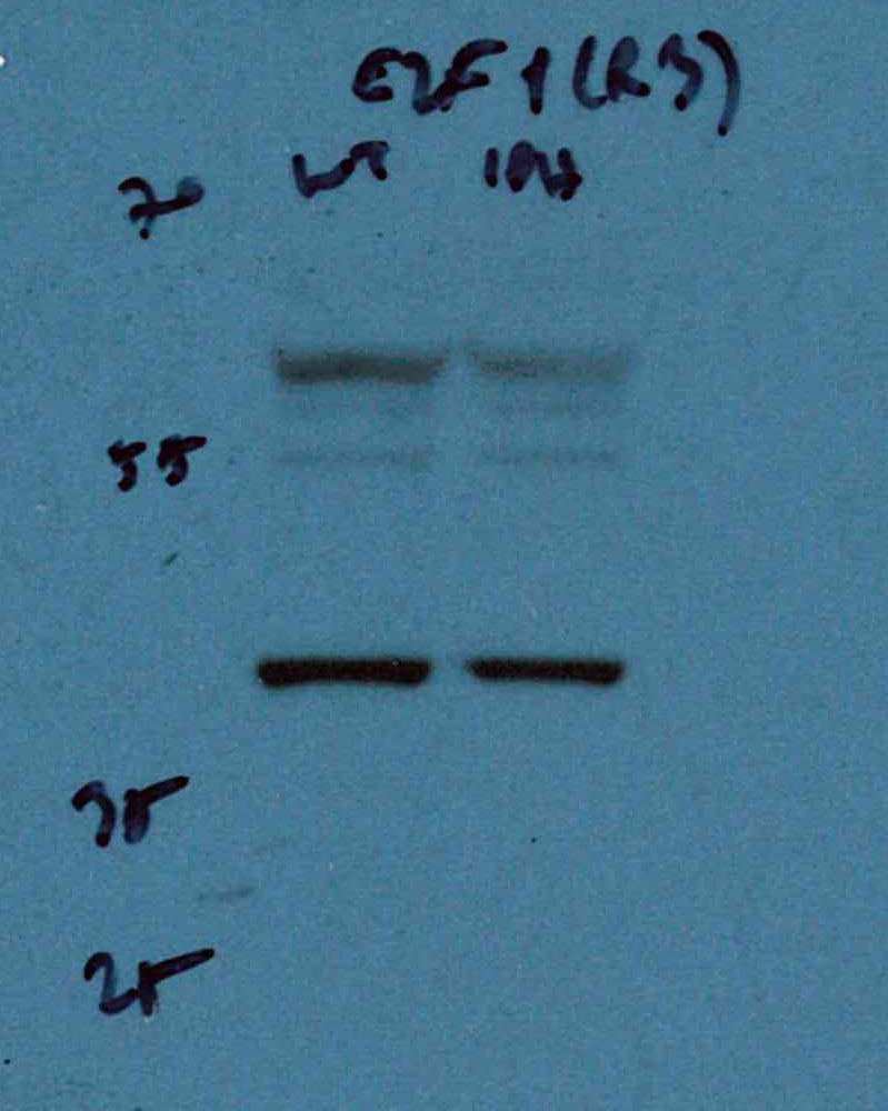
Umut (Verified Customer) (08-30-2023) | It works very well, even though it is diluted a lot, i.e., 1:2000. The total protein concentration of my samples were around 2 ug/ml before adding 1:4 laemmli buffer and denature, and it is able to capture the target at low exposure times.
 |
Juliane (Verified Customer) (05-11-2023) | The antibody worked fine, but only under using ECL sensitve reagent and high exposure when imaging.
 |




