
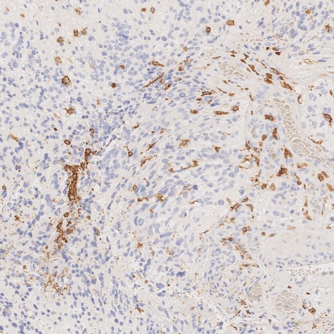
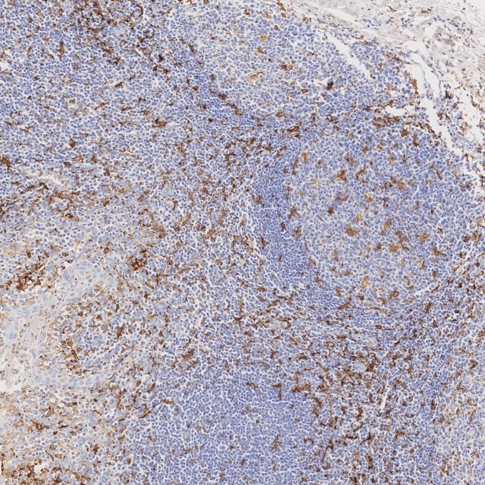
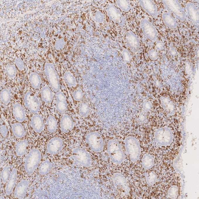
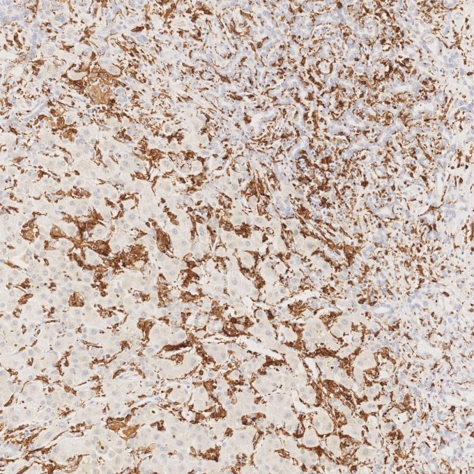
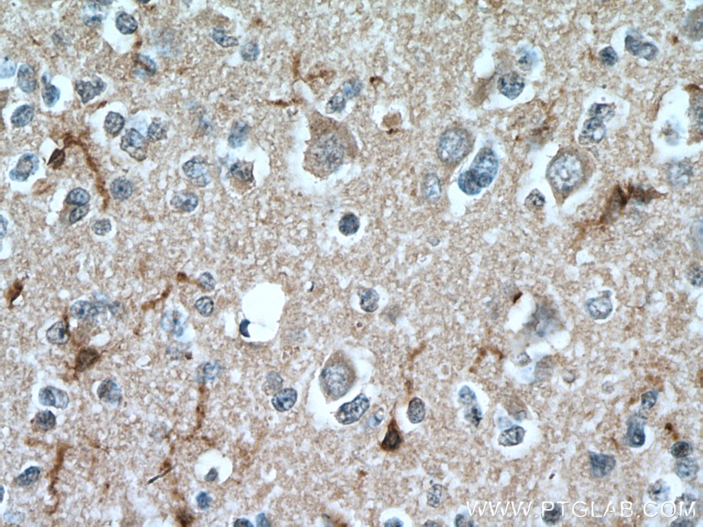

Tested Applications
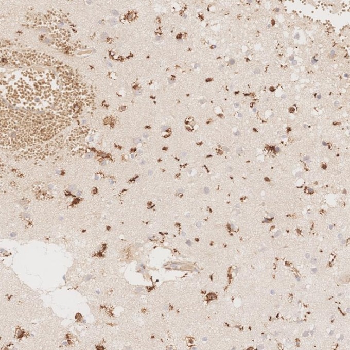
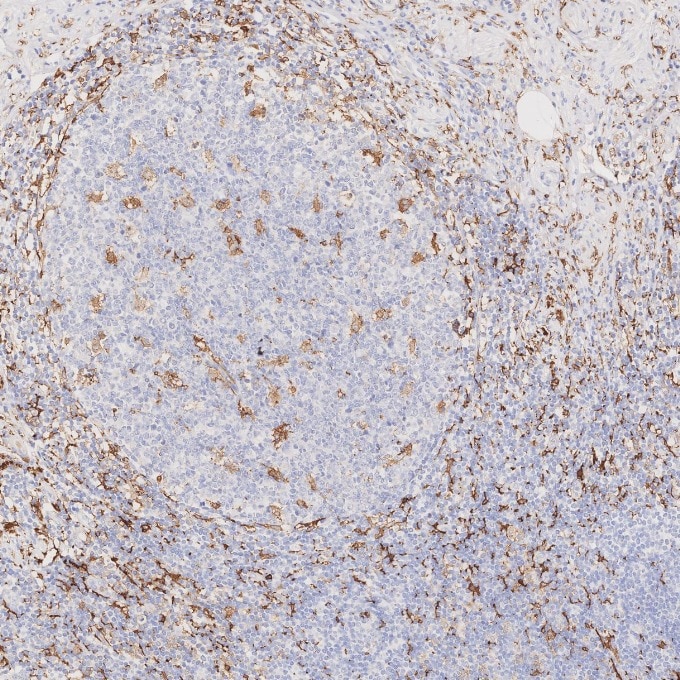
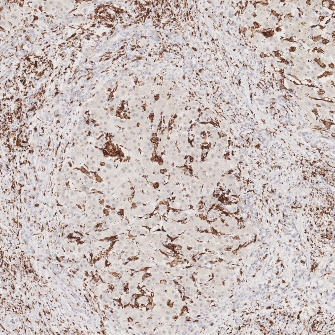
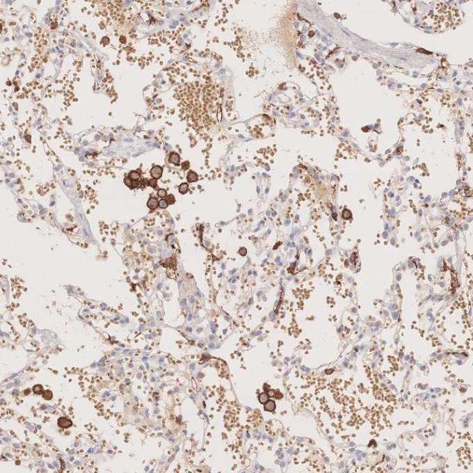
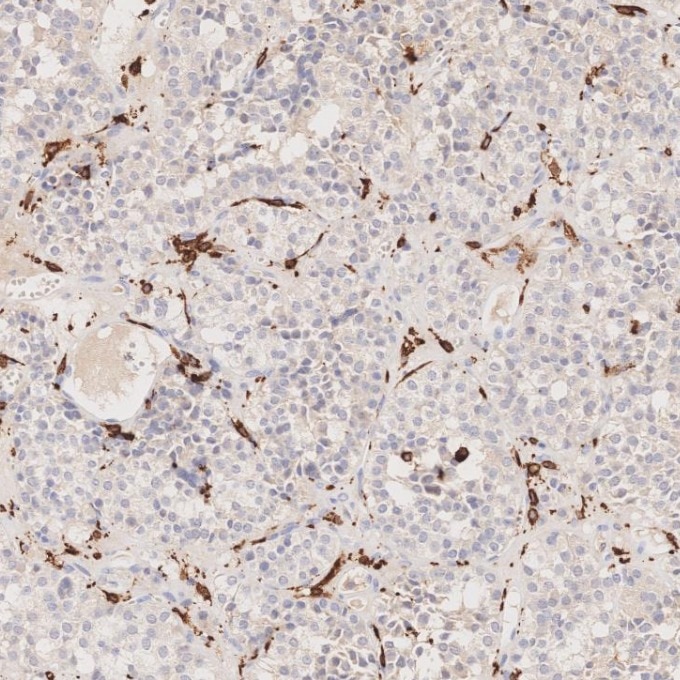
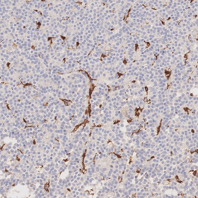
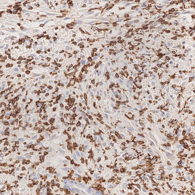
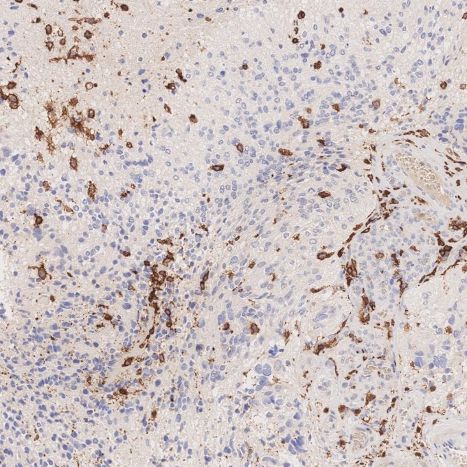
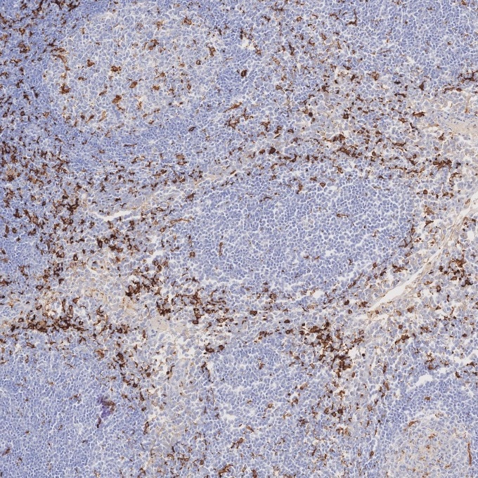
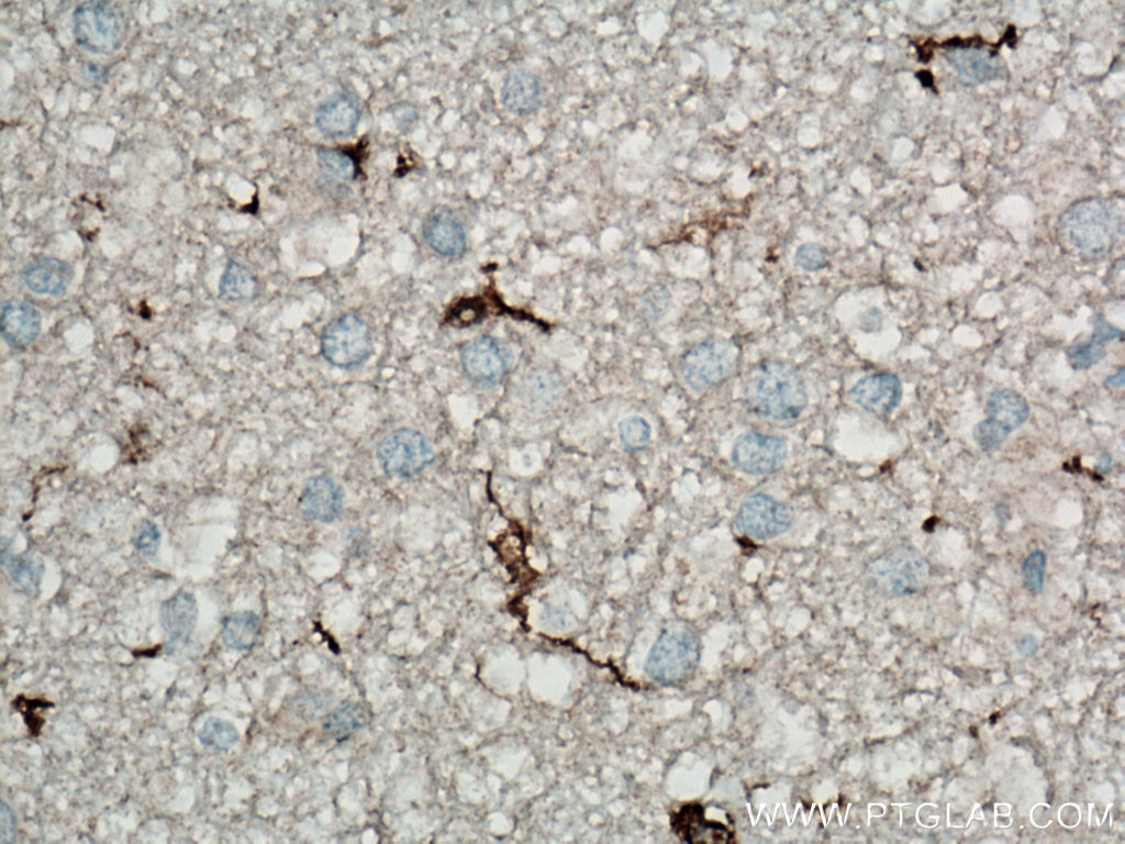
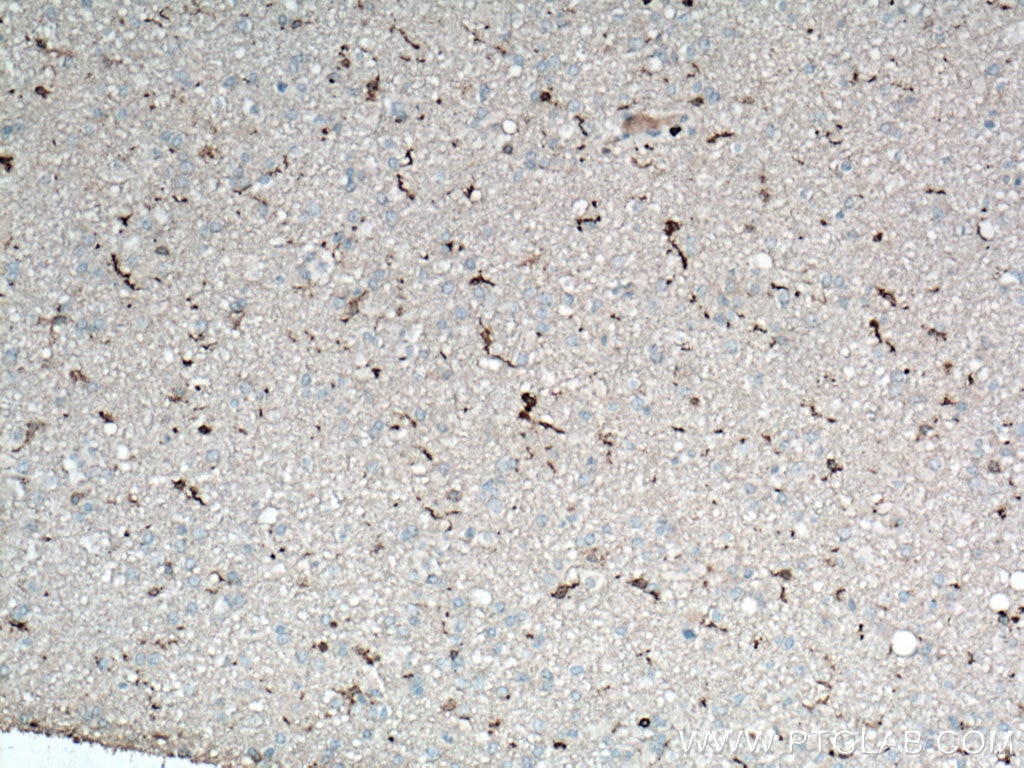
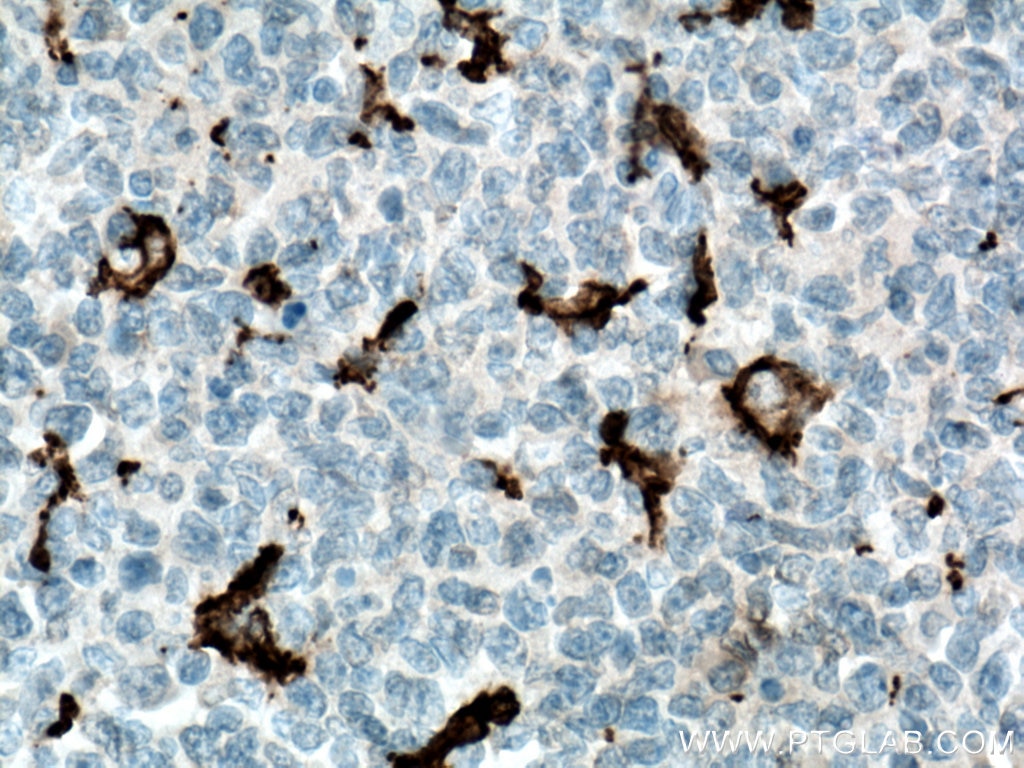
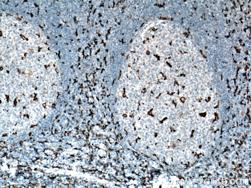
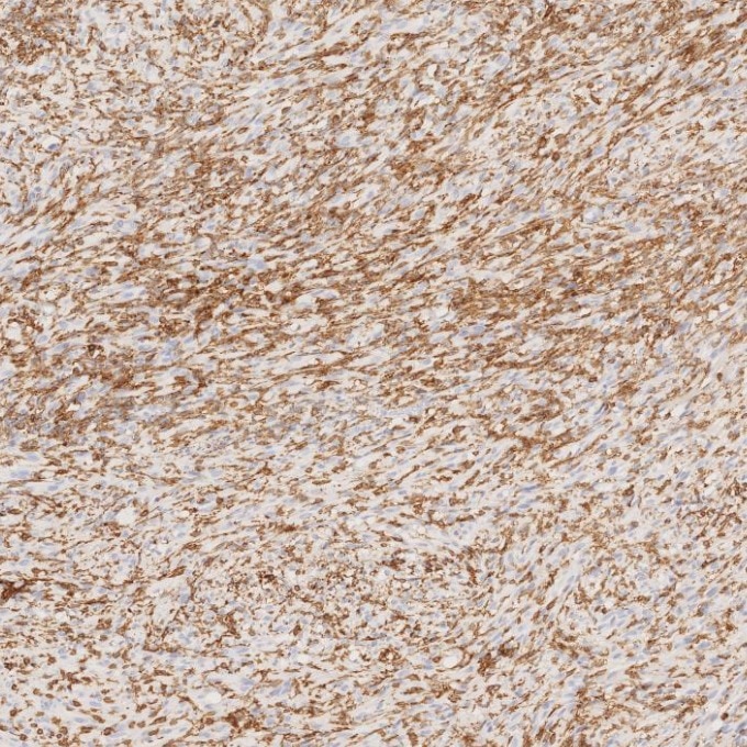
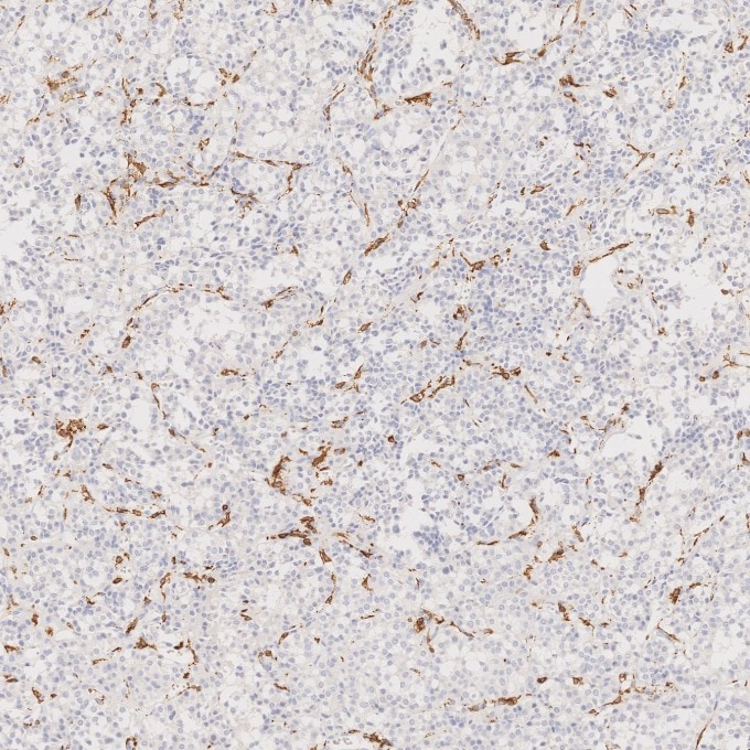
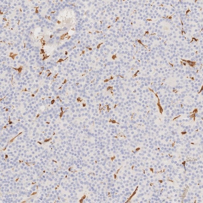
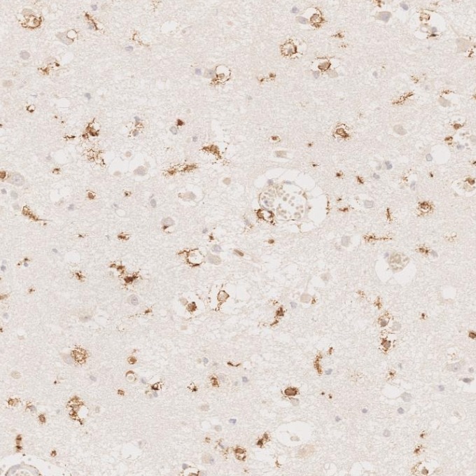
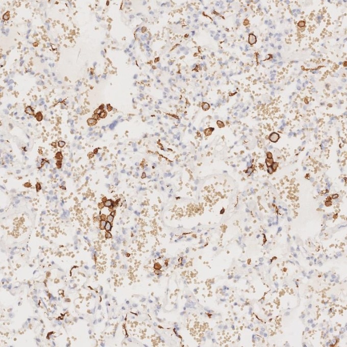
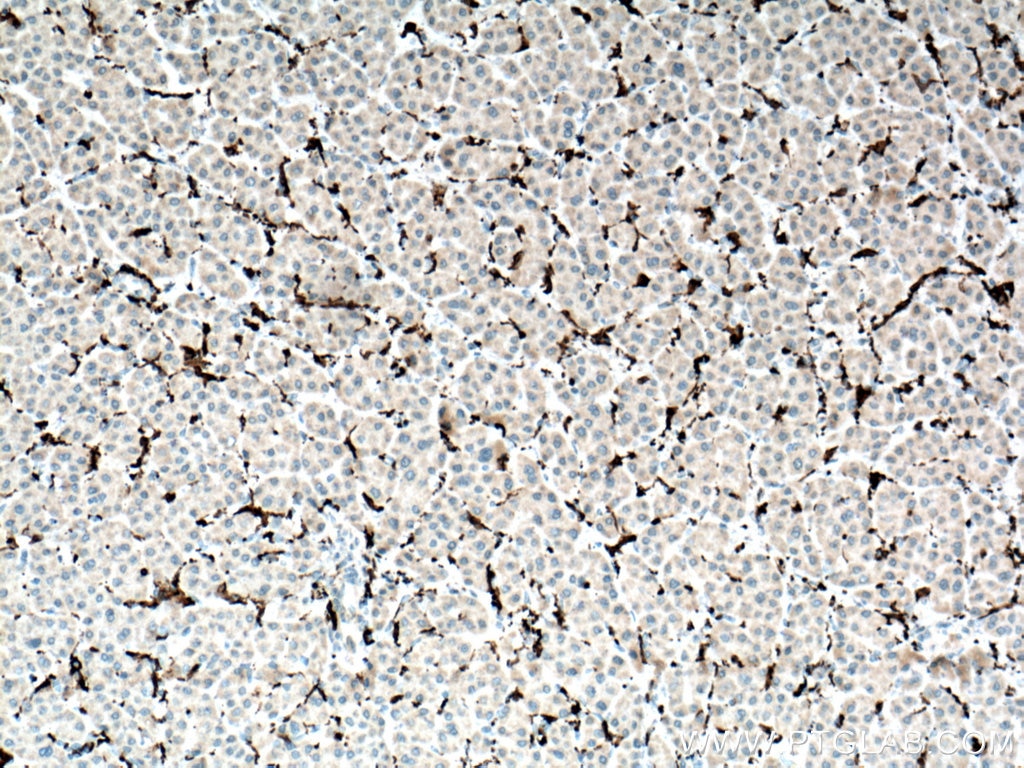
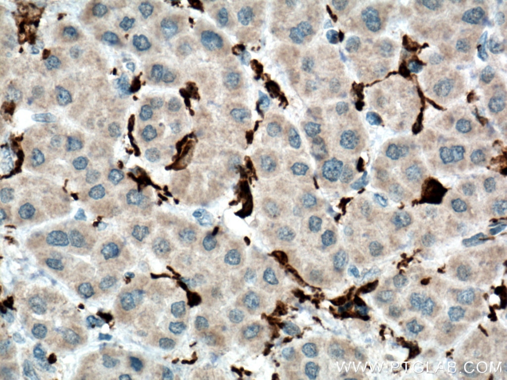
| Positive IHC detected in | human brain tissue, human liver tissue, human tonsillitis tissue, human lung tissue, human liver cancer tissue, human gliomas tissue, human brain tissue, human pituitary adenoma tissue, human appendicitis tissue Note: suggested antigen retrieval with TE buffer pH 9.0; (*) Alternatively, antigen retrieval may be performed with citrate buffer pH 6.0 |
Recommended dilution
| Application | Dilution |
|---|---|
| Immunohistochemistry (IHC) | IHC : 1:1000-1:4000 |
| It is recommended that this reagent should be titrated in each testing system to obtain optimal results. | |
| Sample-dependent, Check data in validation data gallery. | |
Product Information
66827-1-Ig targets human IBA1 in WB, IHC, IF, ELISA applications and shows reactivity with human samples.
| Tested Reactivity | human |
| Cited Reactivity | human |
| Host / Isotype | Mouse / IgG2a |
| Class | Monoclonal |
| Type | Antibody |
| Immunogen |
CatNo: Ag28236 Product name: Recombinant human IBA1 protein Source: e coli.-derived, PET28a Tag: 6*His Domain: 1-147 aa of BC009474 Sequence: MSQTRDLQGGKAFGLLKAQQEERLDEINKQFLDDPKYSSDEDLPSKLEGFKEKYMEFDLNGNGDIDIMSLKRMLEKLGVPKTHLELKKLIGEVSSGSGETFSYPDFLRMMLGKRSAILKMILMYEEKAREKEKPTGPPAKKAISELP Predict reactive species |
| Full Name | allograft inflammatory factor 1 |
| Calculated Molecular Weight | 17 kDa |
| GenBank Accession Number | BC009474 |
| Gene Symbol | IBA1 |
| Gene ID (NCBI) | 199 |
| ENSEMBL Gene ID | ENSG00000204472 |
| RRID | AB_2882170 |
| Conjugate | Unconjugated |
| Form | Liquid |
| Purification Method | Protein A purification |
| UNIPROT ID | P55008 |
| Storage Buffer | PBS with 0.02% sodium azide and 50% glycerol, pH 7.3. |
| Storage Conditions | Store at -20°C. Stable for one year after shipment. Aliquoting is unnecessary for -20oC storage. 20ul sizes contain 0.1% BSA. |
Background Information
IBA1 is a 143 amino acid cytoplasmic, inflammation response scaffold protein. It is constitutively expressed in monocytes and macrophages and is known to be involved in macrophage activation. It is a marker of activated macrophage. Expression of IBA1 is up-regulated in activated microglia following facial nerve axotomy, ischemia, and several brain diseases, thereby implicating it in the activated phenotypes of microglia.
Protocols
| Product Specific Protocols | |
|---|---|
| IHC protocol for human IBA1 antibody 66827-1-Ig | Download protocol |
| Standard Protocols | |
|---|---|
| Click here to view our Standard Protocols |
Publications
What published studies show
66827-1-Ig (Iba1) has accumulated 63 citations published in journals such as Cancer Cell, Nature Communications, Journal of Neuroinflammation, Redox Biology, Cell Death & Disease, Theranostics, and Neural Regeneration Research, among others. Associated research fields include neuroinflammation, microglial biology, neurodegeneration (Alzheimer's, Parkinson's disease), ischemic stroke, spinal cord injury, epilepsy, neuropathic pain, cancer, and programmed cell death (ferroptosis/pyroptosis). Primary applications are IF, IHC, and WB.
| Species | Application | Title |
|---|---|---|
Cancer Cell Distinct roles of TREM2 in central nervous system cancers and peripheral cancers | ||
Nat Commun Fatal iatrogenic cerebral β-amyloid-related arteritis in a woman treated with lecanemab for Alzheimer's disease | ||
Redox Biol CXCR4-BTK axis mediate pyroptosis and lipid peroxidation in early brain injury after subarachnoid hemorrhage via NLRP3 inflammasome and NF-κB pathway | ||
Theranostics An AIM2 inflammasome biomimetic mineralization inhibitor for vascular dementia therapy | ||
Cell Death Dis ChemR23 activation attenuates cognitive impairment in chronic cerebral hypoperfusion by inhibiting NLRP3 inflammasome-induced neuronal pyroptosis | ||
J Neuroinflammation TRPM7 contributes to pyroptosis and its involvement in status epilepticus |
Reviews
What customers say
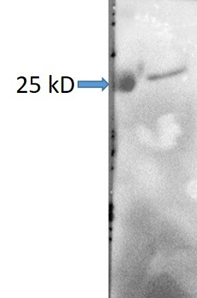
Customer feedback is mixed. One user (5/5) reported excellent immunofluorescence staining in iPSC-derived macrophages and microglia at 1:500 dilution for confocal microscopy. Another user (2/5) noted a problematic result in Western blot on postmortem human frontal cortex tissue at 1:1000 dilution, observing a clear band at 25 kD that did not correspond to the expected molecular weight. Overall, the antibody performs well for IF applications but may show non-specific or incorrect molecular weight bands in WB.
The reviews below have been submitted by verified Proteintech customers who received an incentive for providing their feedback.
Diogo (Verified Customer) (06-25-2026) | Excellent staining in iPSC-derived macrophages and microglia for confocal microscopy
|
Nuc (Verified Customer) (10-24-2023) | There is a clear band at 25 kD which is not the right MW.
 |


























