

Tested Applications
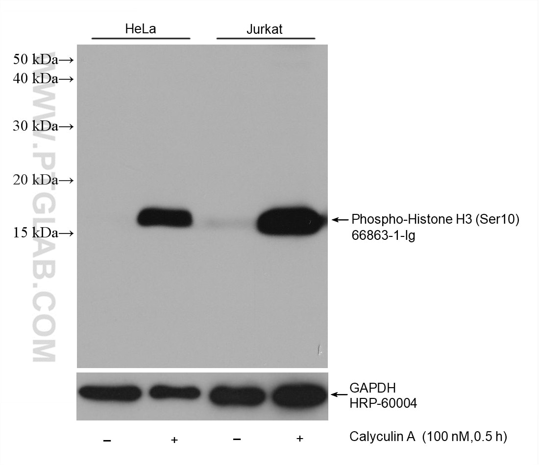
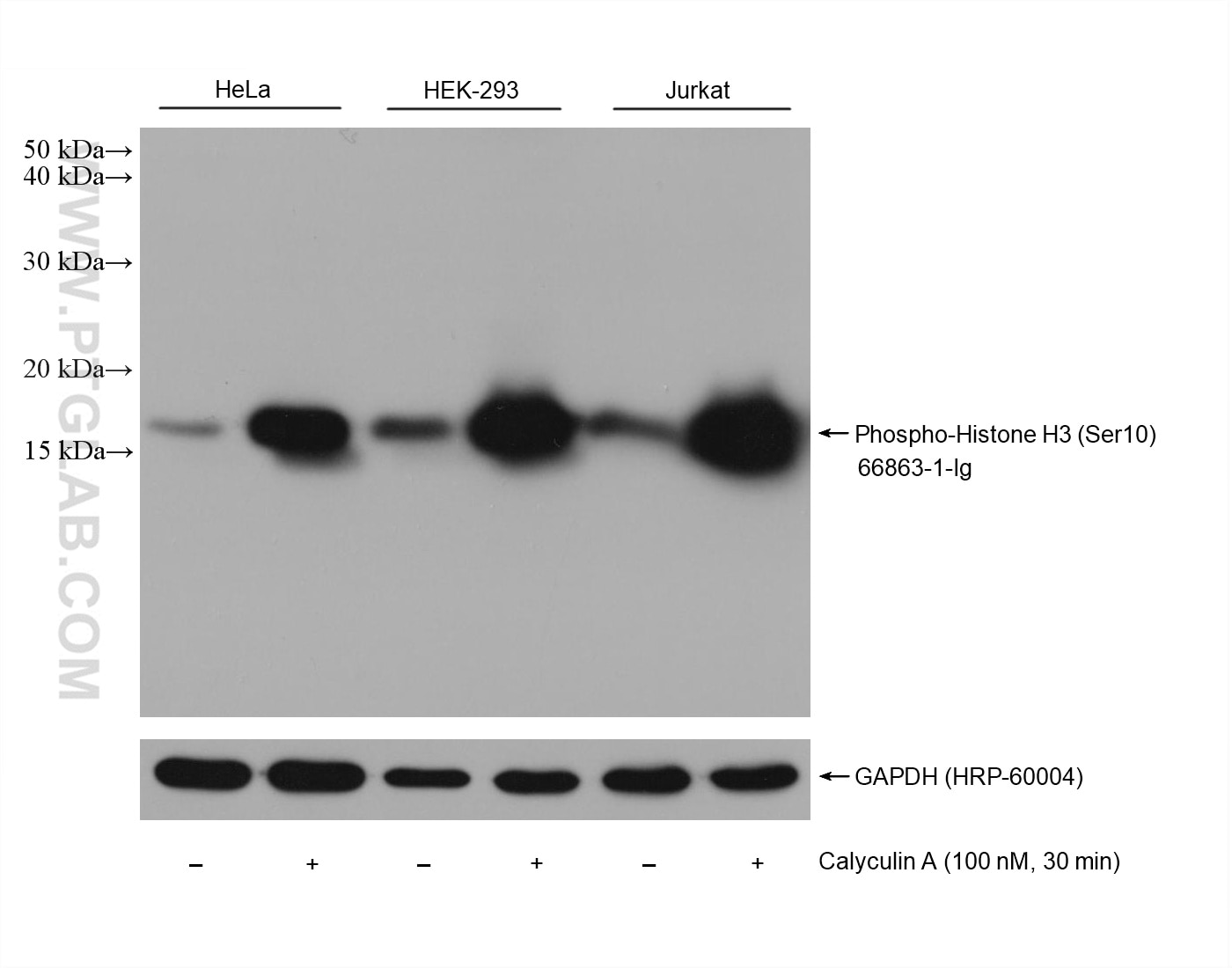
| Positive WB detected in | HeLa cells, HEK-293 cells, Jurkat cells |
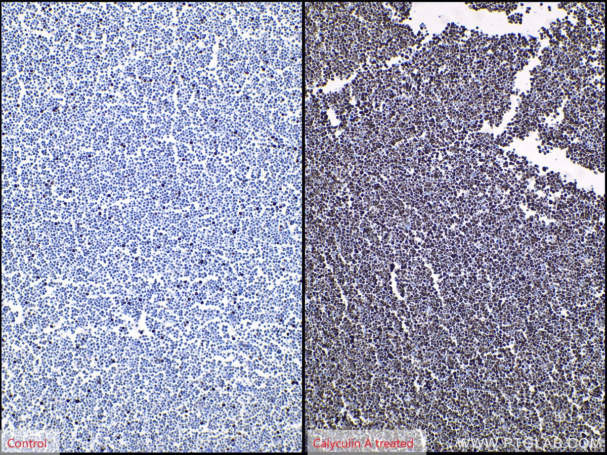
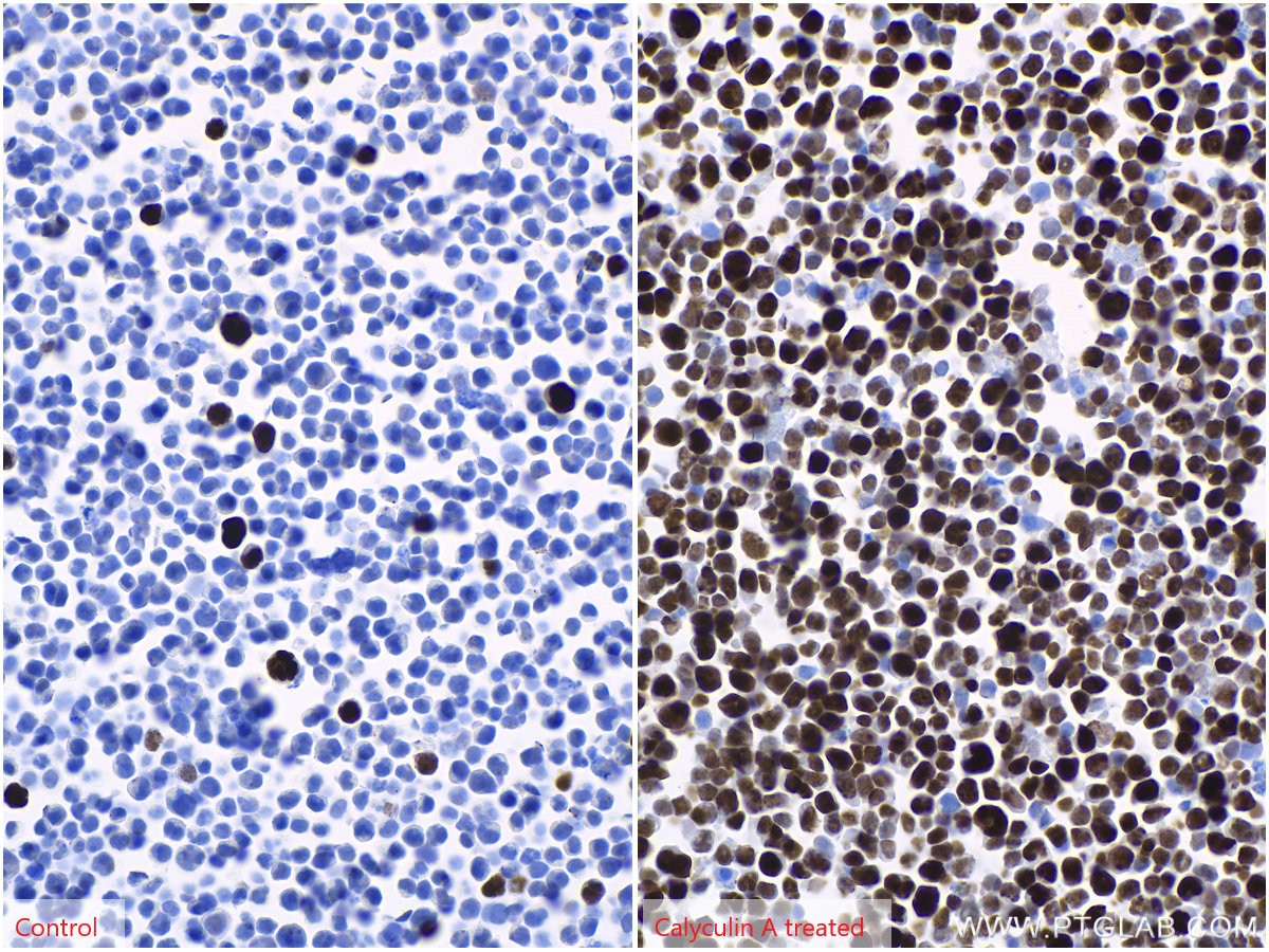
| Positive IHC detected in | Jurkat cells Note: suggested antigen retrieval with TE buffer pH 9.0; (*) Alternatively, antigen retrieval may be performed with citrate buffer pH 6.0 |
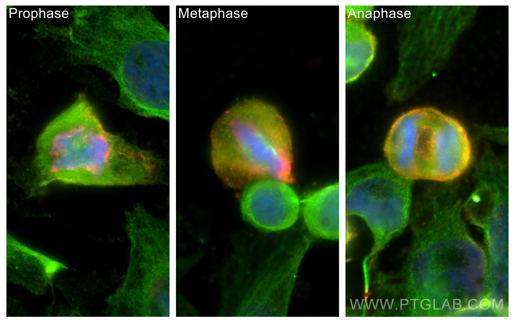
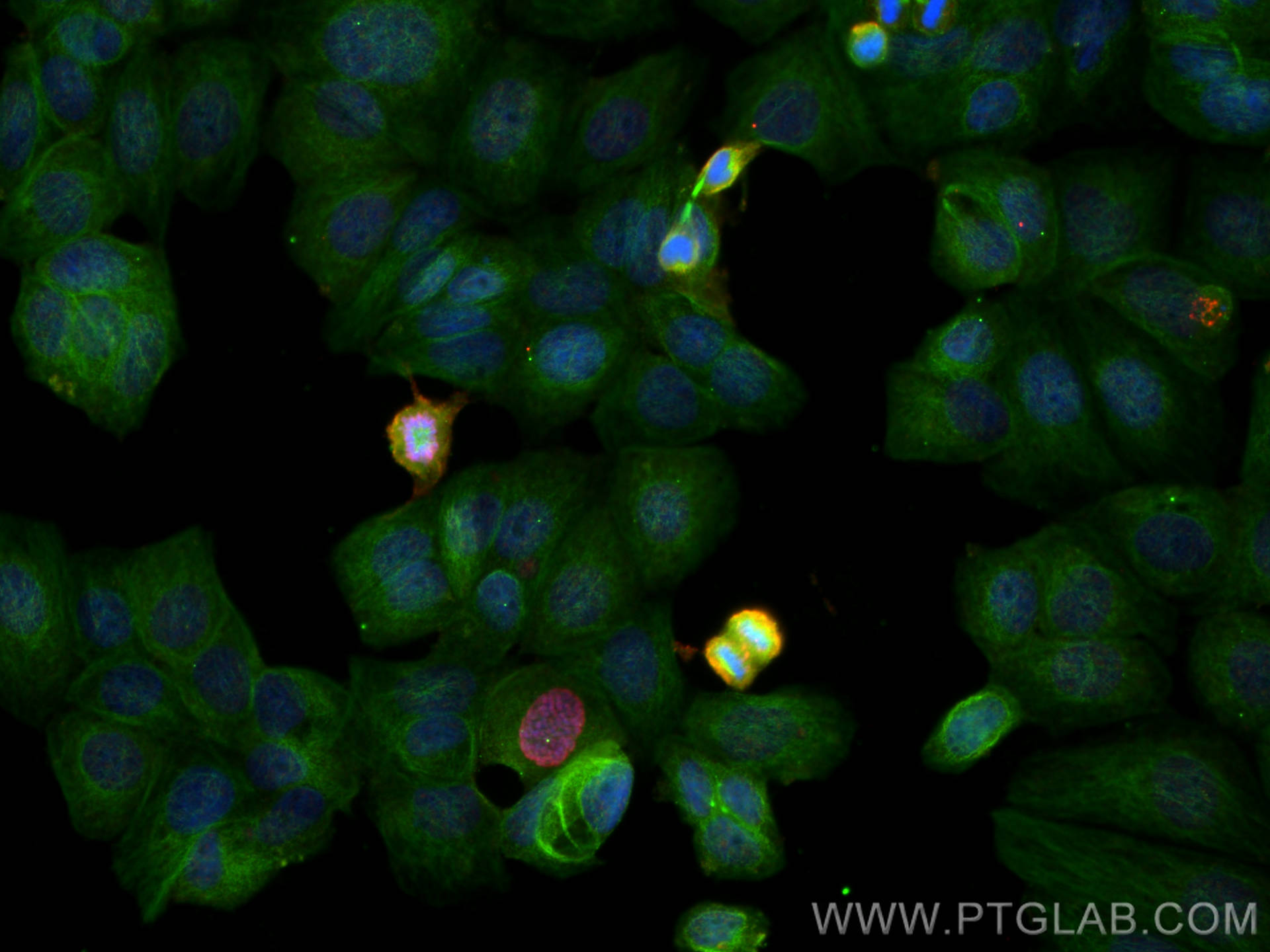
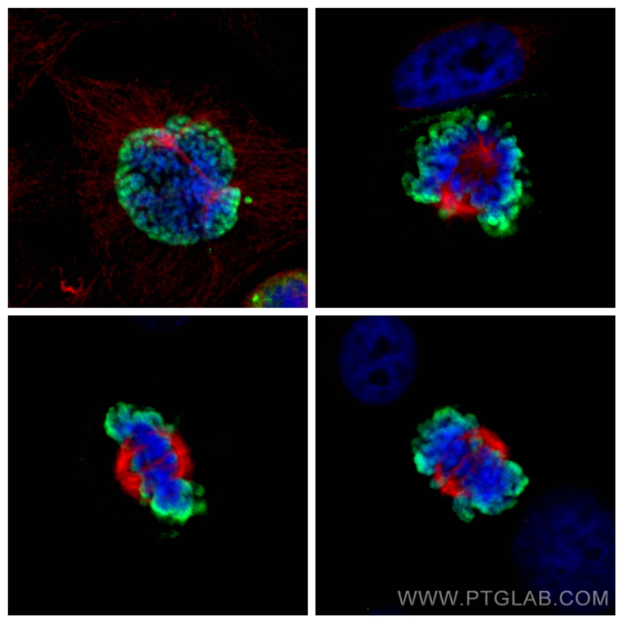
| Positive IF/ICC detected in | HeLa cells, MCF-7 cells |
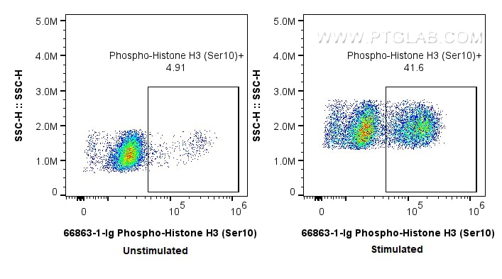
| Positive FC (Intra) detected in | nocodazole treated HeLa cells |
Recommended dilution
| Application | Dilution |
|---|---|
| Western Blot (WB) | WB : 1:5000-1:50000 |
| Immunohistochemistry (IHC) | IHC : 1:1000-1:4000 |
| Immunofluorescence (IF)/ICC | IF/ICC : 1:500-1:2000 |
| Flow Cytometry (FC) (INTRA) | FC (INTRA) : 0.25 ug per 10^6 cells in a 100 µl suspension |
| It is recommended that this reagent should be titrated in each testing system to obtain optimal results. | |
| Sample-dependent, Check data in validation data gallery. | |
Product Information
66863-1-Ig targets Phospho-Histone H3 (Ser10) in WB, IHC, IF/ICC, FC (Intra), ELISA applications and shows reactivity with human samples.
| Tested Reactivity | human |
| Cited Reactivity | human, mouse, rat, chicken |
| Host / Isotype | Mouse / IgG1 |
| Class | Monoclonal |
| Type | Antibody |
| Immunogen |
Peptide Predict reactive species |
| Full Name | histone cluster 1, H3a |
| Calculated Molecular Weight | 15 kDa |
| Observed Molecular Weight | 15-17 kDa |
| GenBank Accession Number | NM_003529 |
| Gene Symbol | HIST1H3A |
| Gene ID (NCBI) | 8350 |
| RRID | AB_2882201 |
| Conjugate | Unconjugated |
| Form | Liquid |
| Purification Method | Protein G purification |
| UNIPROT ID | P68431 |
| Storage Buffer | PBS with 0.02% sodium azide and 50% glycerol, pH 7.3. |
| Storage Conditions | Store at -20°C. Stable for one year after shipment. Aliquoting is unnecessary for -20oC storage. 20ul sizes contain 0.1% BSA. |
Background Information
Phospho-histone-H3 (PHH3) is a core histone protein, which in its phosphorylated state forms the principal constituents of eukaryotic chromatin, with histone H3 being phosphorylated at serine (Ser) 10 or Ser28 as well as its phosphorylation of Ser10 being strongly correlated with the late G2 to M-phase transition in mammalian mitotic cells. On the basis of previous research, a few cell line- and animal model-based researches have displayed an increase in phosphorylation of histone H3 at Ser10 (H3S10ph), the only histone marker that is involved in carcinogenesis and cellular transformation. Histone H3 phosphorylation on serine-10 is specific to mitosis and phosphorylated histone H3 (PHH3) proliferation markers (as counts defined per area or as indices defined per cell numbers) are increasingly being used to evaluate proliferation in various tumors.
Protocols
| Product Specific Protocols | |
|---|---|
| IF protocol for Phospho-Histone H3 (Ser10) antibody 66863-1-Ig | Download protocol |
| IHC protocol for Phospho-Histone H3 (Ser10) antibody 66863-1-Ig | Download protocol |
| WB protocol for Phospho-Histone H3 (Ser10) antibody 66863-1-Ig | Download protocol |
| Standard Protocols | |
|---|---|
| Click here to view our Standard Protocols |
Publications
What published studies show
The Phospho-Histone H3 (Ser10) Monoclonal antibody (4C7G2), catalog number 66863-1-Ig, has 34 citations. These appear across journals including Molecular Cancer, Nature Communications, Nature Metabolism, National Science Review, Cell Reports, Cell Death & Discovery, EMBO Reports, Oncogene, Acta Biomaterialia, Theranostics, Journal of Nanobiotechnology, Journal of Translational Medicine, British Journal of Cancer, Frontiers in Immunology, Cells, iScience, and bioRxiv. Research fields span cancer biology, cell cycle/mitosis, neurobiology, development, immunology, and metabolism, with applications in WB, IHC, and IF.
| Species | Application | Title |
|---|---|---|
Mol Cancer Deubiquitination of CDC6 by OTUD6A promotes tumour progression and chemoresistance | ||
Nat Commun Medium from human iPSC-derived primitive macrophages promotes adult cardiomyocyte proliferation and cardiac regeneration | ||
Natl Sci Rev Epigenomic landscapes during prefrontal cortex development and aging in rhesus | ||
J Nanobiotechnology Hepatocyte-derived tissue extracellular vesicles safeguard liver regeneration and support regenerative therapy | ||
Mater Today Bio Aptamer-functionalized exosomes combined with doxorubicin suppress GBM progression and enhance chemoradiosensitivity by promoting pyroptosis. |
Reviews
What customers say
All four reviews are 5-star, focused on Western Blot. Users report strong, clear band intensity even at high dilutions (1:3,000 to 1:10,000), with overnight incubation at 4°C. The antibody reliably detects phosphorylated Histone H3 (S10) across multiple cell lines, including MDA-MB-231, HeLa, mESCs, U2OS, HCT116, RPE1, and HEK293. Reviewers describe it as excellent, highly sensitive, and satisfying, with one noting strong signal despite a 1:10,000 dilution. Overall, users recommend it for assessing cell cycle synchronisation and general WB applications.
The reviews below have been submitted by verified Proteintech customers who received an incentive for providing their feedback.
Md Asad (Verified Customer) (02-24-2026) | Excellent performance. Would recommend using this antibody.
|
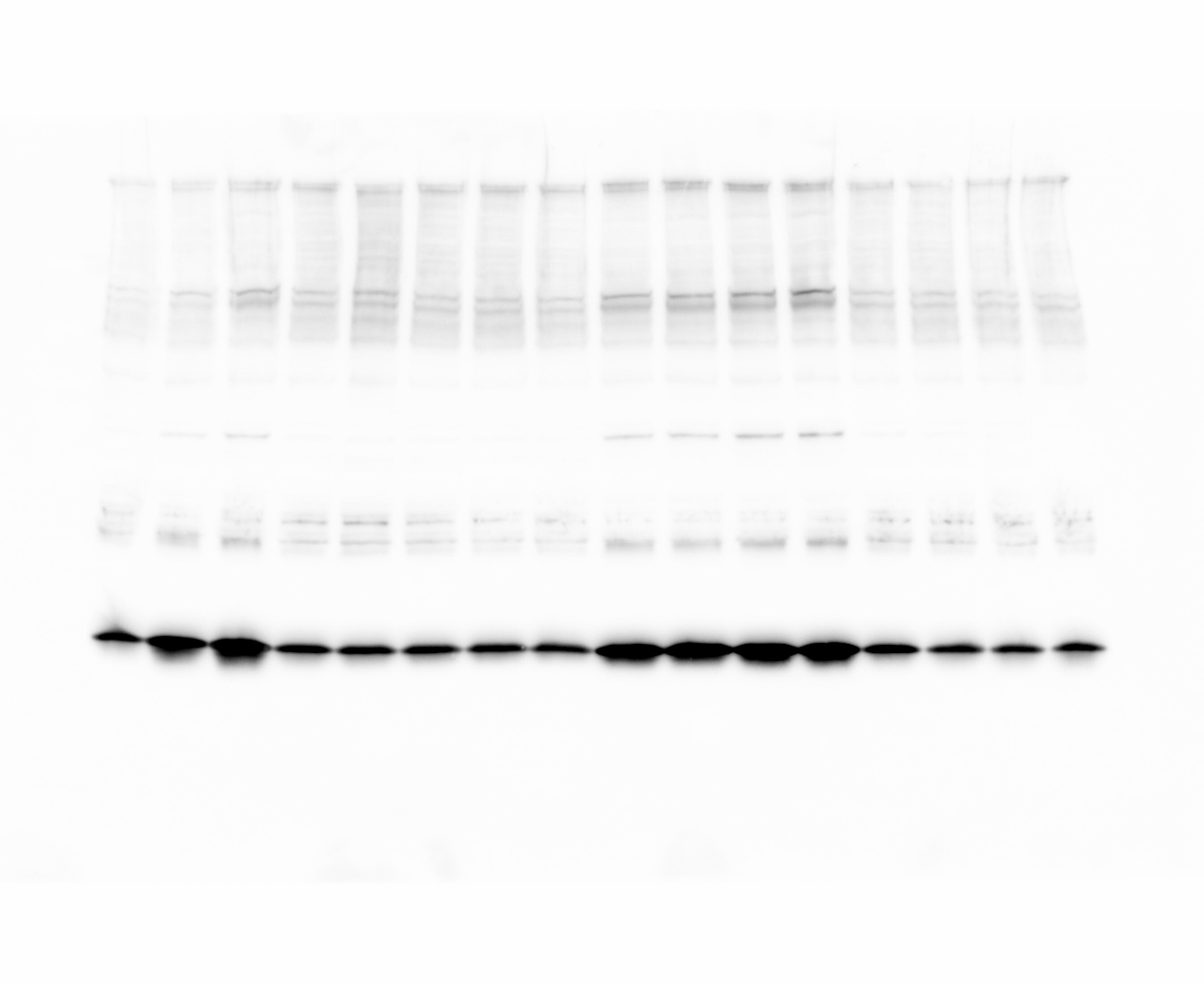
Charlotte (Verified Customer) (09-19-2024) | Blocking and dilution in seablock. Antibody very strong given the dilution and the bands intensity. Very satisfied.
 |
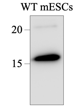
Tsimafei (Verified Customer) (08-04-2024) | Membrane was incubated at 4C, ON
 |
Wissem (Verified Customer) (06-27-2024) | This antibody works great at detecting phosphorylation of Histone H3 at S10; conditions - 1:10,000 overnight at 4C; used to assess cell cycle synchronisation in n extracts from U2OS, HCT116, HeLa, RPE1 and HEK293 cells
|









