
- Home
- Products
- Primary Antibodies
- Beta Catenin Monoclonal antibody, PBS Only (66379-1-PBS)
Proteintech Guarantee
The Proteintech guarantee covers Proteintech antibodies in any species and any application, including those not listed on the datasheet. If the antibody doesn’t perform, you can receive a hassle-free refund or credit note.

WB analysis using 66379-1-Ig (same clone as 66379-1-PBS)
Various lysates were subjected to SDS PAGE followed by western blot with 66379-1-Ig (Beta Catenin antibody) at dilution of 1:15000 incubated at room temperature for 1.5 hours. The membrane was stripped and reblotted with HRP-conjugated Lamin B1 Monoclonal antibody (HRP-66095) as loading control. This data was developed using the same antibody clone with 66379-1-PBS in a different storage buffer formulation.
This validation data has been generated in-house and reviewed by Proteintech scientists. Find out more on our antibody validation policy× Various lysates were subjected to SDS PAGE followed by western blot with 66379-1-Ig (Beta Catenin antibody) at dilution of 1:15000 incubated at room temperature for 1.5 hours. The membrane was stripped and reblotted with HRP-conjugated Lamin B1 Monoclonal antibody (HRP-66095) as loading control. This data was developed using the same antibody clone with 66379-1-PBS in a different storage buffer formulation.

WB analysis using 66379-1-Ig (same clone as 66379-1-PBS)
Various lysates were subjected to SDS PAGE followed by western blot with 66379-1-Ig (beta-Catenin antibody) at dilution of 1:6000 incubated at room temperature for 1.5 hours. This data was developed using the same antibody clone with 66379-1-PBS in a different storage buffer formulation.
This validation data has been generated in-house and reviewed by Proteintech scientists. Find out more on our antibody validation policy× Various lysates were subjected to SDS PAGE followed by western blot with 66379-1-Ig (beta-Catenin antibody) at dilution of 1:6000 incubated at room temperature for 1.5 hours. This data was developed using the same antibody clone with 66379-1-PBS in a different storage buffer formulation.

IP experiment of mouse brain using 66379-1-Ig (same clone as 66379-1-PBS)
IP result of anti-Beta Catenin (IP:66379-1-Ig, 4ug; Detection:66379-1-Ig 1:2000) with mouse brain tissue lysate 4400 ug. This data was developed using the same antibody clone with 66379-1-PBS in a different storage buffer formulation.
This validation data has been generated in-house and reviewed by Proteintech scientists. Find out more on our antibody validation policy× IP result of anti-Beta Catenin (IP:66379-1-Ig, 4ug; Detection:66379-1-Ig 1:2000) with mouse brain tissue lysate 4400 ug. This data was developed using the same antibody clone with 66379-1-PBS in a different storage buffer formulation.

IHC staining of human colorectal cancer using 66379-1-Ig (same clone as 66379-1-PBS)
Immunohistochemical analysis of paraffin-embedded human colorectal cancer tissue slide using 66379-1-Ig (Beta Catenin antibody) at dilution of 1:40000. Heat mediated antigen retrieval with Tris-EDTA buffer (pH 9.0). The staining was performed on an autostainer. This data was developed using the same antibody clone with 66379-1-PBS in a different storage buffer formulation.
This validation data has been generated in-house and reviewed by Proteintech scientists. Find out more on our antibody validation policy× Immunohistochemical analysis of paraffin-embedded human colorectal cancer tissue slide using 66379-1-Ig (Beta Catenin antibody) at dilution of 1:40000. Heat mediated antigen retrieval with Tris-EDTA buffer (pH 9.0). The staining was performed on an autostainer. This data was developed using the same antibody clone with 66379-1-PBS in a different storage buffer formulation.

IHC staining of human stomach cancer using 66379-1-Ig (same clone as 66379-1-PBS)
Immunohistochemical analysis of paraffin-embedded human stomach cancer tissue slide using 66379-1-Ig (Beta Catenin antibody) at dilution of 1:40000. Heat mediated antigen retrieval with Tris-EDTA buffer (pH 9.0). The staining was performed on an autostainer. This data was developed using the same antibody clone with 66379-1-PBS in a different storage buffer formulation.
This validation data has been generated in-house and reviewed by Proteintech scientists. Find out more on our antibody validation policy× Immunohistochemical analysis of paraffin-embedded human stomach cancer tissue slide using 66379-1-Ig (Beta Catenin antibody) at dilution of 1:40000. Heat mediated antigen retrieval with Tris-EDTA buffer (pH 9.0). The staining was performed on an autostainer. This data was developed using the same antibody clone with 66379-1-PBS in a different storage buffer formulation.

IHC staining of human appendicitis using 66379-1-Ig (same clone as 66379-1-PBS)
Immunohistochemical analysis of paraffin-embedded human appendicitis tissue slide using 66379-1-Ig (Beta Catenin antibody) at dilution of 1:40000. Heat mediated antigen retrieval with Tris-EDTA buffer (pH 9.0). The staining was performed on an autostainer. This data was developed using the same antibody clone with 66379-1-PBS in a different storage buffer formulation.
This validation data has been generated in-house and reviewed by Proteintech scientists. Find out more on our antibody validation policy× Immunohistochemical analysis of paraffin-embedded human appendicitis tissue slide using 66379-1-Ig (Beta Catenin antibody) at dilution of 1:40000. Heat mediated antigen retrieval with Tris-EDTA buffer (pH 9.0). The staining was performed on an autostainer. This data was developed using the same antibody clone with 66379-1-PBS in a different storage buffer formulation.

IHC staining of human pancreas cancer using 66379-1-Ig (same clone as 66379-1-PBS)
Immunohistochemical analysis of paraffin-embedded human pancreas cancer tissue slide using 66379-1-Ig (Beta Catenin antibody) at dilution of 1:40000. Heat mediated antigen retrieval with Tris-EDTA buffer (pH 9.0). The staining was performed on an autostainer. This data was developed using the same antibody clone with 66379-1-PBS in a different storage buffer formulation.
This validation data has been generated in-house and reviewed by Proteintech scientists. Find out more on our antibody validation policy× Immunohistochemical analysis of paraffin-embedded human pancreas cancer tissue slide using 66379-1-Ig (Beta Catenin antibody) at dilution of 1:40000. Heat mediated antigen retrieval with Tris-EDTA buffer (pH 9.0). The staining was performed on an autostainer. This data was developed using the same antibody clone with 66379-1-PBS in a different storage buffer formulation.

IHC staining of human ovary cancer using 66379-1-Ig (same clone as 66379-1-PBS)
Immunohistochemical analysis of paraffin-embedded human ovary cancer tissue slide using 66379-1-Ig (Beta Catenin antibody) at dilution of 1:40000. Heat mediated antigen retrieval with Tris-EDTA buffer (pH 9.0). The staining was performed on an autostainer. This data was developed using the same antibody clone with 66379-1-PBS in a different storage buffer formulation.
This validation data has been generated in-house and reviewed by Proteintech scientists. Find out more on our antibody validation policy× Immunohistochemical analysis of paraffin-embedded human ovary cancer tissue slide using 66379-1-Ig (Beta Catenin antibody) at dilution of 1:40000. Heat mediated antigen retrieval with Tris-EDTA buffer (pH 9.0). The staining was performed on an autostainer. This data was developed using the same antibody clone with 66379-1-PBS in a different storage buffer formulation.

IHC staining of human breast cancer using 66379-1-Ig (same clone as 66379-1-PBS)
Immunohistochemical analysis of paraffin-embedded human breast cancer tissue slide using 66379-1-Ig (Beta Catenin antibody) at dilution of 1:40000. Heat mediated antigen retrieval with Tris-EDTA buffer (pH 9.0). The staining was performed on an autostainer. This data was developed using the same antibody clone with 66379-1-PBS in a different storage buffer formulation.
This validation data has been generated in-house and reviewed by Proteintech scientists. Find out more on our antibody validation policy× Immunohistochemical analysis of paraffin-embedded human breast cancer tissue slide using 66379-1-Ig (Beta Catenin antibody) at dilution of 1:40000. Heat mediated antigen retrieval with Tris-EDTA buffer (pH 9.0). The staining was performed on an autostainer. This data was developed using the same antibody clone with 66379-1-PBS in a different storage buffer formulation.

IHC staining of human liver cancer using 66379-1-Ig (same clone as 66379-1-PBS)
Immunohistochemical analysis of paraffin-embedded human liver cancer tissue slide using 66379-1-Ig (Beta Catenin antibody) at dilution of 1:40000. Heat mediated antigen retrieval with Tris-EDTA buffer (pH 9.0). The staining was performed on an autostainer. This data was developed using the same antibody clone with 66379-1-PBS in a different storage buffer formulation.
This validation data has been generated in-house and reviewed by Proteintech scientists. Find out more on our antibody validation policy× Immunohistochemical analysis of paraffin-embedded human liver cancer tissue slide using 66379-1-Ig (Beta Catenin antibody) at dilution of 1:40000. Heat mediated antigen retrieval with Tris-EDTA buffer (pH 9.0). The staining was performed on an autostainer. This data was developed using the same antibody clone with 66379-1-PBS in a different storage buffer formulation.

IHC staining of human placenta using 66379-1-Ig (same clone as 66379-1-PBS)
Immunohistochemical analysis of paraffin-embedded human placenta tissue slide using 66379-1-Ig (Beta Catenin antibody) at dilution of 1:40000. Heat mediated antigen retrieval with Tris-EDTA buffer (pH 9.0). The staining was performed on an autostainer. This data was developed using the same antibody clone with 66379-1-PBS in a different storage buffer formulation.
This validation data has been generated in-house and reviewed by Proteintech scientists. Find out more on our antibody validation policy× Immunohistochemical analysis of paraffin-embedded human placenta tissue slide using 66379-1-Ig (Beta Catenin antibody) at dilution of 1:40000. Heat mediated antigen retrieval with Tris-EDTA buffer (pH 9.0). The staining was performed on an autostainer. This data was developed using the same antibody clone with 66379-1-PBS in a different storage buffer formulation.

IHC staining of human stomach cancer using 66379-1-Ig (same clone as 66379-1-PBS)
Immunohistochemical analysis of paraffin-embedded human stomach cancer tissue slide using 66379-1-Ig (Beta Catenin antibody) at dilution of 1:40000. Heat mediated antigen retrieval with Tris-EDTA buffer (pH 9.0). The staining was performed on an autostainer. This data was developed using the same antibody clone with 66379-1-PBS in a different storage buffer formulation.
This validation data has been generated in-house and reviewed by Proteintech scientists. Find out more on our antibody validation policy× Immunohistochemical analysis of paraffin-embedded human stomach cancer tissue slide using 66379-1-Ig (Beta Catenin antibody) at dilution of 1:40000. Heat mediated antigen retrieval with Tris-EDTA buffer (pH 9.0). The staining was performed on an autostainer. This data was developed using the same antibody clone with 66379-1-PBS in a different storage buffer formulation.
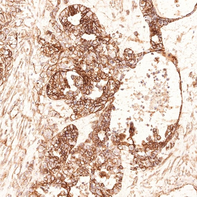
IHC staining of human ovary cancer using 66379-1-Ig (same clone as 66379-1-PBS)
Immunohistochemical analysis of paraffin-embedded human ovary cancer tissue slide using 66379-1-Ig (Beta Catenin antibody) at dilution of 1:40000. Heat mediated antigen retrieval with Tris-EDTA buffer (pH 9.0). The staining was performed on an autostainer. This data was developed using the same antibody clone with 66379-1-PBS in a different storage buffer formulation.
This validation data has been generated in-house and reviewed by Proteintech scientists. Find out more on our antibody validation policy× Immunohistochemical analysis of paraffin-embedded human ovary cancer tissue slide using 66379-1-Ig (Beta Catenin antibody) at dilution of 1:40000. Heat mediated antigen retrieval with Tris-EDTA buffer (pH 9.0). The staining was performed on an autostainer. This data was developed using the same antibody clone with 66379-1-PBS in a different storage buffer formulation.

IHC staining of mouse colon using 66379-1-Ig (same clone as 66379-1-PBS)
Immunohistochemical analysis of paraffin-embedded mouse colon tissue slide using 66379-1-Ig (Beta Catenin antibody) at dilution of 1:20000 (under 10x lens). Heat mediated antigen retrieval with Tris-EDTA buffer (pH 9.0). This data was developed using the same antibody clone with 66379-1-PBS in a different storage buffer formulation.
This validation data has been generated in-house and reviewed by Proteintech scientists. Find out more on our antibody validation policy× Immunohistochemical analysis of paraffin-embedded mouse colon tissue slide using 66379-1-Ig (Beta Catenin antibody) at dilution of 1:20000 (under 10x lens). Heat mediated antigen retrieval with Tris-EDTA buffer (pH 9.0). This data was developed using the same antibody clone with 66379-1-PBS in a different storage buffer formulation.

IHC staining of mouse colon using 66379-1-Ig (same clone as 66379-1-PBS)
Immunohistochemical analysis of paraffin-embedded mouse colon tissue slide using 66379-1-Ig (Beta Catenin antibody) at dilution of 1:20000 (under 40x lens). Heat mediated antigen retrieval with Tris-EDTA buffer (pH 9.0). This data was developed using the same antibody clone with 66379-1-PBS in a different storage buffer formulation.
This validation data has been generated in-house and reviewed by Proteintech scientists. Find out more on our antibody validation policy× Immunohistochemical analysis of paraffin-embedded mouse colon tissue slide using 66379-1-Ig (Beta Catenin antibody) at dilution of 1:20000 (under 40x lens). Heat mediated antigen retrieval with Tris-EDTA buffer (pH 9.0). This data was developed using the same antibody clone with 66379-1-PBS in a different storage buffer formulation.

IHC staining of rat colon using 66379-1-Ig (same clone as 66379-1-PBS)
Immunohistochemical analysis of paraffin-embedded rat colon tissue slide using 66379-1-Ig (Beta Catenin antibody) at dilution of 1:20000 (under 10x lens). Heat mediated antigen retrieval with Tris-EDTA buffer (pH 9.0). This data was developed using the same antibody clone with 66379-1-PBS in a different storage buffer formulation.
This validation data has been generated in-house and reviewed by Proteintech scientists. Find out more on our antibody validation policy× Immunohistochemical analysis of paraffin-embedded rat colon tissue slide using 66379-1-Ig (Beta Catenin antibody) at dilution of 1:20000 (under 10x lens). Heat mediated antigen retrieval with Tris-EDTA buffer (pH 9.0). This data was developed using the same antibody clone with 66379-1-PBS in a different storage buffer formulation.

IHC staining of rat colon using 66379-1-Ig (same clone as 66379-1-PBS)
Immunohistochemical analysis of paraffin-embedded rat colon tissue slide using 66379-1-Ig (Beta Catenin antibody) at dilution of 1:20000 (under 40x lens). Heat mediated antigen retrieval with Tris-EDTA buffer (pH 9.0). This data was developed using the same antibody clone with 66379-1-PBS in a different storage buffer formulation.
This validation data has been generated in-house and reviewed by Proteintech scientists. Find out more on our antibody validation policy× Immunohistochemical analysis of paraffin-embedded rat colon tissue slide using 66379-1-Ig (Beta Catenin antibody) at dilution of 1:20000 (under 40x lens). Heat mediated antigen retrieval with Tris-EDTA buffer (pH 9.0). This data was developed using the same antibody clone with 66379-1-PBS in a different storage buffer formulation.

IHC staining of human ovary cancer using 66379-1-Ig (same clone as 66379-1-PBS)
Immunohistochemical analysis of paraffin-embedded human ovary cancer tissue slide using 66379-1-Ig (Beta Catenin antibody) at dilution of 1:40000 (under 10x lens). Heat mediated antigen retrieval with Tris-EDTA buffer (pH 9.0). This data was developed using the same antibody clone with 66379-1-PBS in a different storage buffer formulation.
This validation data has been generated in-house and reviewed by Proteintech scientists. Find out more on our antibody validation policy× Immunohistochemical analysis of paraffin-embedded human ovary cancer tissue slide using 66379-1-Ig (Beta Catenin antibody) at dilution of 1:40000 (under 10x lens). Heat mediated antigen retrieval with Tris-EDTA buffer (pH 9.0). This data was developed using the same antibody clone with 66379-1-PBS in a different storage buffer formulation.

IHC staining of human breast cancer using 66379-1-Ig (same clone as 66379-1-PBS)
Immunohistochemical analysis of paraffin-embedded human breast cancer tissue slide using 66379-1-Ig (Beta Catenin antibody) at dilution of 1:40000. Heat mediated antigen retrieval with Tris-EDTA buffer (pH 9.0). This data was developed using the same antibody clone with 66379-1-PBS in a different storage buffer formulation.
This validation data has been generated in-house and reviewed by Proteintech scientists. Find out more on our antibody validation policy× Immunohistochemical analysis of paraffin-embedded human breast cancer tissue slide using 66379-1-Ig (Beta Catenin antibody) at dilution of 1:40000. Heat mediated antigen retrieval with Tris-EDTA buffer (pH 9.0). This data was developed using the same antibody clone with 66379-1-PBS in a different storage buffer formulation.

IHC staining of human appendicitis using 66379-1-Ig (same clone as 66379-1-PBS)
Immunohistochemical analysis of paraffin-embedded human appendicitis tissue slide using 66379-1-Ig (Beta Catenin antibody) at dilution of 1:40000. Heat mediated antigen retrieval with Tris-EDTA buffer (pH 9.0). This data was developed using the same antibody clone with 66379-1-PBS in a different storage buffer formulation.
This validation data has been generated in-house and reviewed by Proteintech scientists. Find out more on our antibody validation policy× Immunohistochemical analysis of paraffin-embedded human appendicitis tissue slide using 66379-1-Ig (Beta Catenin antibody) at dilution of 1:40000. Heat mediated antigen retrieval with Tris-EDTA buffer (pH 9.0). This data was developed using the same antibody clone with 66379-1-PBS in a different storage buffer formulation.

IHC staining of human cervical cancer using 66379-1-Ig (same clone as 66379-1-PBS)
Immunohistochemical analysis of paraffin-embedded human cervical cancer tissue slide using 66379-1-Ig (Beta Catenin antibody) at dilution of 1:40000. Heat mediated antigen retrieval with Tris-EDTA buffer (pH 9.0). This data was developed using the same antibody clone with 66379-1-PBS in a different storage buffer formulation.
This validation data has been generated in-house and reviewed by Proteintech scientists. Find out more on our antibody validation policy× Immunohistochemical analysis of paraffin-embedded human cervical cancer tissue slide using 66379-1-Ig (Beta Catenin antibody) at dilution of 1:40000. Heat mediated antigen retrieval with Tris-EDTA buffer (pH 9.0). This data was developed using the same antibody clone with 66379-1-PBS in a different storage buffer formulation.

IHC staining of human liver cancer using 66379-1-Ig (same clone as 66379-1-PBS)
Immunohistochemical analysis of paraffin-embedded human liver cancer tissue slide using 66379-1-Ig (Beta Catenin antibody) at dilution of 1:40000. Heat mediated antigen retrieval with Tris-EDTA buffer (pH 9.0). This data was developed using the same antibody clone with 66379-1-PBS in a different storage buffer formulation.
This validation data has been generated in-house and reviewed by Proteintech scientists. Find out more on our antibody validation policy× Immunohistochemical analysis of paraffin-embedded human liver cancer tissue slide using 66379-1-Ig (Beta Catenin antibody) at dilution of 1:40000. Heat mediated antigen retrieval with Tris-EDTA buffer (pH 9.0). This data was developed using the same antibody clone with 66379-1-PBS in a different storage buffer formulation.

IHC staining of human pancreas cancer using 66379-1-Ig (same clone as 66379-1-PBS)
Immunohistochemical analysis of paraffin-embedded human pancreas cancer tissue slide using 66379-1-Ig (Beta Catenin antibody) at dilution of 1:40000. Heat mediated antigen retrieval with Tris-EDTA buffer (pH 9.0). This data was developed using the same antibody clone with 66379-1-PBS in a different storage buffer formulation.
This validation data has been generated in-house and reviewed by Proteintech scientists. Find out more on our antibody validation policy× Immunohistochemical analysis of paraffin-embedded human pancreas cancer tissue slide using 66379-1-Ig (Beta Catenin antibody) at dilution of 1:40000. Heat mediated antigen retrieval with Tris-EDTA buffer (pH 9.0). This data was developed using the same antibody clone with 66379-1-PBS in a different storage buffer formulation.

IHC staining of human ovary cancer using 66379-1-Ig (same clone as 66379-1-PBS)
Immunohistochemical analysis of paraffin-embedded human ovary cancer tissue slide using 66379-1-Ig (Beta Catenin antibody) at dilution of 1:40000. Heat mediated antigen retrieval with Tris-EDTA buffer (pH 9.0). This data was developed using the same antibody clone with 66379-1-PBS in a different storage buffer formulation.
This validation data has been generated in-house and reviewed by Proteintech scientists. Find out more on our antibody validation policy× Immunohistochemical analysis of paraffin-embedded human ovary cancer tissue slide using 66379-1-Ig (Beta Catenin antibody) at dilution of 1:40000. Heat mediated antigen retrieval with Tris-EDTA buffer (pH 9.0). This data was developed using the same antibody clone with 66379-1-PBS in a different storage buffer formulation.

IHC staining of human colorectal cancer using 66379-1-Ig (same clone as 66379-1-PBS)
Immunohistochemical analysis of paraffin-embedded human colorectal cancer tissue slide using 66379-1-Ig (Beta Catenin antibody) at dilution of 1:40000. Heat mediated antigen retrieval with Tris-EDTA buffer (pH 9.0). This data was developed using the same antibody clone with 66379-1-PBS in a different storage buffer formulation.
This validation data has been generated in-house and reviewed by Proteintech scientists. Find out more on our antibody validation policy× Immunohistochemical analysis of paraffin-embedded human colorectal cancer tissue slide using 66379-1-Ig (Beta Catenin antibody) at dilution of 1:40000. Heat mediated antigen retrieval with Tris-EDTA buffer (pH 9.0). This data was developed using the same antibody clone with 66379-1-PBS in a different storage buffer formulation.

IHC staining of human stomach cancer using 66379-1-Ig (same clone as 66379-1-PBS)
Immunohistochemical analysis of paraffin-embedded human stomach cancer tissue slide using 66379-1-Ig (Beta Catenin antibody) at dilution of 1:40000. Heat mediated antigen retrieval with Tris-EDTA buffer (pH 9.0). This data was developed using the same antibody clone with 66379-1-PBS in a different storage buffer formulation.
This validation data has been generated in-house and reviewed by Proteintech scientists. Find out more on our antibody validation policy× Immunohistochemical analysis of paraffin-embedded human stomach cancer tissue slide using 66379-1-Ig (Beta Catenin antibody) at dilution of 1:40000. Heat mediated antigen retrieval with Tris-EDTA buffer (pH 9.0). This data was developed using the same antibody clone with 66379-1-PBS in a different storage buffer formulation.

IHC staining of human breast cancer using 66379-1-Ig (same clone as 66379-1-PBS)
Immunohistochemical analysis of paraffin-embedded human breast cancer tissue slide using 66379-1-Ig (Beta Catenin antibody) at dilution of 1:20000 (under 10x lens). Heat mediated antigen retrieval with Tris-EDTA buffer (pH 9.0). This data was developed using the same antibody clone with 66379-1-PBS in a different storage buffer formulation.
This validation data has been generated in-house and reviewed by Proteintech scientists. Find out more on our antibody validation policy× Immunohistochemical analysis of paraffin-embedded human breast cancer tissue slide using 66379-1-Ig (Beta Catenin antibody) at dilution of 1:20000 (under 10x lens). Heat mediated antigen retrieval with Tris-EDTA buffer (pH 9.0). This data was developed using the same antibody clone with 66379-1-PBS in a different storage buffer formulation.

IHC staining of human breast cancer using 66379-1-Ig (same clone as 66379-1-PBS)
Immunohistochemical analysis of paraffin-embedded human breast cancer tissue slide using 66379-1-Ig (Beta Catenin antibody) at dilution of 1:20000 (under 40x lens). Heat mediated antigen retrieval with Tris-EDTA buffer (pH 9.0). This data was developed using the same antibody clone with 66379-1-PBS in a different storage buffer formulation.
This validation data has been generated in-house and reviewed by Proteintech scientists. Find out more on our antibody validation policy× Immunohistochemical analysis of paraffin-embedded human breast cancer tissue slide using 66379-1-Ig (Beta Catenin antibody) at dilution of 1:20000 (under 40x lens). Heat mediated antigen retrieval with Tris-EDTA buffer (pH 9.0). This data was developed using the same antibody clone with 66379-1-PBS in a different storage buffer formulation.

IHC staining of rat colon using 66379-1-Ig (same clone as 66379-1-PBS)
Immunohistochemical analysis of paraffin-embedded rat colon tissue slide using 66379-1-Ig (Beta Catenin antibody) at dilution of 1:20000 (under 10x lens). Heat mediated antigen retrieval with Tris-EDTA buffer (pH 9.0). This data was developed using the same antibody clone with 66379-1-PBS in a different storage buffer formulation.
This validation data has been generated in-house and reviewed by Proteintech scientists. Find out more on our antibody validation policy× Immunohistochemical analysis of paraffin-embedded rat colon tissue slide using 66379-1-Ig (Beta Catenin antibody) at dilution of 1:20000 (under 10x lens). Heat mediated antigen retrieval with Tris-EDTA buffer (pH 9.0). This data was developed using the same antibody clone with 66379-1-PBS in a different storage buffer formulation.

IHC staining of mouse colon using 66379-1-Ig (same clone as 66379-1-PBS)
Immunohistochemical analysis of paraffin-embedded mouse colon tissue slide using 66379-1-Ig (Beta Catenin antibody) at dilution of 1:20000 (under 10x lens). Heat mediated antigen retrieval with Tris-EDTA buffer (pH 9.0). This data was developed using the same antibody clone with 66379-1-PBS in a different storage buffer formulation.
This validation data has been generated in-house and reviewed by Proteintech scientists. Find out more on our antibody validation policy× Immunohistochemical analysis of paraffin-embedded mouse colon tissue slide using 66379-1-Ig (Beta Catenin antibody) at dilution of 1:20000 (under 10x lens). Heat mediated antigen retrieval with Tris-EDTA buffer (pH 9.0). This data was developed using the same antibody clone with 66379-1-PBS in a different storage buffer formulation.

IHC staining of human colon using 66379-1-Ig (same clone as 66379-1-PBS)
Immunohistochemical analysis of paraffin-embedded human colon tissue slide using 66379-1-Ig (Beta Catenin antibody) at dilution of 1:20000 (under 10x lens). Heat mediated antigen retrieval with Tris-EDTA buffer (pH 9.0). This data was developed using the same antibody clone with 66379-1-PBS in a different storage buffer formulation.
This validation data has been generated in-house and reviewed by Proteintech scientists. Find out more on our antibody validation policy× Immunohistochemical analysis of paraffin-embedded human colon tissue slide using 66379-1-Ig (Beta Catenin antibody) at dilution of 1:20000 (under 10x lens). Heat mediated antigen retrieval with Tris-EDTA buffer (pH 9.0). This data was developed using the same antibody clone with 66379-1-PBS in a different storage buffer formulation.

IHC staining of human colon using 66379-1-Ig (same clone as 66379-1-PBS)
Immunohistochemical analysis of paraffin-embedded human colon tissue slide using 66379-1-Ig (Beta Catenin antibody) at dilution of 1:20000 (under 40x lens). Heat mediated antigen retrieval with Tris-EDTA buffer (pH 9.0). This data was developed using the same antibody clone with 66379-1-PBS in a different storage buffer formulation.
This validation data has been generated in-house and reviewed by Proteintech scientists. Find out more on our antibody validation policy× Immunohistochemical analysis of paraffin-embedded human colon tissue slide using 66379-1-Ig (Beta Catenin antibody) at dilution of 1:20000 (under 40x lens). Heat mediated antigen retrieval with Tris-EDTA buffer (pH 9.0). This data was developed using the same antibody clone with 66379-1-PBS in a different storage buffer formulation.

IHC staining of human skin cancer using 66379-1-Ig (same clone as 66379-1-PBS)
Immunohistochemical analysis of paraffin-embedded human skin cancer tissue slide using 66379-1-Ig (beta-Catenin antibody) at dilution of 1:10000 (under 10x lens). Heat mediated antigen retrieval with Tris-EDTA buffer (pH 9.0). This data was developed using the same antibody clone with 66379-1-PBS in a different storage buffer formulation.
This validation data has been generated in-house and reviewed by Proteintech scientists. Find out more on our antibody validation policy× Immunohistochemical analysis of paraffin-embedded human skin cancer tissue slide using 66379-1-Ig (beta-Catenin antibody) at dilution of 1:10000 (under 10x lens). Heat mediated antigen retrieval with Tris-EDTA buffer (pH 9.0). This data was developed using the same antibody clone with 66379-1-PBS in a different storage buffer formulation.

IF Staining of human colon cancer using 66379-1-Ig (same clone as 66379-1-PBS)
Immunofluorescent analysis of (4% PFA) fixed paraffin-embedded human colon cancer tissue using Beta Catenin antibody (66379-1-Ig, Clone: 1B8A1 ) at dilution of 1:400 and CoraLite®488-Conjugated Goat Anti-Mouse IgG(H+L) (SA00013-1). Heat mediated antigen retrieval with Tris-EDTA buffer (pH 9.0). This data was developed using the same antibody clone with 66379-1-PBS in a different storage buffer formulation.
This validation data has been generated in-house and reviewed by Proteintech scientists. Find out more on our antibody validation policy× Immunofluorescent analysis of (4% PFA) fixed paraffin-embedded human colon cancer tissue using Beta Catenin antibody (66379-1-Ig, Clone: 1B8A1 ) at dilution of 1:400 and CoraLite®488-Conjugated Goat Anti-Mouse IgG(H+L) (SA00013-1). Heat mediated antigen retrieval with Tris-EDTA buffer (pH 9.0). This data was developed using the same antibody clone with 66379-1-PBS in a different storage buffer formulation.

IF Staining of mouse liver using 66379-1-Ig (same clone as 66379-1-PBS)
Immunofluorescent analysis of (4% PFA) fixed paraffin-embedded mouse liver tissue using Beta Catenin antibody (66379-1-Ig, Clone: 1B8A1 ) at dilution of 1:400 and CoraLite®647-conjugated F(ab')2 Fragment Goat Anti-Mouse IgG (H+L) (SA00014-10), Glutamine Synthetase antibody (11037-2-AP, green). Heat mediated antigen retrieval with Tris-EDTA buffer (pH 9.0). This data was developed using the same antibody clone with 66379-1-PBS in a different storage buffer formulation.
This validation data has been generated in-house and reviewed by Proteintech scientists. Find out more on our antibody validation policy× Immunofluorescent analysis of (4% PFA) fixed paraffin-embedded mouse liver tissue using Beta Catenin antibody (66379-1-Ig, Clone: 1B8A1 ) at dilution of 1:400 and CoraLite®647-conjugated F(ab')2 Fragment Goat Anti-Mouse IgG (H+L) (SA00014-10), Glutamine Synthetase antibody (11037-2-AP, green). Heat mediated antigen retrieval with Tris-EDTA buffer (pH 9.0). This data was developed using the same antibody clone with 66379-1-PBS in a different storage buffer formulation.

IF Staining of human skin cancer using 66379-1-Ig (same clone as 66379-1-PBS)
Immunofluorescent analysis of (4% PFA) fixed human skin cancer tissue using 66379-1-Ig (beta-Catenin antibody) at dilution of 1:100 and CoraLite488-Conjugated AffiniPure Goat Anti-Mouse IgG(H+L). This data was developed using the same antibody clone with 66379-1-PBS in a different storage buffer formulation.
This validation data has been generated in-house and reviewed by Proteintech scientists. Find out more on our antibody validation policy× Immunofluorescent analysis of (4% PFA) fixed human skin cancer tissue using 66379-1-Ig (beta-Catenin antibody) at dilution of 1:100 and CoraLite488-Conjugated AffiniPure Goat Anti-Mouse IgG(H+L). This data was developed using the same antibody clone with 66379-1-PBS in a different storage buffer formulation.

IF Staining of MCF-7 using 66379-1-Ig (same clone as 66379-1-PBS)
Immunofluorescent analysis of (4% PFA) fixed MCF-7 cells using Beta Catenin antibody (66379-1-Ig, Clone: 1B8A1 ) at dilution of 1:1500 and CoraLite®488-Conjugated AffiniPure Goat Anti-Mouse IgG(H+L), Alpha Tubulin antibody (11224-1-AP, red). This data was developed using the same antibody clone with 66379-1-PBS in a different storage buffer formulation.
This validation data has been generated in-house and reviewed by Proteintech scientists. Find out more on our antibody validation policy× Immunofluorescent analysis of (4% PFA) fixed MCF-7 cells using Beta Catenin antibody (66379-1-Ig, Clone: 1B8A1 ) at dilution of 1:1500 and CoraLite®488-Conjugated AffiniPure Goat Anti-Mouse IgG(H+L), Alpha Tubulin antibody (11224-1-AP, red). This data was developed using the same antibody clone with 66379-1-PBS in a different storage buffer formulation.

FC experiment of MCF-7 using 66379-1-Ig (same clone as 66379-1-PBS)
1X10^6 MCF-7 cells were intracellularly stained with 0.5 ug Anti-Human Beta Catenin (66379-1-Ig, Clone:1B8A1) (red) labeled with FlexAble CoraLite® Plus 555 Antibody Labeling Kit for Mouse IgG1 (KFA022), or 0.5 ug Control Antibody. Cells were fixed with 4% PFA and permeabilized with Flow Cytometry Perm Buffer. This data was developed using the same antibody clone with 66379-1-PBS in a different storage buffer formulation.
This validation data has been generated in-house and reviewed by Proteintech scientists. Find out more on our antibody validation policy× 1X10^6 MCF-7 cells were intracellularly stained with 0.5 ug Anti-Human Beta Catenin (66379-1-Ig, Clone:1B8A1) (red) labeled with FlexAble CoraLite® Plus 555 Antibody Labeling Kit for Mouse IgG1 (KFA022), or 0.5 ug Control Antibody. Cells were fixed with 4% PFA and permeabilized with Flow Cytometry Perm Buffer. This data was developed using the same antibody clone with 66379-1-PBS in a different storage buffer formulation.

Product Information
66379-1-PBS targets Beta Catenin in WB, IHC, IHC-Autostainer, IF/ICC, IF-P, FC (Intra), IP, ELISA applications and shows reactivity with human, mouse, rat, pig samples.
| Tested Reactivity | human, mouse, rat, pig |
| Host / Isotype | Mouse / IgG1 |
| Class | Monoclonal |
| Type | Antibody |
| Immunogen |
Peptide Predict reactive species |
| Full Name | catenin (cadherin-associated protein), beta 1, 88kDa |
| Calculated Molecular Weight | 781 aa, 86 kDa |
| Observed Molecular Weight | 92 kDa |
| GenBank Accession Number | NM_001904 |
| Gene Symbol | Beta Catenin |
| Gene ID (NCBI) | 1499 |
| ENSEMBL Gene ID | ENSG00000168036 |
| RRID | AB_2857358 |
| Conjugate | Unconjugated |
| Form | Liquid |
| Purification Method | Protein G purification |
| UNIPROT ID | P35222 |
| Storage Buffer | PBS only, pH 7.3. |
| Storage Conditions | Store at -80°C. |
Background Information
β-Catenin, also known as CTNNB1, is an evolutionarily conserved, multifunctional intracellular protein. β-Catenin was originally identified in cell adherens junctions (AJs) where it functions to bridge the cytoplasmic domain of cadherins to a-catenin and the actin cytoskeleton. Besides its essential role in the AJs, β-catenin is also a key downstream component of the canonical Wnt pathway that plays diverse and critical roles in embryonic development and adult tissue homeostasis. The Wnt/β-catenin pathway is also involved in the activation of other intracellular messengers such as calcium fluxes, JNK, and SRC kinases. Deregulation of β-catenin activity is associated with multiple diseases including cancers. (PMID: 22617422; 18334222)





