
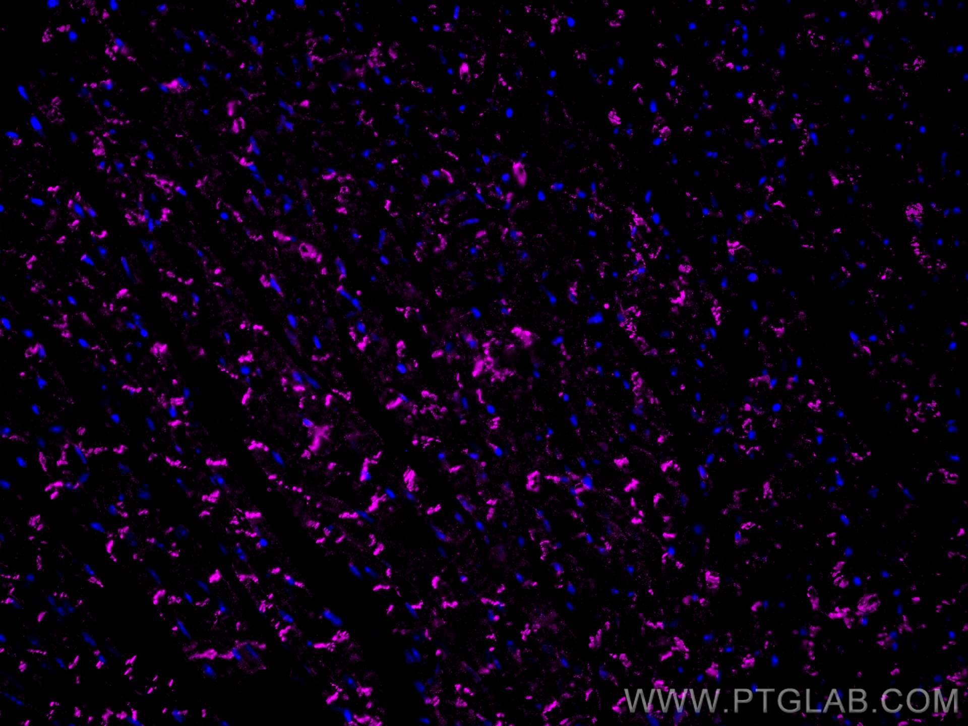

Tested Applications
| Positive IF-P detected in | mouse heart tissue |
Recommended dilution
| Application | Dilution |
|---|---|
| Immunofluorescence (IF)-P | IF-P : 1:50-1:500 |
| It is recommended that this reagent should be titrated in each testing system to obtain optimal results. | |
| Sample-dependent, Check data in validation data gallery. | |
Product Information
CL647-66219 targets N-cadherin in IF-P applications and shows reactivity with human, pig, mouse, rat, rabbit samples.
| Tested Reactivity | human, pig, mouse, rat, rabbit |
| Host / Isotype | Mouse / IgG1 |
| Class | Monoclonal |
| Type | Antibody |
| Immunogen |
CatNo: Ag4996 Product name: Recombinant human N-cadherin protein Source: e coli.-derived, PET28a Tag: 6*His Domain: 33-350 aa of BC036470 Sequence: KTGFPEDVYSAVLSKDVHEGQPLLNVKFSNCNGKRKVQYESSEPADFKVDEDGMVYAVRSFPLSSEHAKFLIYAQDKETQEKWQVAVKLSLKPTLTEESVKESAEVEEIVFPRQFSKHSGHLQRQKRDWVISPINLPENSRGPFPQELVRIRSDRDKNLSLRYSVTGPGADQPPTGIFIINPISGQLSVTKPLDREQIARFHLRAHAVDINGNQVENPIDIVINVIDMNDNRPEFLHQVWNGTVPEGSKPGTYVMTVTAIDADDPNALNGMLRYRIVSQAPSTPSPNMFTINNETGDIITVAAGLDREKVQQYTLIIQ Predict reactive species |
| Full Name | cadherin 2, type 1, N-cadherin (neuronal) |
| Calculated Molecular Weight | 906 aa, 100 kDa |
| Observed Molecular Weight | 130 kDa |
| GenBank Accession Number | BC036470 |
| Gene Symbol | N-cadherin |
| Gene ID (NCBI) | 1000 |
| RRID | AB_3084820 |
| Conjugate | CoraLite® Plus 647 Fluorescent Dye |
| Excitation/Emission Maxima Wavelengths | 654 nm / 674 nm |
| Excitation Laser | Red Laser (633 nm) |
| Form | Liquid |
| Purification Method | Protein G purification |
| UNIPROT ID | P19022 |
| Storage Buffer | PBS with 50% glycerol, 0.05% Proclin300, 0.5% BSA, pH 7.3. |
| Storage Conditions | Store at -20°C. Avoid exposure to light. Stable for one year after shipment. Aliquoting is unnecessary for -20oC storage. |
Background Information
Cadherins are a family of transmembrane glycoproteins that mediate calcium-dependent cell-cell adhesion and play an important role in the maintenance of normal tissue architecture. N-cadherin (neural cadherin), also known as CDH2 (cadherin 2), is a 130-kDa transmembrane protein and a classical member of the cadherin superfamily which also include E-, P-, R-, and B-cadherins. Expression of N-cadherin has been reported on various cell types including neurons, endothelial cells and cardiac myocytes (PMID: 11282032; 9508779; 8125202). N-cadherin has functions in early brain morphogenesis, synaptogenesis and synaptic plasticity (PMID: 23321619). The N-cadherin ectodomain can be cleaved, leading to the generation of a 90 kD N-terminal fragment (PMID: 16998833; 17028923).
Protocols
| Product Specific Protocols | |
|---|---|
| IF protocol for CL Plus 647 N-cadherin antibody CL647-66219 | Download protocol |
| Standard Protocols | |
|---|---|
| Click here to view our Standard Protocols |
Reviews
What customers say
The user review for CL647-66219 rates it 4 out of 5. Used mainly for IF staining as a CoraLite® Plus 647-conjugated N-cadherin mouse monoclonal antibody, it produced a clear and specific fluorescence signal with good membrane localization and low background staining. The reviewer found it suitable for visualizing N-cadherin expression, easy to use, and consistent under their experimental conditions. It's described as a reliable reagent for IF applications, with the caveat that optimizing antibody dilution and imaging settings may be beneficial depending on cell type.
The reviews below have been submitted by verified Proteintech customers who received an incentive for providing their feedback.
Abi (Verified Customer) (07-28-2026) | I used The CoraLite® Plus 647-conjugated N-cadherin mouse monoclonal antibody mainly for IF staining. The antibody produced a clear and specific fluorescence signal with good membrane localisation with low background staining, making it suitable for visualising N-cadherin expression. The antibody was easy to use and provided consistent results under our experimental conditions. Overall, a reliable reagent for IF applications, although optimisation of antibody dilution and imaging settings may be beneficial depending on the cell type.
|


