

Tested Applications
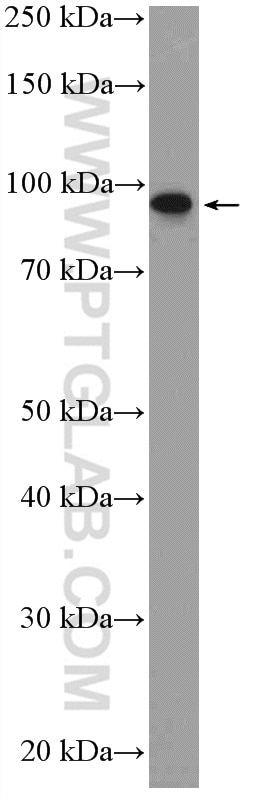
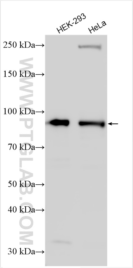
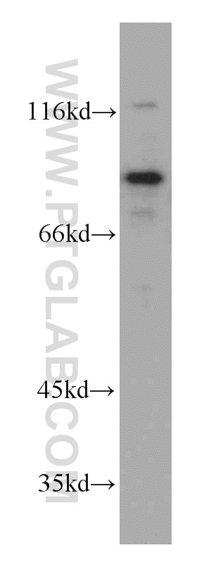
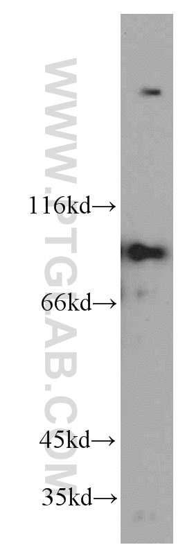
| Positive WB detected in | HEK-293 cells, MCF-7 cells, K-562 cells, HeLa cells |
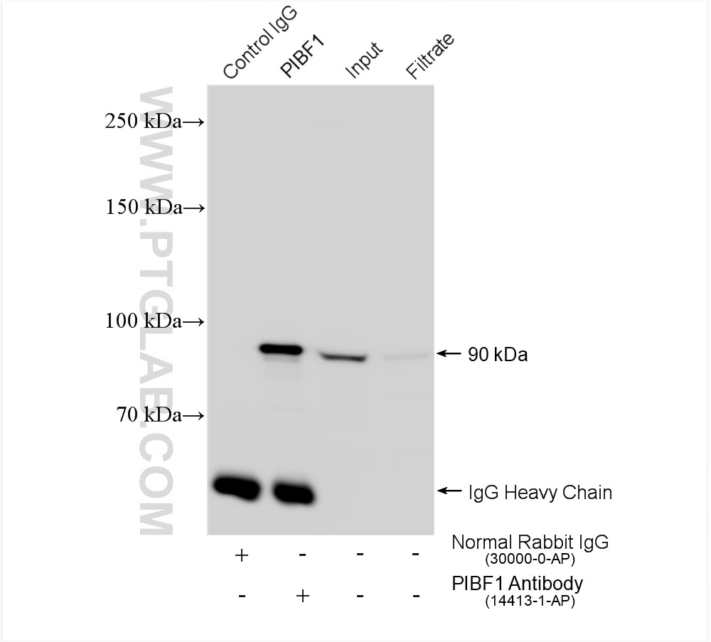
| Positive IP detected in | HEK-293 cells |
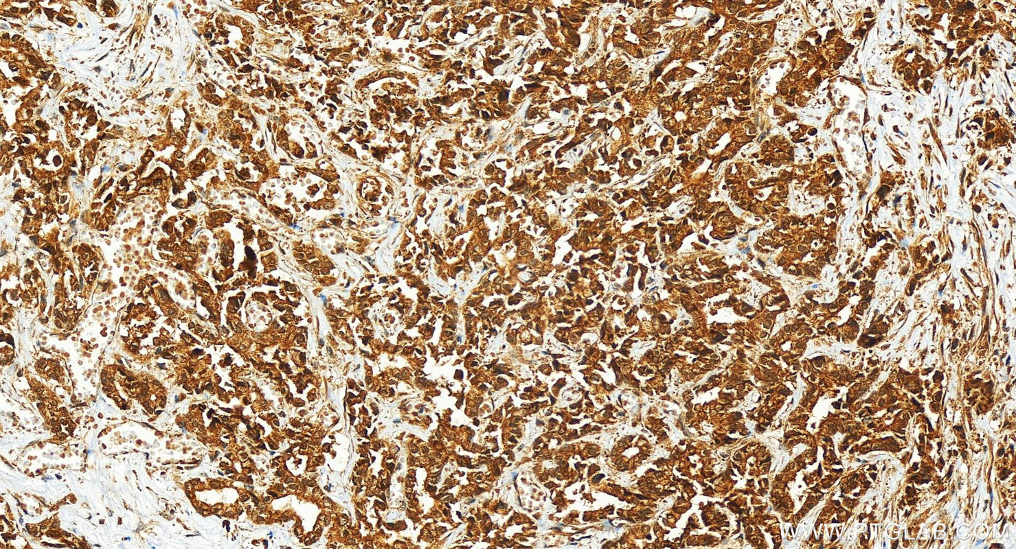
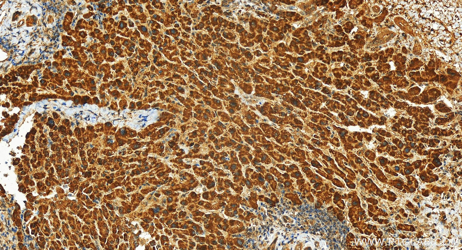
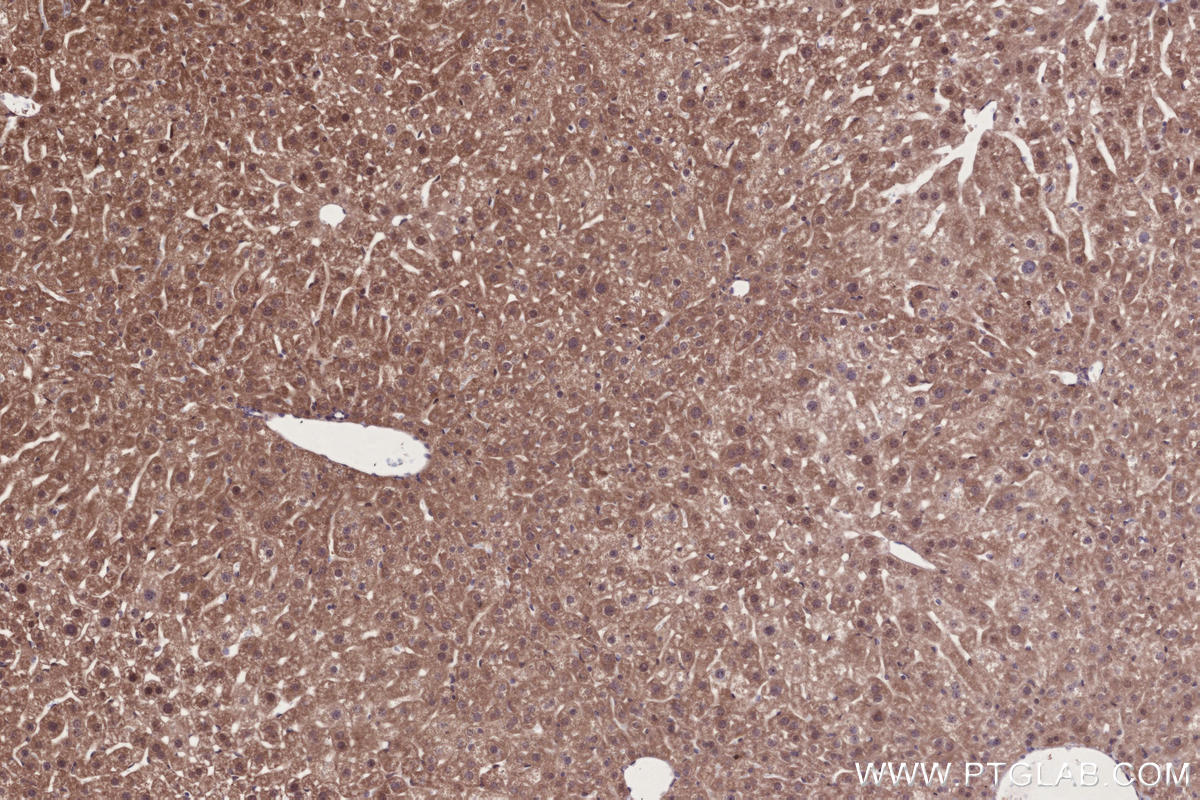
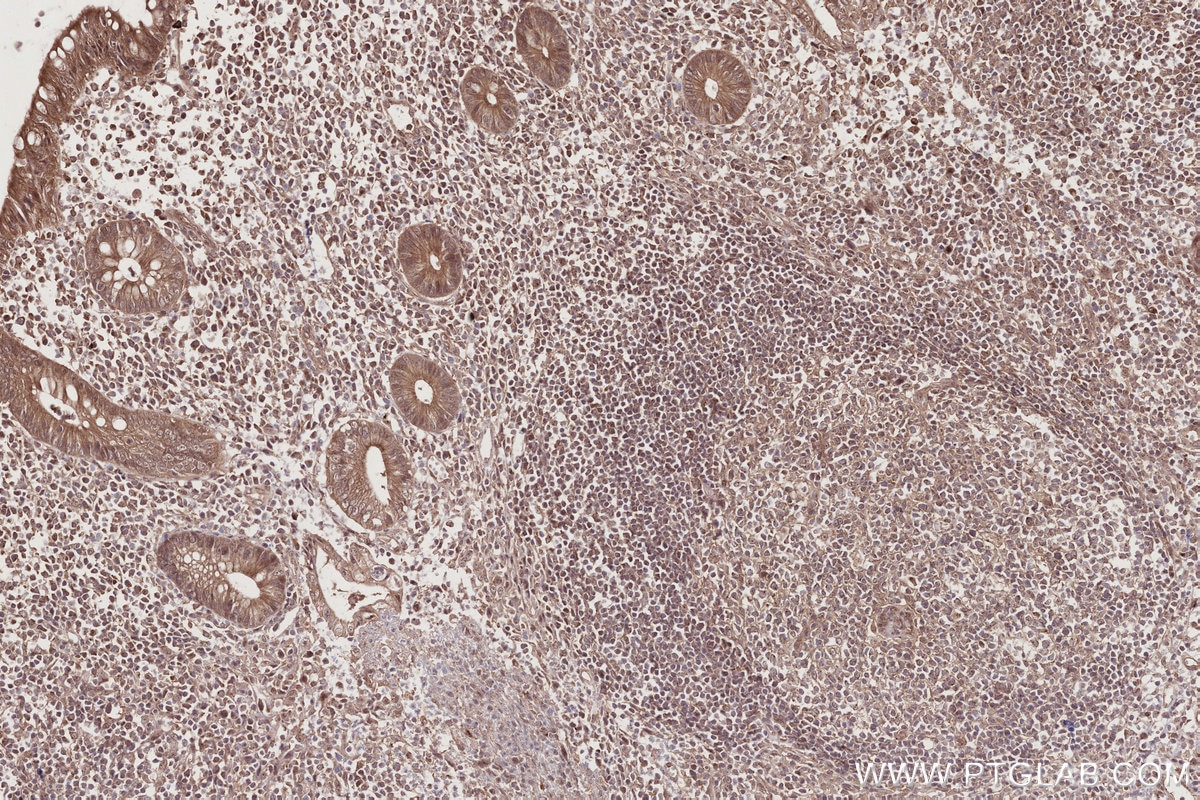
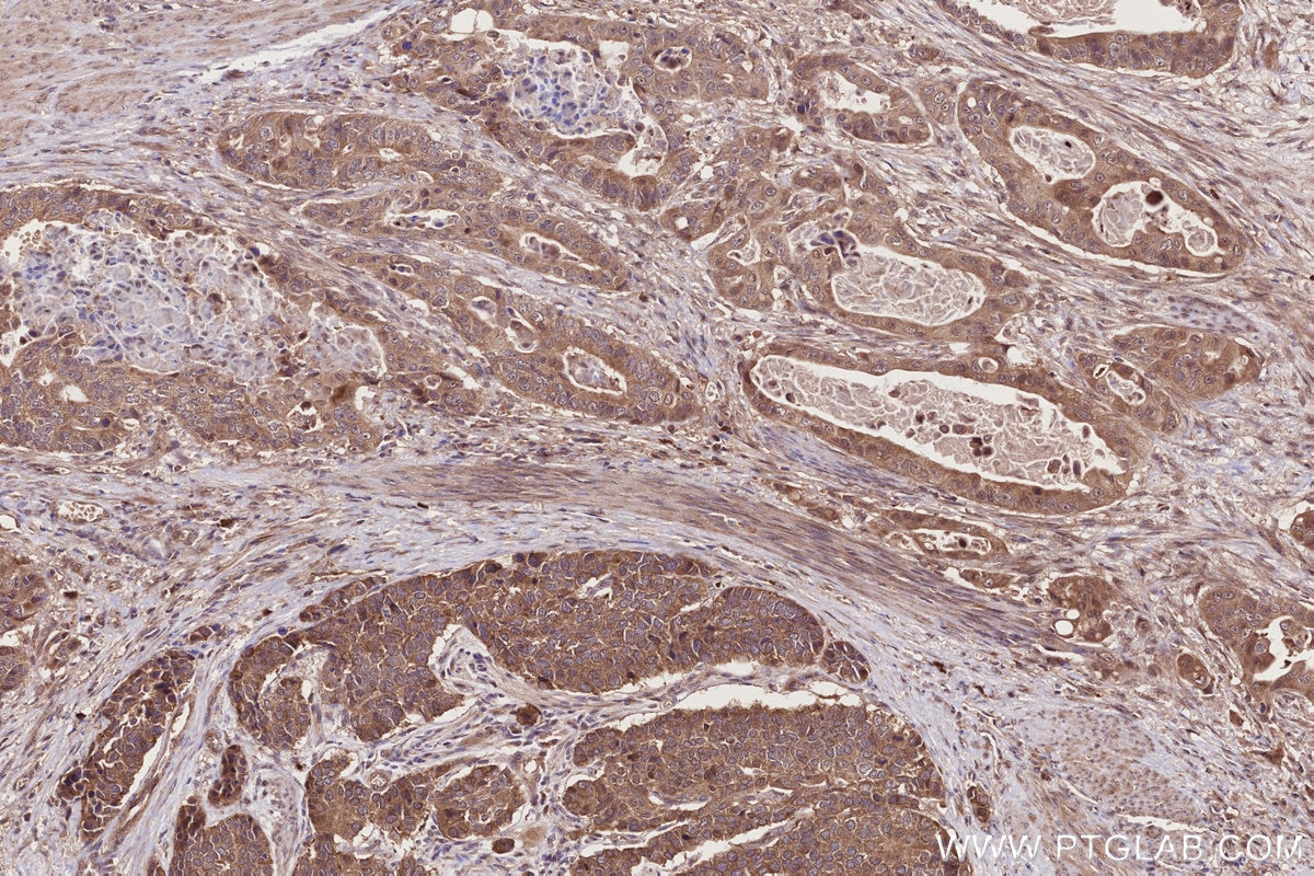
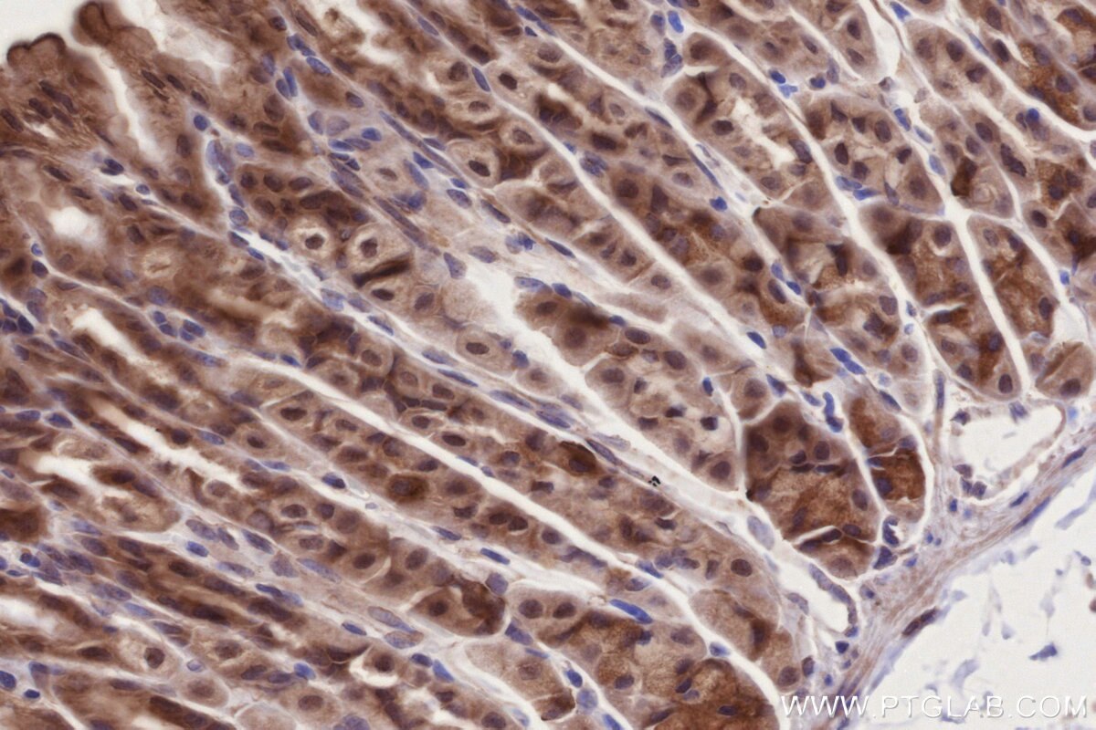
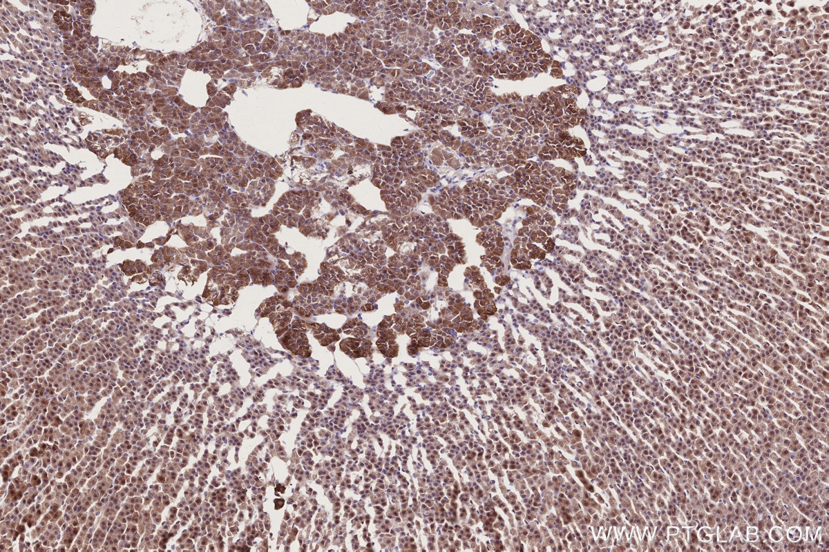
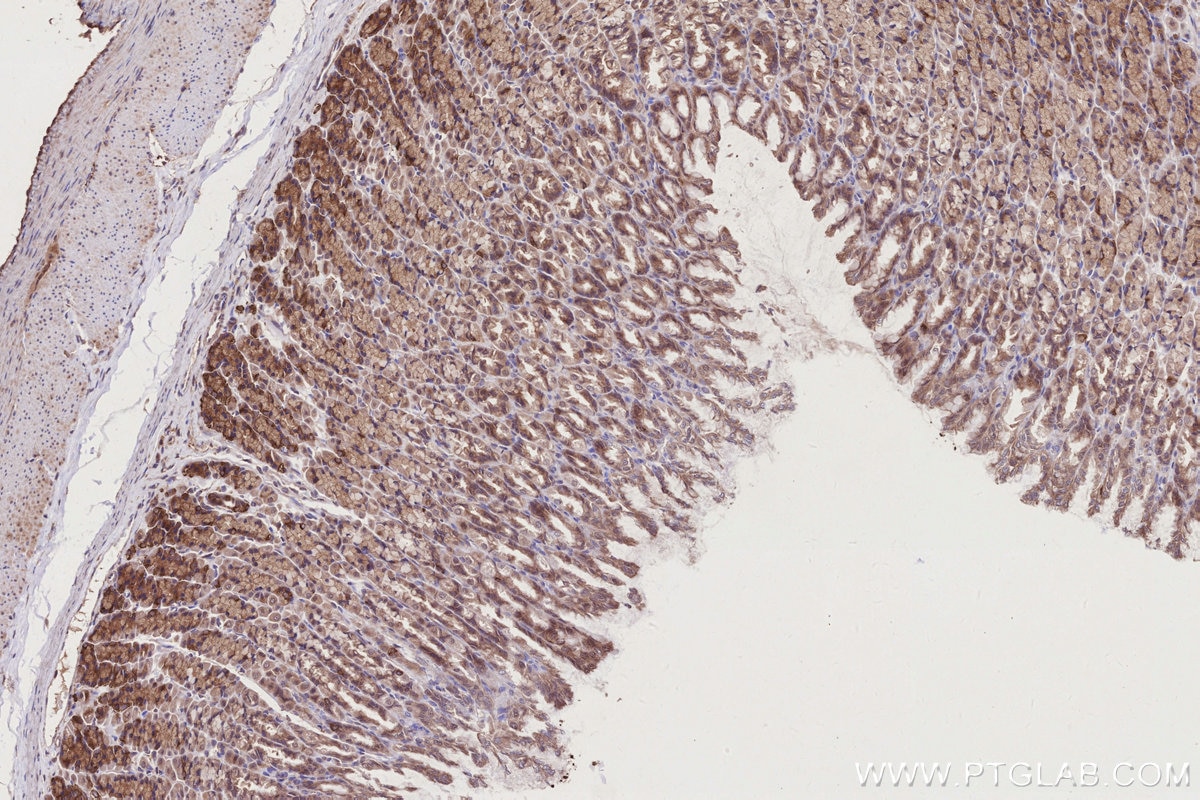
| Positive IHC detected in | human intrahepatic cholangiocarcinoma tissue, rat adrenal gland tissue, human appendicitis tissue, rat stomach tissue, mouse stomach tissue, mouse liver tissue, human stomach cancer tissue Note: suggested antigen retrieval with TE buffer pH 9.0; (*) Alternatively, antigen retrieval may be performed with citrate buffer pH 6.0 |
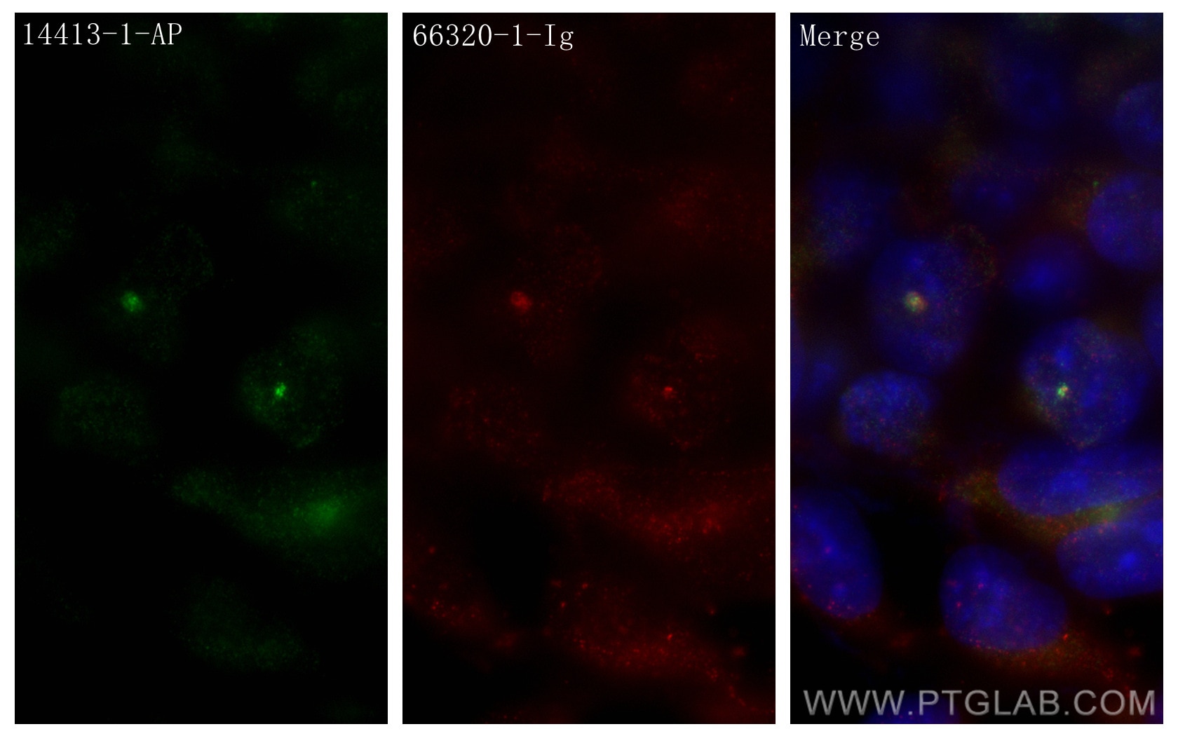
| Positive IF/ICC detected in | HEK-293 cells |
Recommended dilution
| Application | Dilution |
|---|---|
| Western Blot (WB) | WB : 1:1000-1:6000 |
| Immunoprecipitation (IP) | IP : 0.5-4.0 ug for 1.0-3.0 mg of total protein lysate |
| Immunohistochemistry (IHC) | IHC : 1:50-1:500 |
| Immunofluorescence (IF)/ICC | IF/ICC : 1:200-1:800 |
| It is recommended that this reagent should be titrated in each testing system to obtain optimal results. | |
| Sample-dependent, Check data in validation data gallery. | |
Product Information
14413-1-AP targets PIBF1 in WB, IHC, IF/ICC, IP, ELISA applications and shows reactivity with human, mouse, rat samples.
| Tested Reactivity | human, mouse, rat |
| Cited Reactivity | human |
| Host / Isotype | Rabbit / IgG |
| Class | Polyclonal |
| Type | Antibody |
| Immunogen |
CatNo: Ag5755 Product name: Recombinant human PIBF1 protein Source: e coli.-derived, PGEX-4T Tag: GST Domain: 452-757 aa of BC051911 Sequence: KVTEFLHQSKLKSFESERVQLLQEETARNLTQCQLECEKYQKKLEVLTKEFYSLQASSEKRITELQAQNSEHQARLDIYEKLEKELDEIIMQTAEIENEDEAERVLFSYGYGANVPTTAKRRLKQSVHLARRVLQLEKQNSLILKDLEHRKDQVTQLSQELDRANSLLNQTQQPYRYLIESVRQRDSKIDSLTESIAQLEKDVSNLNKEKSALLQTKNQMALDLEQLLNHREELAAMKQILVKMHSKHSENSLLLTKTEPKHVTENQKSKTLNVPKEHEDNIFTPKPTLFTKKEAPEWSKKQKMKT Predict reactive species |
| Full Name | progesterone immunomodulatory binding factor 1 |
| Calculated Molecular Weight | 90 kDa |
| Observed Molecular Weight | 90 kDa |
| GenBank Accession Number | BC051911 |
| Gene Symbol | PIBF1 |
| Gene ID (NCBI) | 10464 |
| RRID | AB_10858916 |
| Conjugate | Unconjugated |
| Form | Liquid |
| Purification Method | Antigen affinity purification |
| UNIPROT ID | Q8WXW3 |
| Storage Buffer | PBS with 0.02% sodium azide and 50% glycerol, pH 7.3. |
| Storage Conditions | Store at -20°C. Stable for one year after shipment. Aliquoting is unnecessary for -20oC storage. 20ul sizes contain 0.1% BSA. |
Background Information
PIBF1 is induced by the steroid hormone progesterone and plays a role in the maintenance of pregnancy. PIBF1 regulates multiple facets of the immune system to promote normal pregnancy including cytokine synthesis, natural killer (NK) cell activity, and arachidonic acid metabolism. Low serum levels of this protein have been associated with spontaneous pre-term labor in humans. PIBF1 may promote the proliferation, migration and invasion of glioma.
Protocols
| Product Specific Protocols | |
|---|---|
| IF protocol for PIBF1 antibody 14413-1-AP | Download protocol |
| IHC protocol for PIBF1 antibody 14413-1-AP | Download protocol |
| IP protocol for PIBF1 antibody 14413-1-AP | Download protocol |
| WB protocol for PIBF1 antibody 14413-1-AP | Download protocol |
| Standard Protocols | |
|---|---|
| Click here to view our Standard Protocols |
Publications
What published studies show
The PIBF1 Polyclonal antibody (Cat# 14413-1-AP) has 10 total citations. These appear in journals including ACS Nano, EMBO Journal, Cell Reports, PLoS Biology, Journal of Cell Biology, EMBO Reports, Virology Journal, bioRxiv, and eLife. The research fields span centriole biology and duplication, ciliogenesis, centrosome organization, viral infection mechanisms (Zika and HSV), and multiplexed nanoscopy imaging.
| Species | Application | Title |
|---|---|---|
EMBO J Human SFI1 and Centrin form a complex critical for centriole architecture and ciliogenesis | ||
Cell Rep CCDC57 Cooperates with Microtubules and Microcephaly Protein CEP63 and Regulates Centriole Duplication and Mitotic Progression. | ||
PLoS Biol The luminal ring protein C2CD3 acts as a radial in-to-out organizer of the distal centriole and appendages. | ||
PLoS Biol The evolutionary conserved proteins CEP90, FOPNL, and OFD1 recruit centriolar distal appendage proteins to initiate their assembly
| ||
J Cell Biol A ciliopathy complex builds distal appendages to initiate ciliogenesis.
|
Reviews
What customers say
Users consistently rate this antibody positively (4–5 stars) across Western blot and immunofluorescence applications. One reviewer confirmed it performs well in both WB and IF. Another provided a detailed IF protocol on HEK293 cells, noting clear centrosome staining at 1:200 dilution. A third user reported reliable WB results at 1:1000 dilution on RPE1 and HEK293 samples, and also used it for immunoprecipitation. Overall, the antibody is regarded as specific and effective at standard dilutions for multiple applications.
The reviews below have been submitted by verified Proteintech customers who received an incentive for providing their feedback.
Nicolas (Verified Customer) (03-02-2026) | works both for blot and immunofluorescence
|
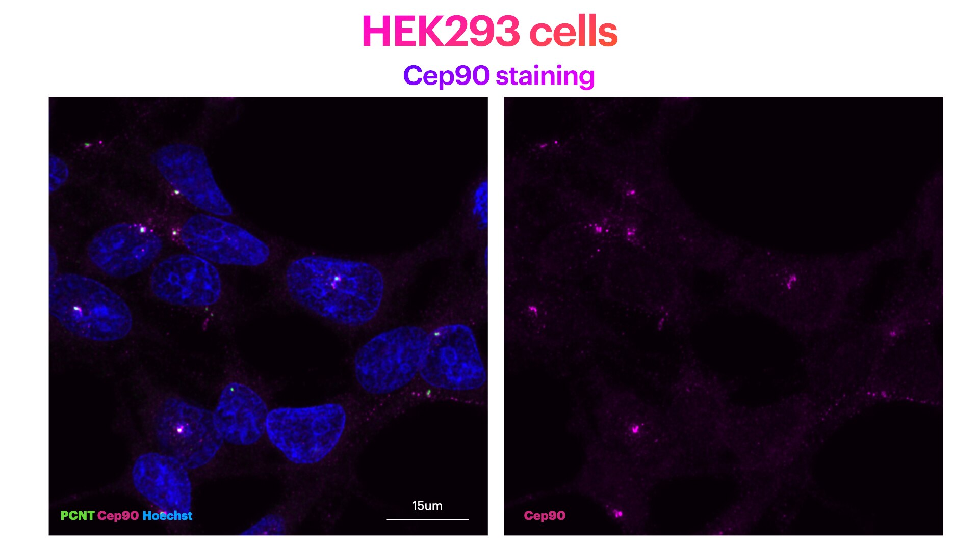
Elisa (Verified Customer) (06-20-2022) | HEK293 cells stained for Hoechst (DNA marker, in green), Cep90 (mother centriole distal appendage marker, in magenta) and PCNT (pericentriolar matrix marker, in green). HEK293 cells were plated on Poly-lysine coated coverslips and fixed in cold methanol for 2' at -20C. Cells were then rehydrated with PBS for 5'. Membrane permeabilisation was then performed with 0.1% Triton + 0.1% Tween +0.01%SDS in PBS for 5'. Cells were finally incubated with blocking buffer (5% BSA+ 0.1% Tween in PBS) for 30' at RT. Primary antibody was diluted in blocking buffer 1:200 and incubated for 1h at room temperature. Alexa-555-Anti-rabbit was used as secondary antibody (1:600 dilution) (1h at room temperature). Cep90 antibody recognises clearly dots at the centrosome (identified by the presence of PCNT).
 |
Pierrick (Verified Customer) (10-24-2019) | Antibody mostly used in wester blot and work well at 1/1000 dilution on RPE1 and HEK293 sample
|















