




Tested Applications
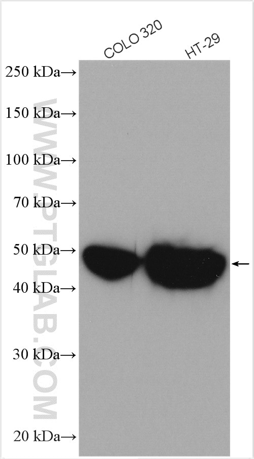
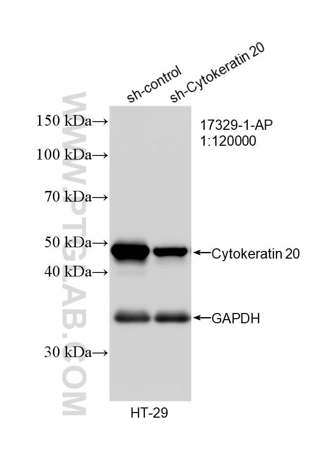
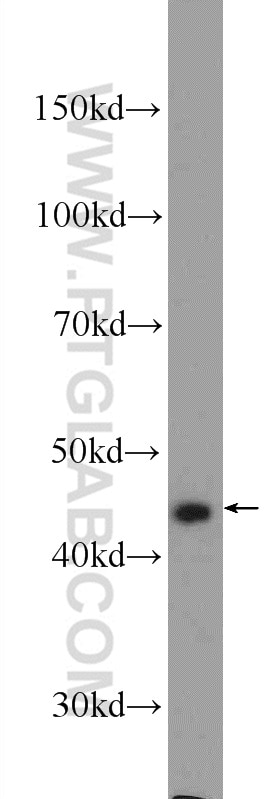
| Positive WB detected in | COLO 320 cells, HeLa cells, HT-29 cells |
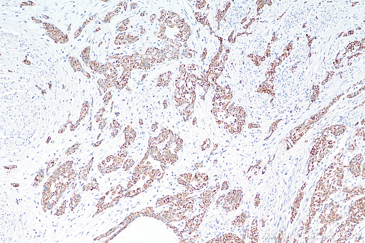
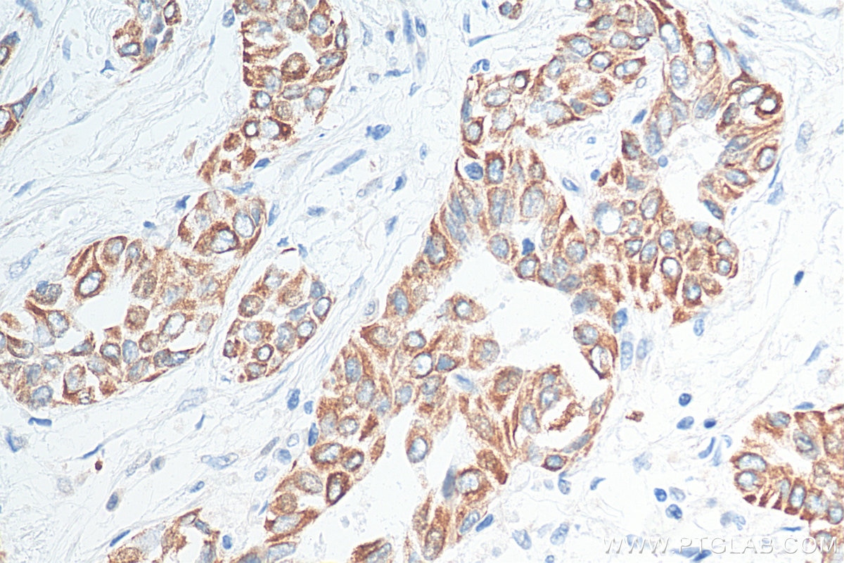
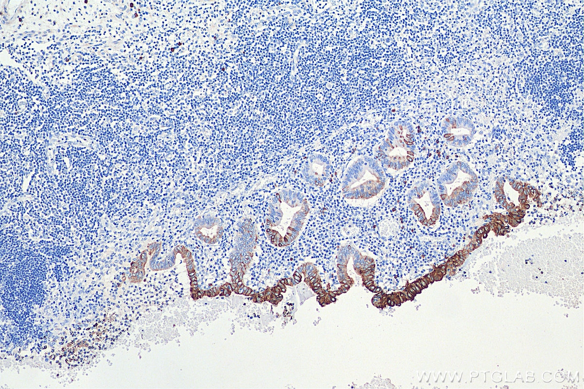
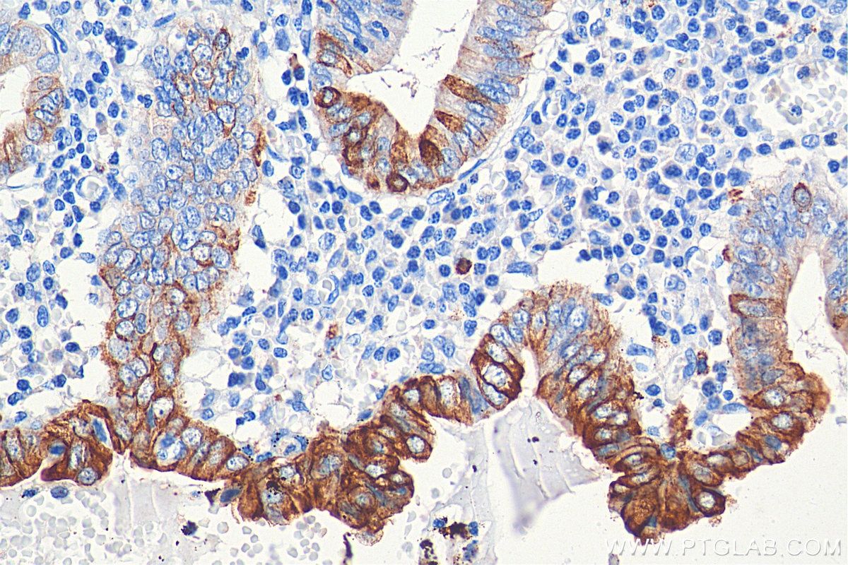
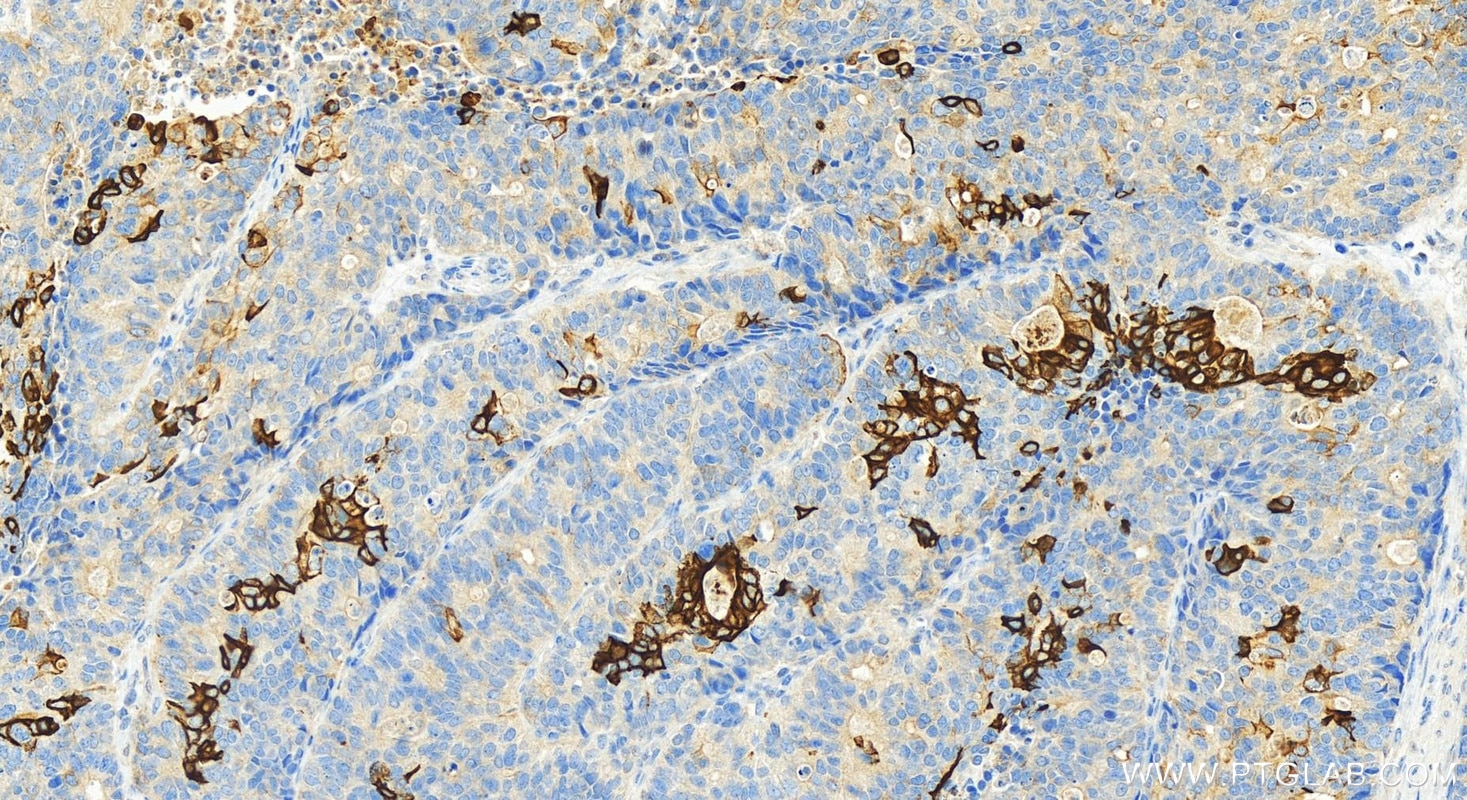
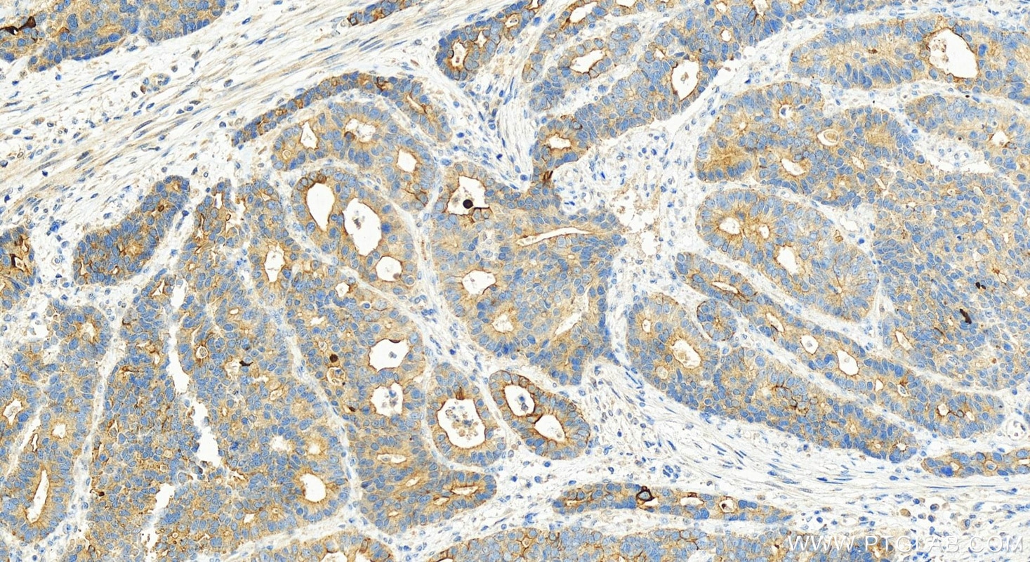
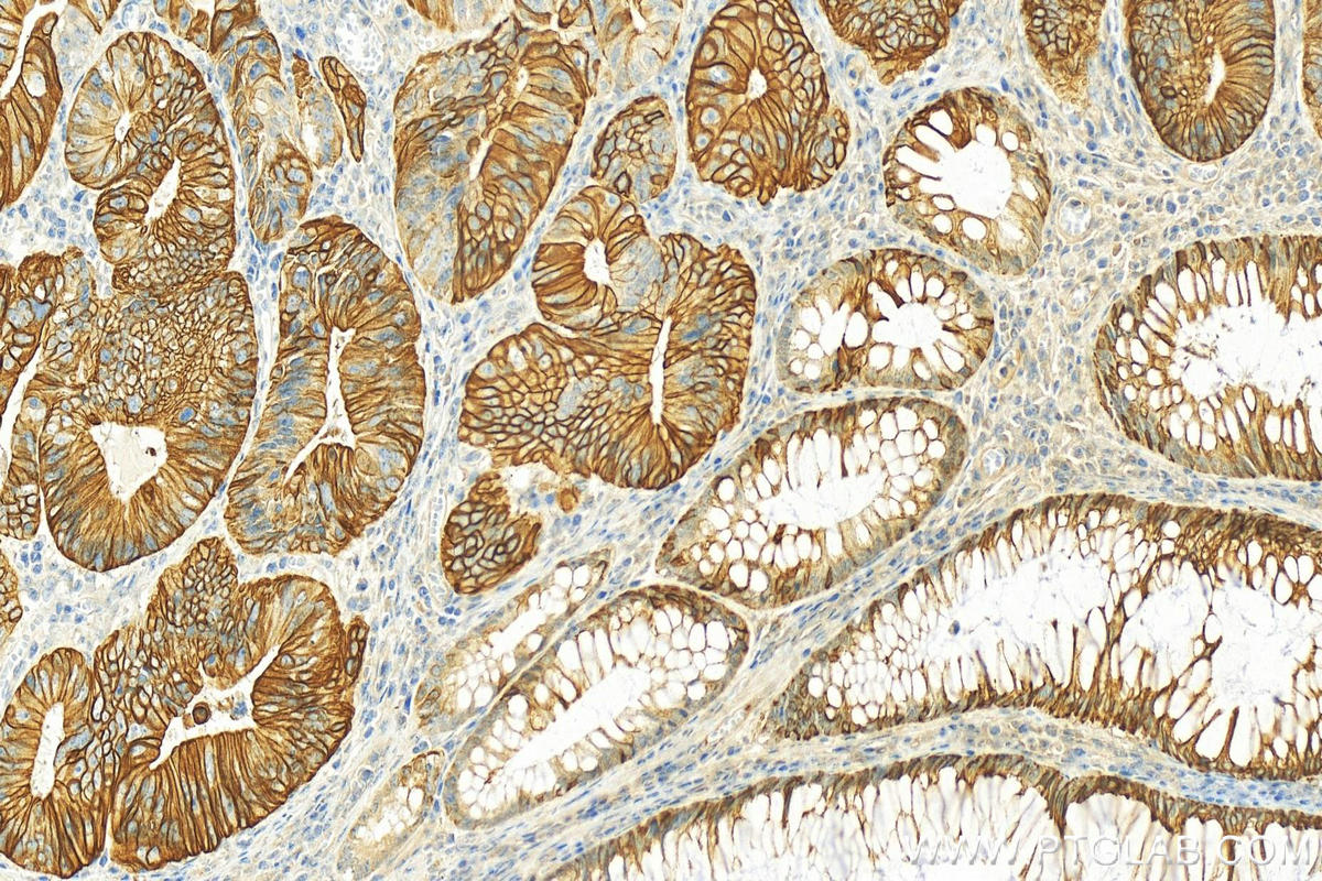
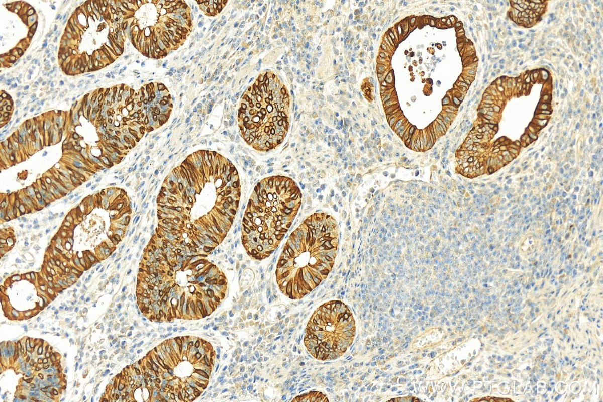
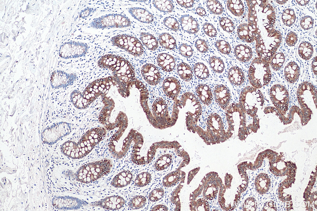
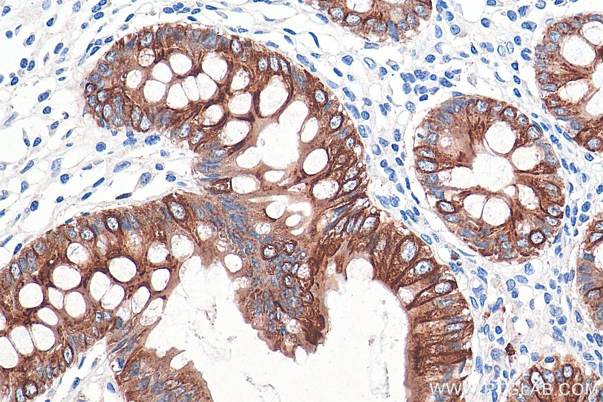
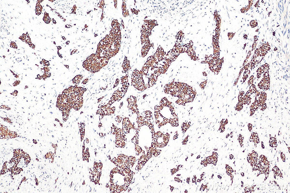
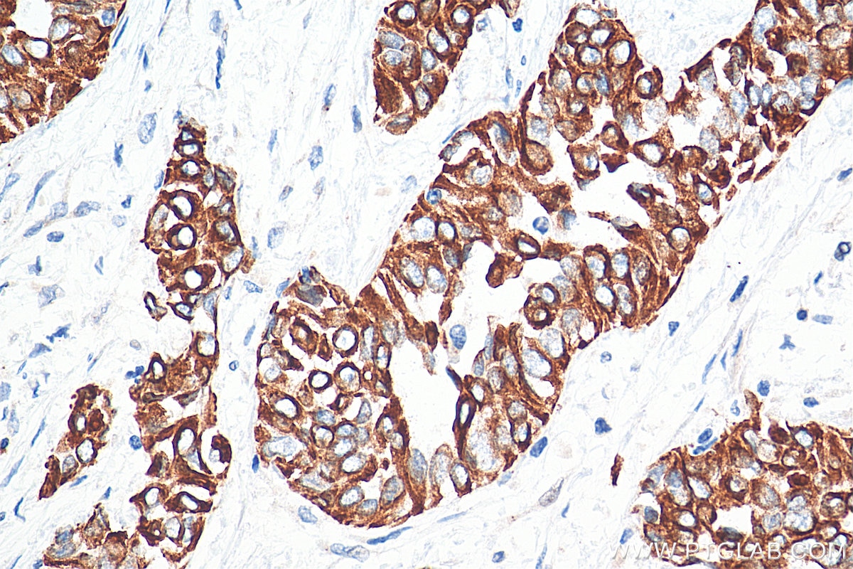
| Positive IHC detected in | human colon tissue, human appendicitis tissue, human colon cancer tissue, human stomach cancer tissue, human urothelial carcinoma tissue Note: suggested antigen retrieval with TE buffer pH 9.0; (*) Alternatively, antigen retrieval may be performed with citrate buffer pH 6.0 |
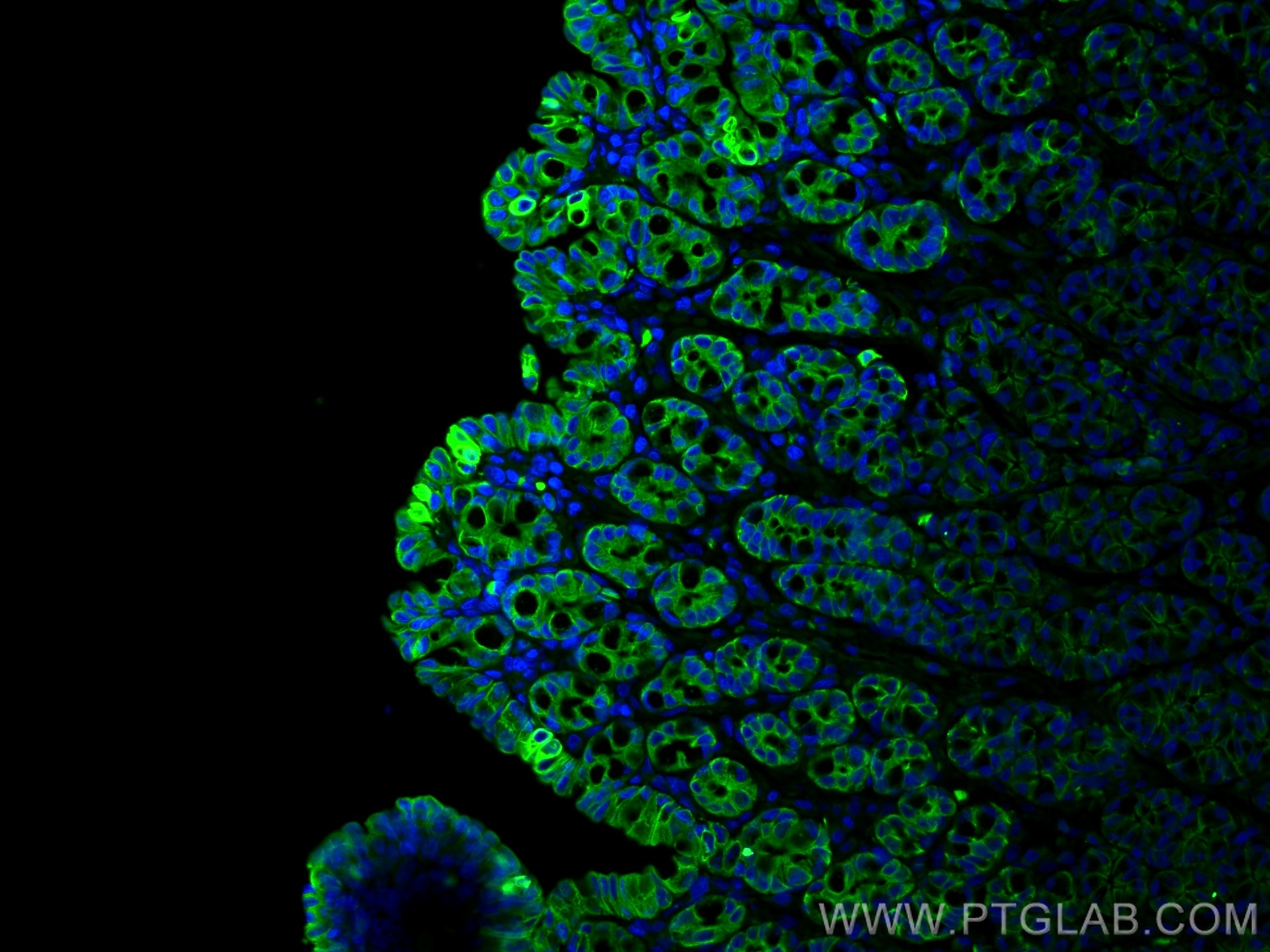
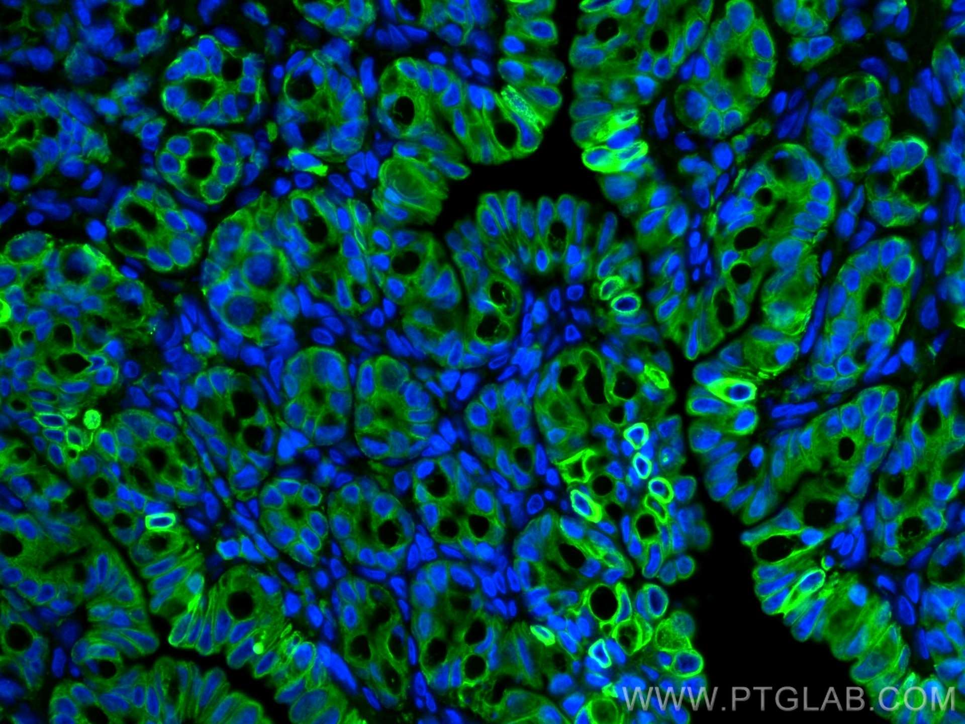
| Positive IF-P detected in | Rabbit colon tissue, human colon cancer tissue |
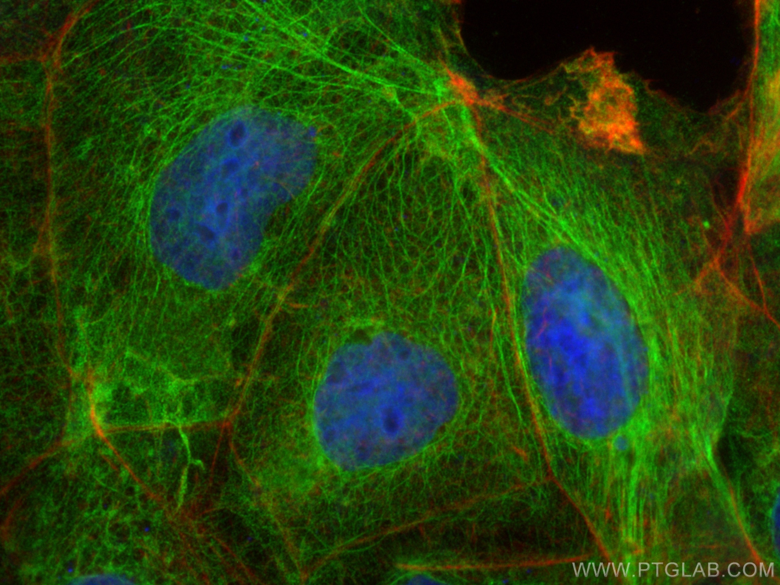
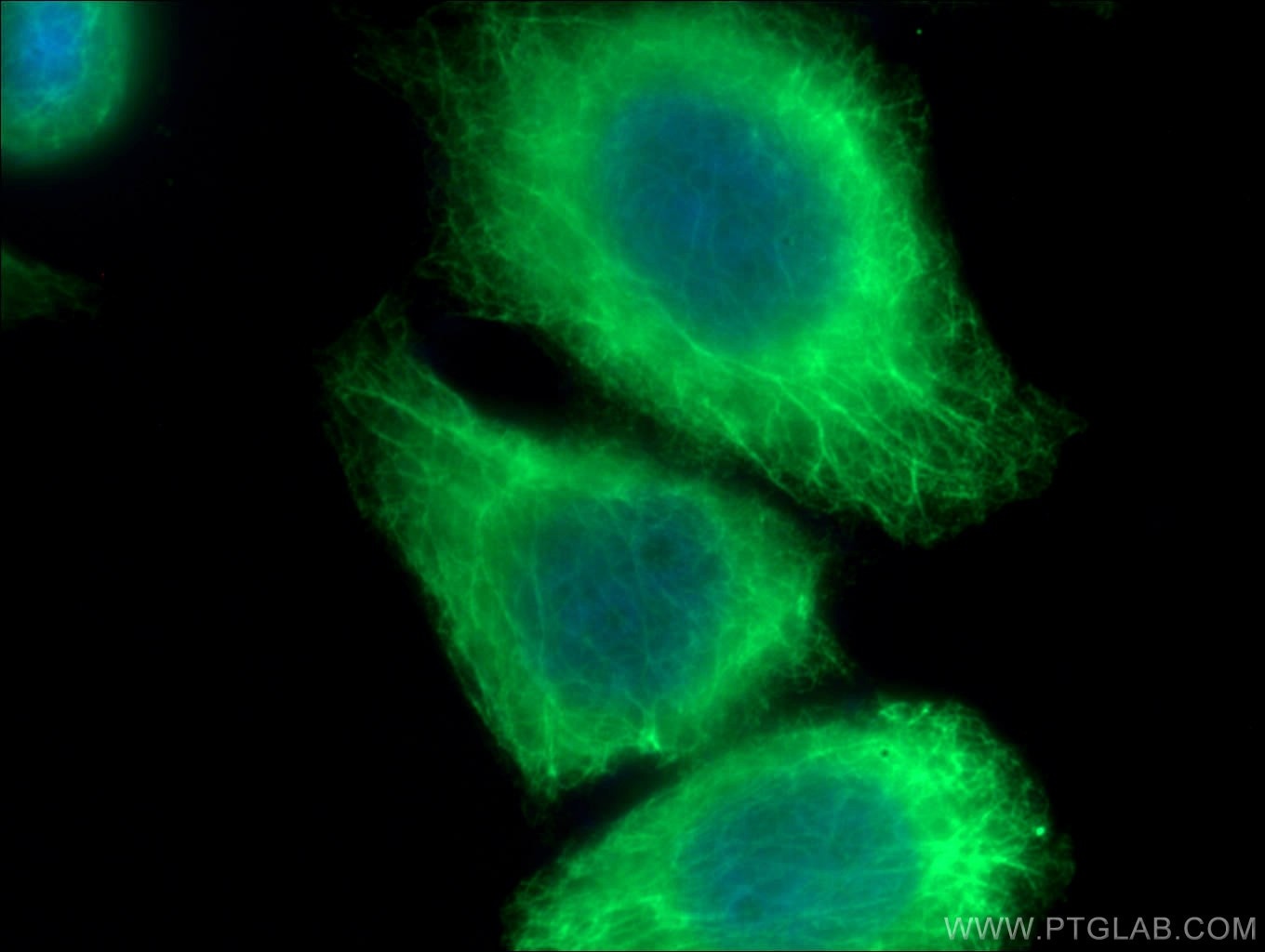
| Positive IF/ICC detected in | Caco-2 cells, HeLa cells |
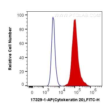
| Positive FC (Intra) detected in | HT-29 cells |
Recommended dilution
| Application | Dilution |
|---|---|
| Western Blot (WB) | WB : 1:2000-1:16000 |
| Immunohistochemistry (IHC) | IHC : 1:800-1:3200 |
| Immunofluorescence (IF)-P | IF-P : 1:500-1:2000 |
| Immunofluorescence (IF)/ICC | IF/ICC : 1:200-1:800 |
| Flow Cytometry (FC) (INTRA) | FC (INTRA) : 0.20 ug per 10^6 cells in a 100 µl suspension |
| It is recommended that this reagent should be titrated in each testing system to obtain optimal results. | |
| Sample-dependent, Check data in validation data gallery. | |
Product Information
17329-1-AP targets Cytokeratin 20 in WB, IHC, IF/ICC, IF-P, FC (Intra), ELISA applications and shows reactivity with human, mouse, rat, rabbit samples.
| Tested Reactivity | human, mouse, rat, rabbit |
| Cited Reactivity | human, mouse |
| Host / Isotype | Rabbit / IgG |
| Class | Polyclonal |
| Type | Antibody |
| Immunogen |
CatNo: Ag11308 Product name: Recombinant human KRT20 protein Source: e coli.-derived, PGEX-4T Tag: GST Domain: 74-424 aa of BC031559 Sequence: MQNLNDRLASYLEKVRTLEQSNSKLEVQIKQWYETNAPRAGRDYSAYYRQIEELRSQIKDAQLQNARCVLQIDNAKLAAEDFRLKYETERGIRLTVEADLQGLNKVFDDLTLHKTDLEIQIEELNKDLALLKKEHQEEVDGLHKHLGNTVNVEVDAAPGLNLGVIMNEMRQKYEVMAQKNLQEAKEQFERQTAVLQQQVTVNTEELKGTEVQLTELRRTSQSLEIELQSHLSMKESLEHTLEETKARYSSQLANLQSLLSSLEAQLMQIRSNMERQNNEYHILLDIKTRLEQEIATYRRLLEGEDVKTTEYQLSTLEERDIKKTRKIKTVVQEVVDGKVVSSEVKEVEENI Predict reactive species |
| Full Name | keratin 20 |
| Calculated Molecular Weight | 424 aa, 48 kDa |
| Observed Molecular Weight | 46 kDa |
| GenBank Accession Number | BC031559 |
| Gene Symbol | Cytokeratin 20 |
| Gene ID (NCBI) | 54474 |
| RRID | AB_2133592 |
| Conjugate | Unconjugated |
| Form | Liquid |
| Purification Method | Antigen affinity purification |
| UNIPROT ID | P35900 |
| Storage Buffer | PBS with 0.02% sodium azide and 50% glycerol, pH 7.3. |
| Storage Conditions | Store at -20°C. Stable for one year after shipment. Aliquoting is unnecessary for -20oC storage. 20ul sizes contain 0.1% BSA. |
Background Information
Keratins are a large family of proteins that form the intermediate filament cytoskeleton of epithelial cells, which are classified into two major sequence types. Type I keratins are a group of acidic intermediate filament proteins, including K9-K23, and the hair keratins Ha1-Ha8. Type II keratins are the basic or neutral courterparts to the acidic type I keratins, including K1-K8, and the hair keratins, Hb1-Hb6. Keratin 20 is a type I cytokeratin. It is a major cellular protein of mature enterocytes and goblet cells and is specifically found in the gastric and intestinal mucosa.
Protocols
| Product Specific Protocols | |
|---|---|
| FC protocol for Cytokeratin 20 antibody 17329-1-AP | Download protocol |
| IF protocol for Cytokeratin 20 antibody 17329-1-AP | Download protocol |
| IHC protocol for Cytokeratin 20 antibody 17329-1-AP | Download protocol |
| WB protocol for Cytokeratin 20 antibody 17329-1-AP | Download protocol |
| Standard Protocols | |
|---|---|
| Click here to view our Standard Protocols |
Publications
What published studies show
The Cytokeratin 20 (KRT20) polyclonal antibody (Cat# 17329-1-AP) has 24 citations published in journals including Nature Communications, Nature Cell Biology, Nature Immunology, Cell Research, Neuron, Molecular Cancer, Cell Reports, Advanced Science, Biomaterials, Cancer Letters, Molecular Medicine, Communications Biology, iScience, and others. Research fields span intestinal and gastric stem cell biology, colorectal and gastric cancer, bladder cancer, epithelial differentiation, gut microbiota, metastasis, and organoid models, with cited applications in IHC, IF, and WB.
| Species | Application | Title |
|---|---|---|
Mol Cancer DC - SIGNR by influencing the lncRNA HNRNPKP2 upregulates the expression of CXCR4 in gastric cancer liver metastasis. | ||
Cell Res Gut microbiota drives macrophage-dependent self-renewal of intestinal stem cells via niche enteric serotonergic neurons. | ||
Nat Immunol IL-13 secreted by ILC2s promotes the self-renewal of intestinal stem cells through circular RNA circPan3. | ||
Nat Cell Biol LncGata6 maintains stemness of intestinal stem cells and promotes intestinal tumorigenesis. | ||
Nat Commun Colorectal cancer co-opts an epidermal wound healing program during metastasis to generate disseminated tumor cells. | ||
Nat Commun Single-cell transcriptomics uncovers EGFR signaling-mediated gastric progenitor cell differentiation in stomach homeostasis |
Reviews
What customers say
Both reviewers gave this antibody a 5-star rating for immunohistochemistry. One used it at 1:100 dilution with pH 9 TE buffer for DAB staining on human lung cancer patient-derived tissues and reported complete satisfaction. The other applied it at 1:1500 dilution on human colorectal carcinoma tissue using an automated staining system with a 30-minute primary antibody incubation, polymer-based HRP detection, and heat-mediated Tris-EDTA (pH 9.0) antigen retrieval, scanning at 20x. Overall, users found the antibody reliable and effective for IHC applications across different tissue types and protocols.
The reviews below have been submitted by verified Proteintech customers who received an incentive for providing their feedback.
Nikolett (Verified Customer) (03-31-2026) | I used this antibody to characterize my human lung cancer patient derived tissues. For the IHC DAB staining I applied in a 1:100 dilution using pH9 TE buffer. I'm absolutely satisfied.
|
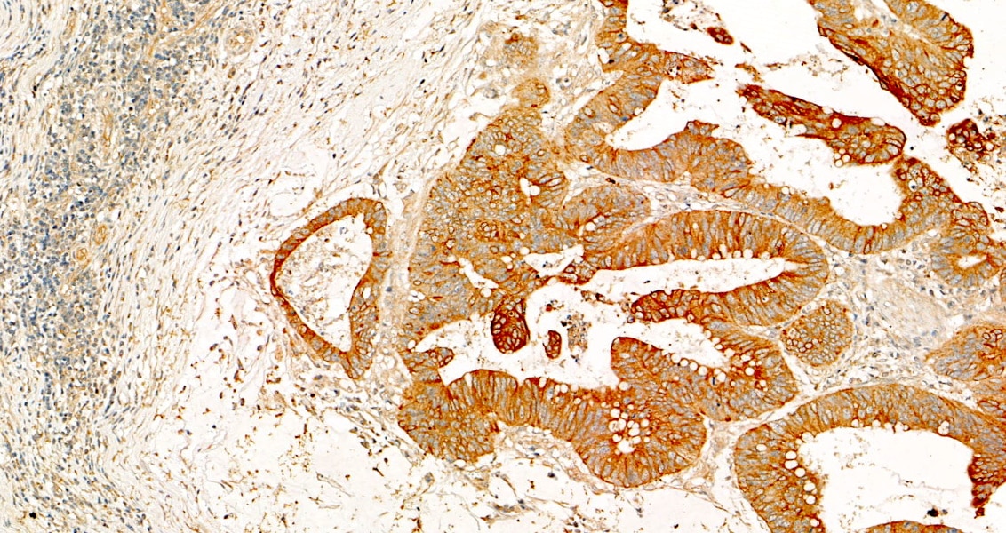
Felletár (Verified Customer) (05-09-2025) | Immunohistochemical staining was performed using a novel, pre-commercial automated staining-covering and scanning system. The protocol included a 30-minute incubation with the primary antibody and utilized a polymer-based horseradish peroxidase (HRP) detection system. Heat mediated antigen retrieval with Tris-EDTA buffer (pH 9.0). scanning under 20x lens.
 |
























