



Tested Applications
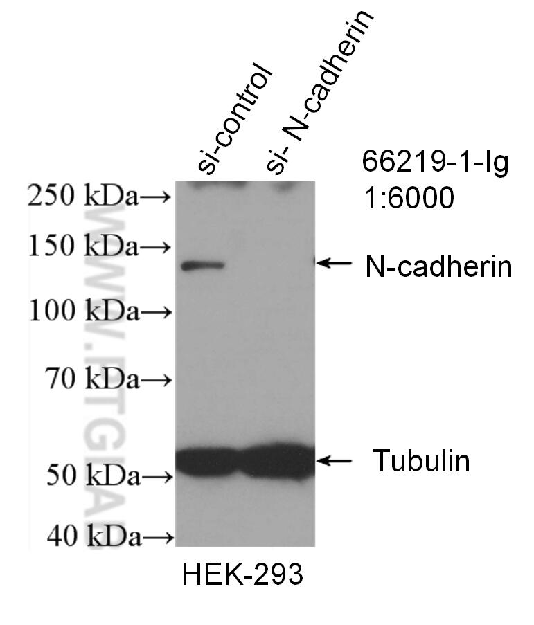
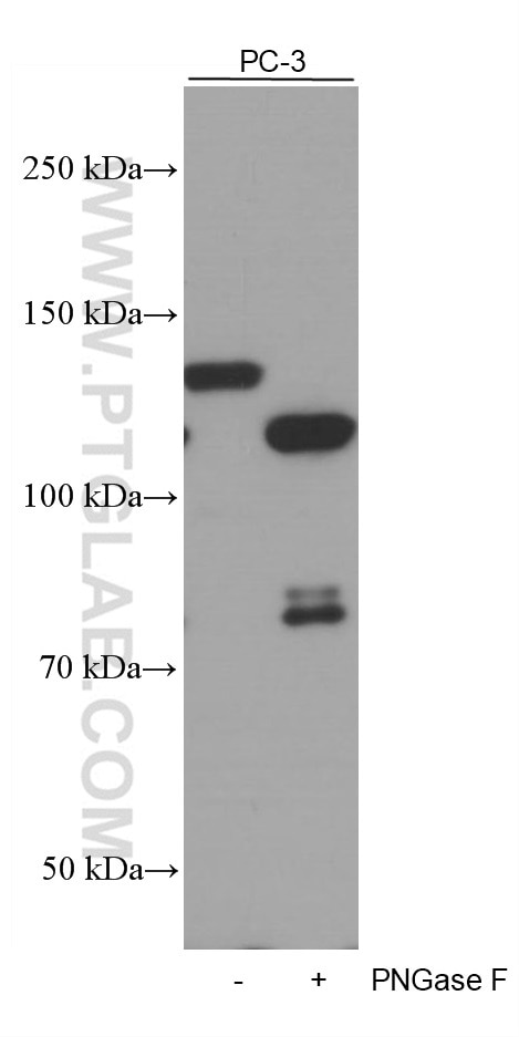
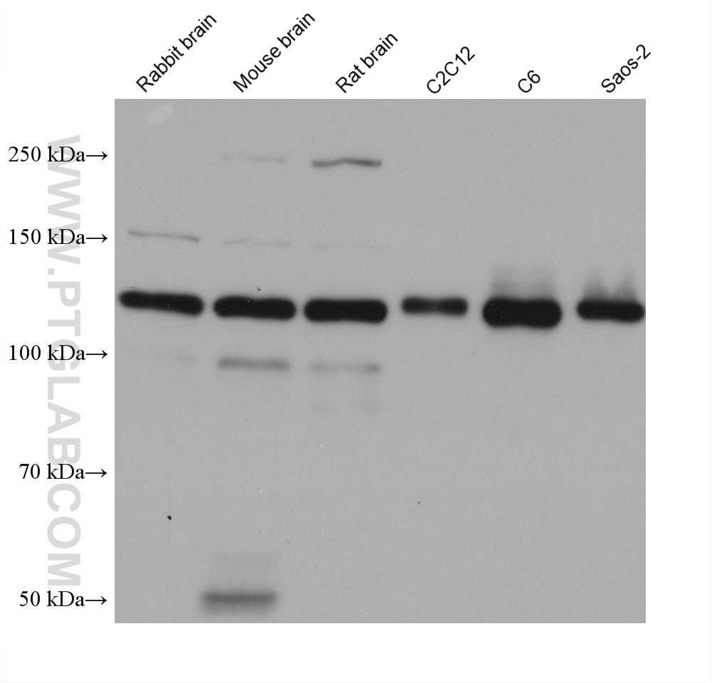
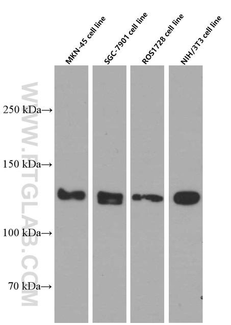
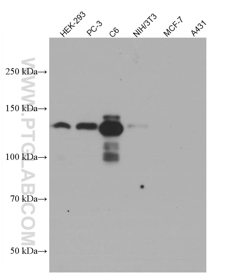
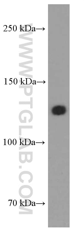
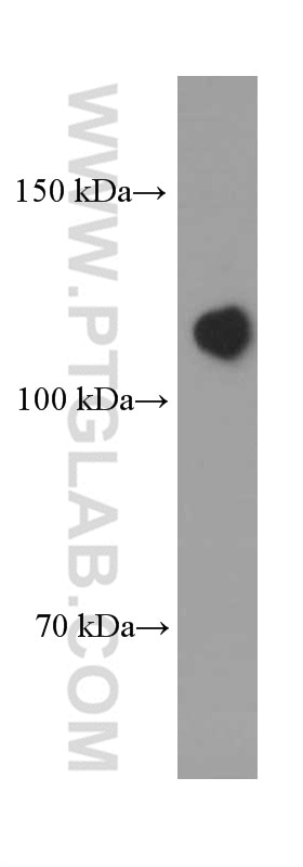
| Positive WB detected in | rabbit brain tissue, pig brain tissue, human heart tissue, MKN-45 cells, HEK-293 cells, PC-3 cells, mouse brain tissue, rat brain tissue, C2C12 cells, C6 cells, Saos-2 cells, SGC-7901 cells, ROS1728 cells, NIH/3T3 cells |
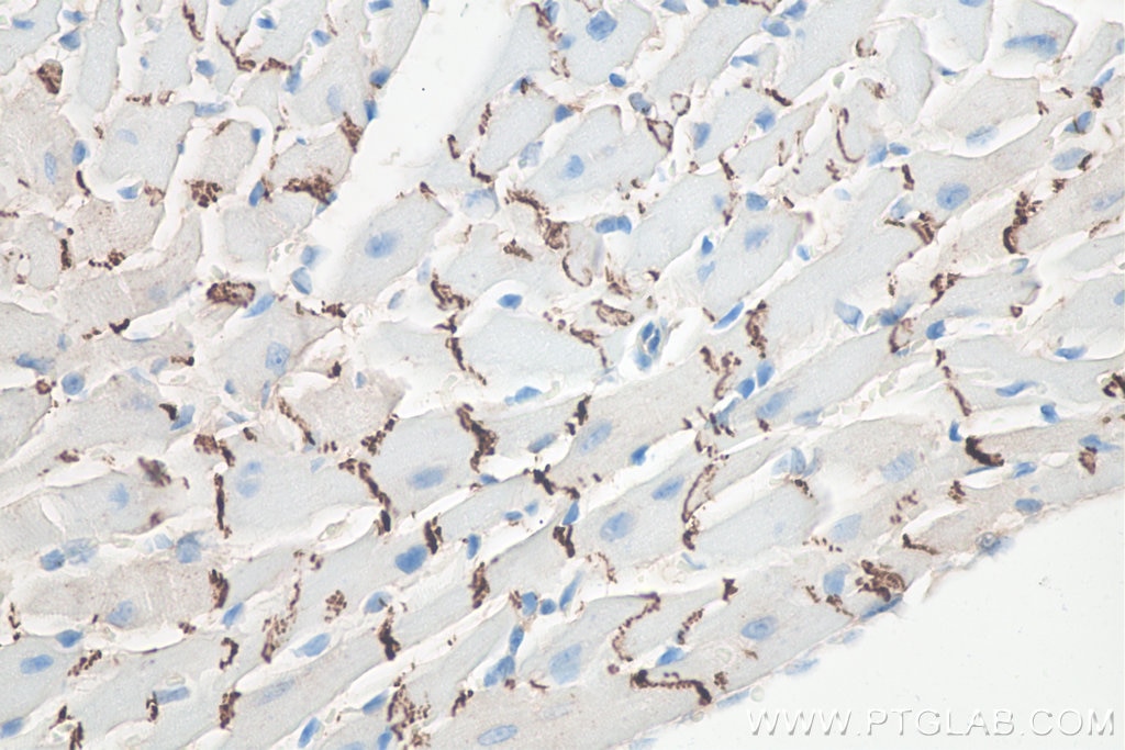
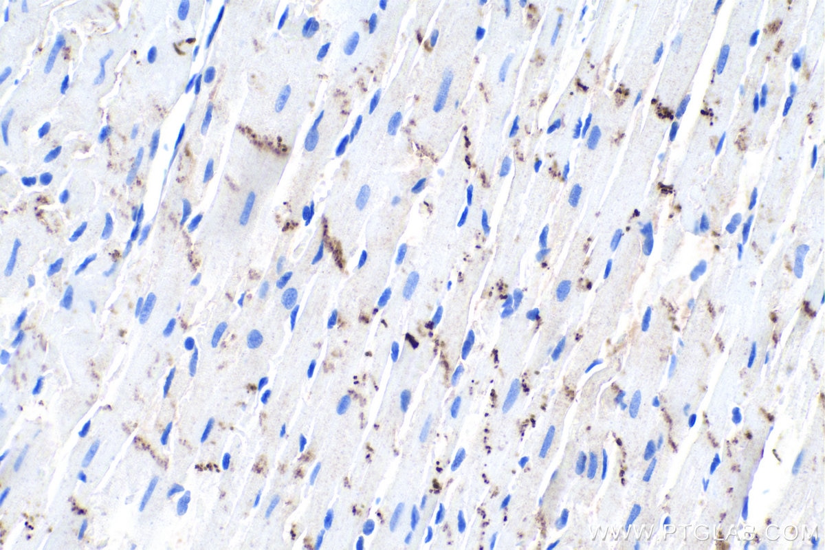
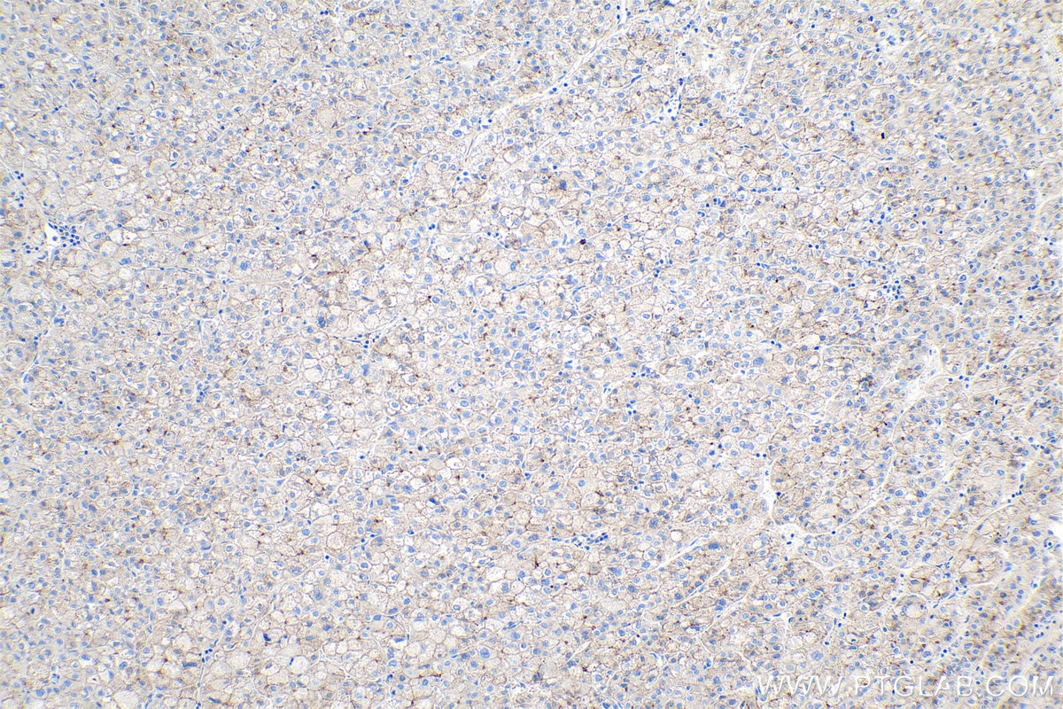
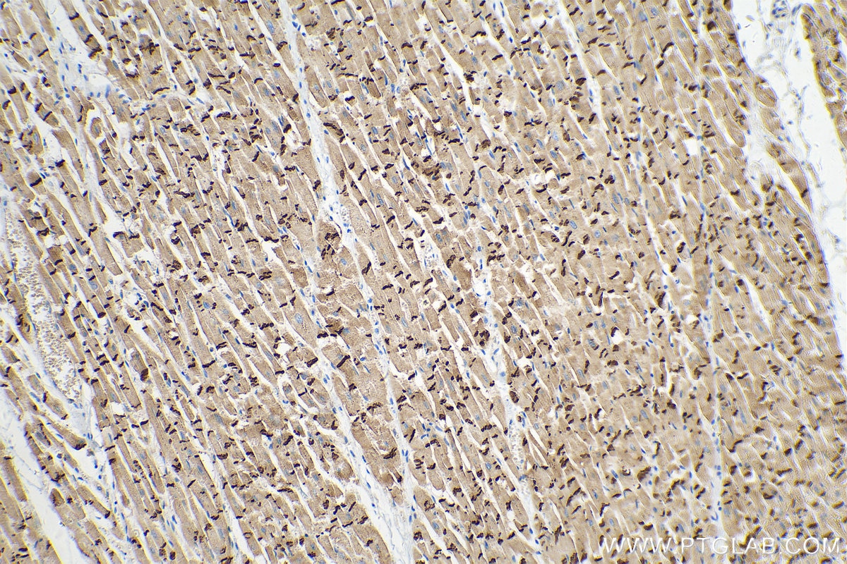
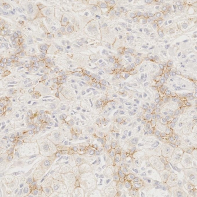
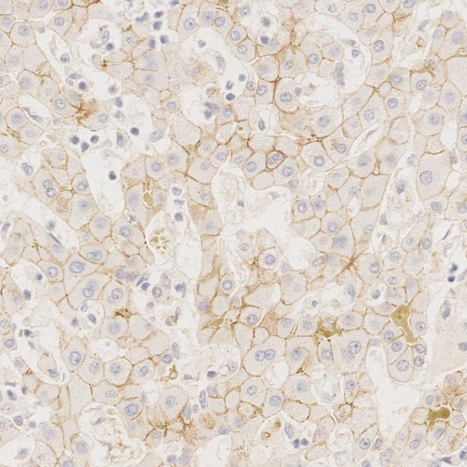
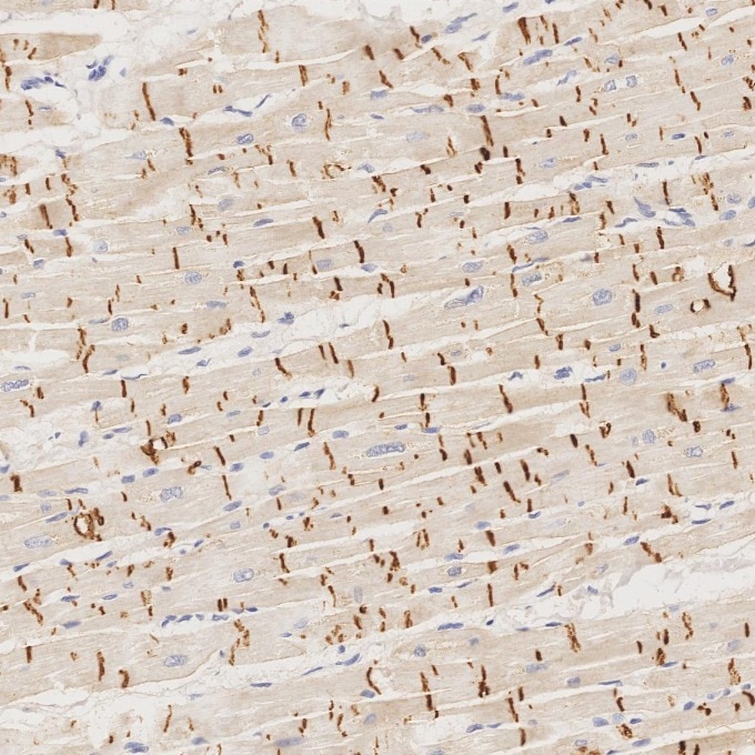
| Positive IHC-Autostainer detected in | human heart tissue, human liver tissue, human brain tissue |
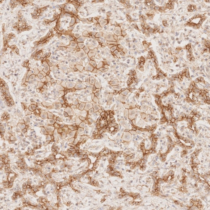
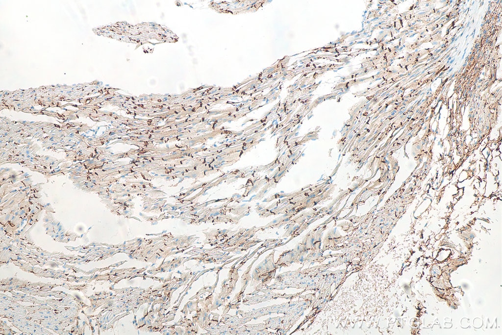
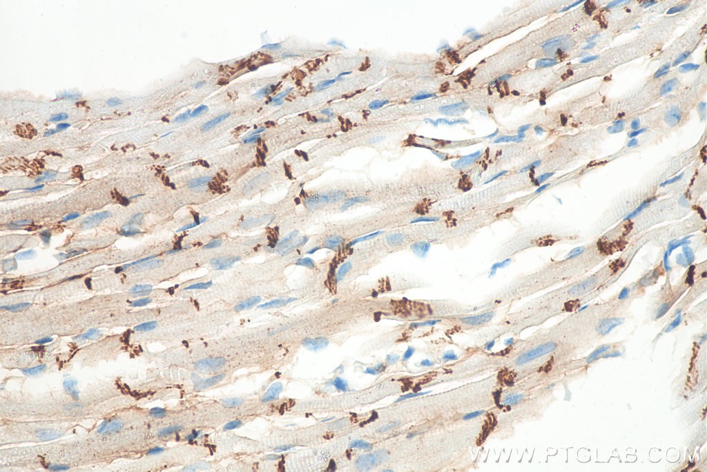
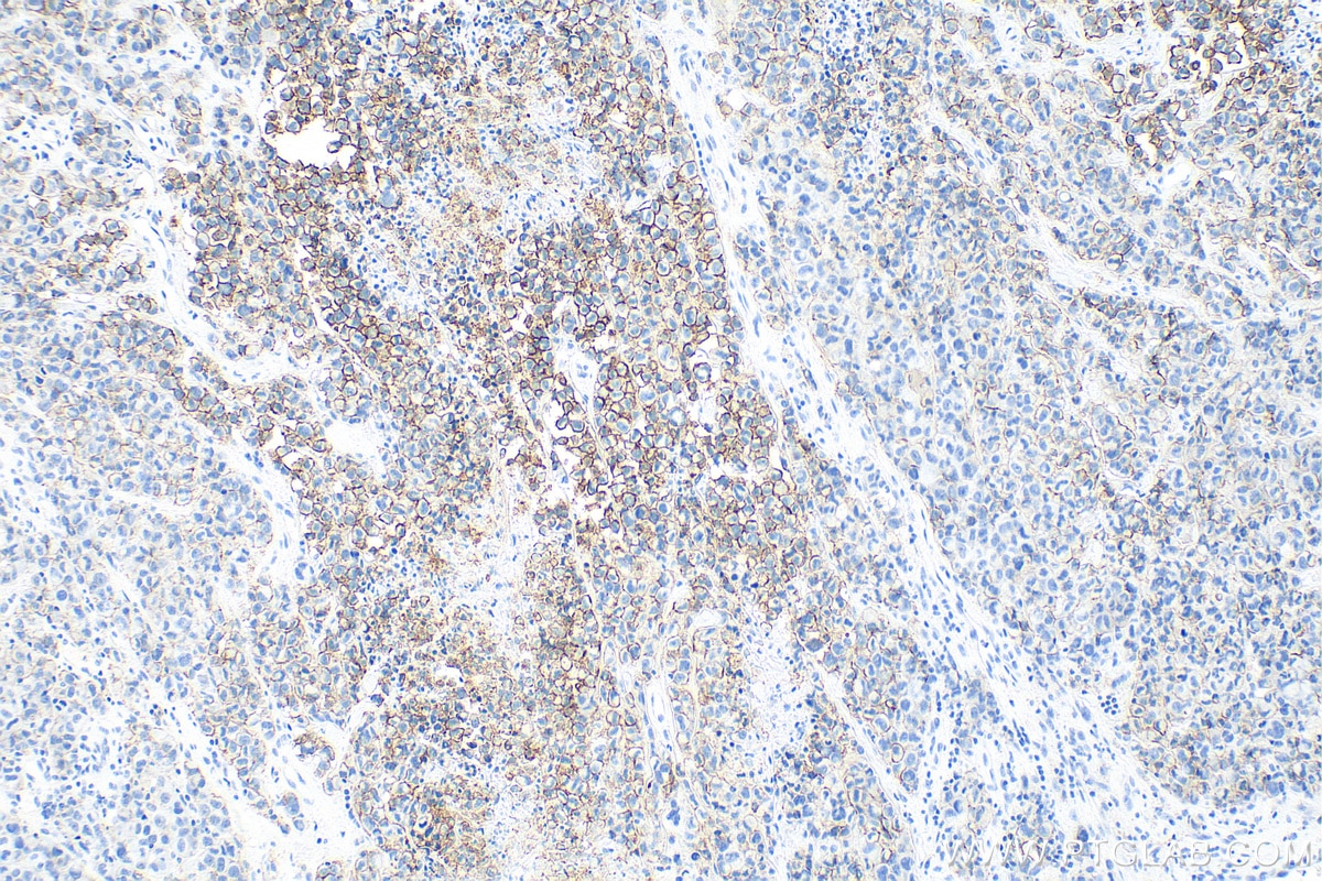
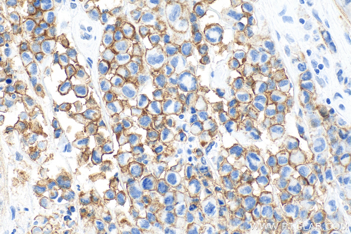
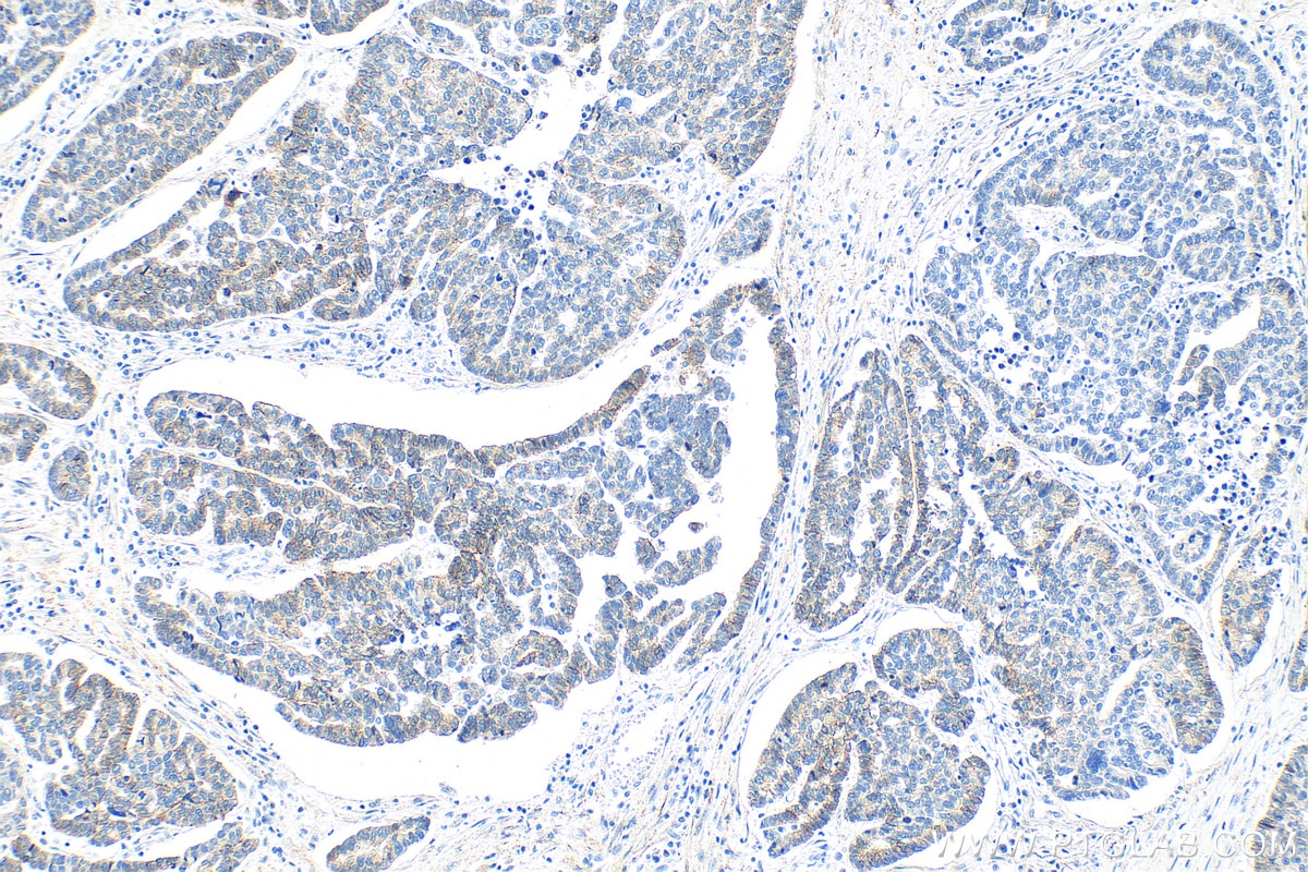
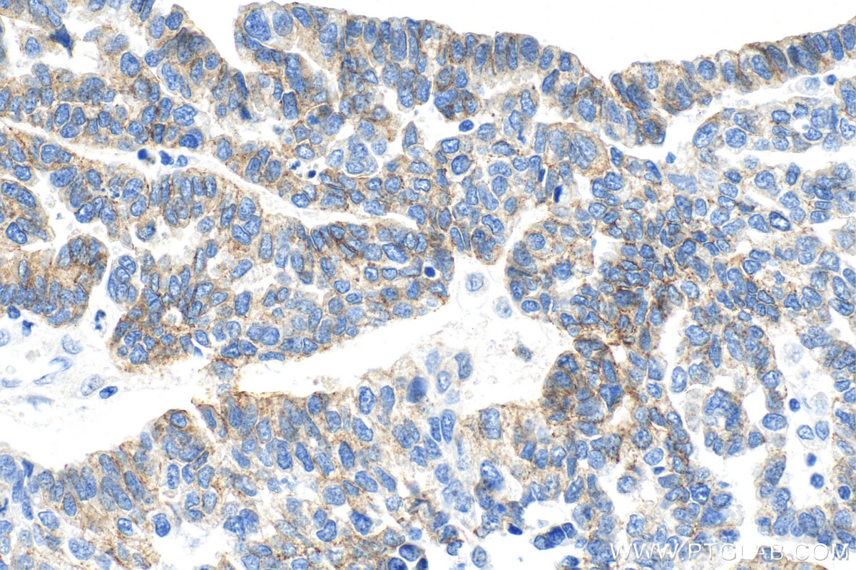
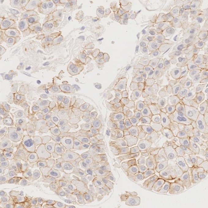
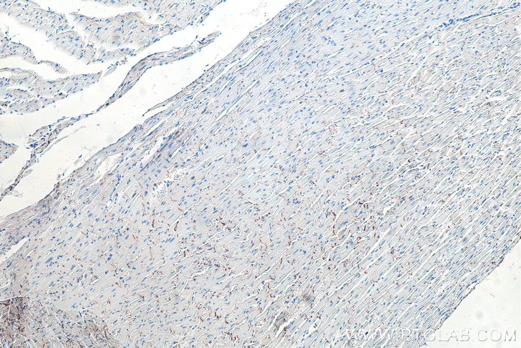
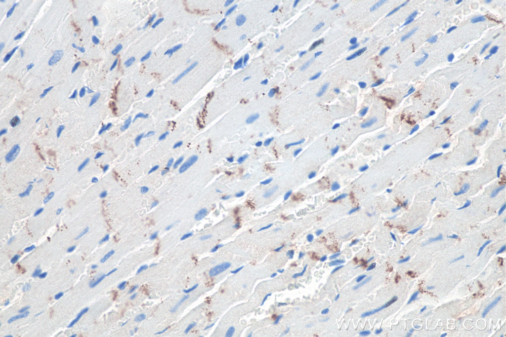
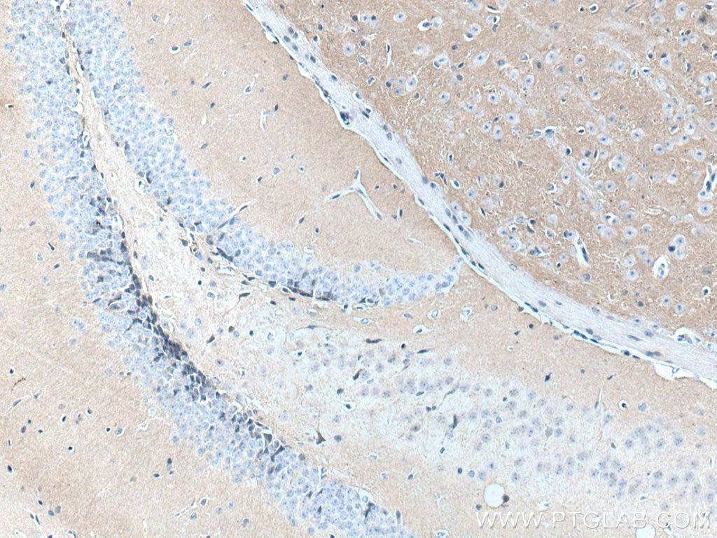
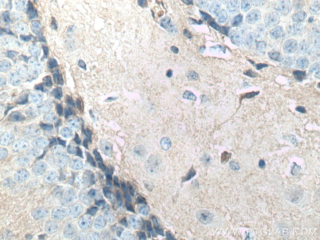
| Positive IHC detected in | mouse heart tissue, Cholangiocarcinoma tissue, human heart tissue, human malignant melanoma tissue, human ovary tumor tissue, mouse brain tissue, human liver tissue, human liver cancer tissue, rat heart tissue Note: suggested antigen retrieval with TE buffer pH 9.0; (*) Alternatively, antigen retrieval may be performed with citrate buffer pH 6.0 |
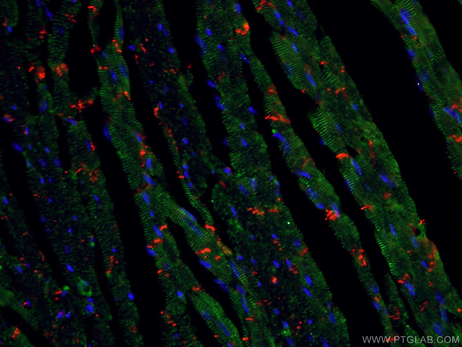
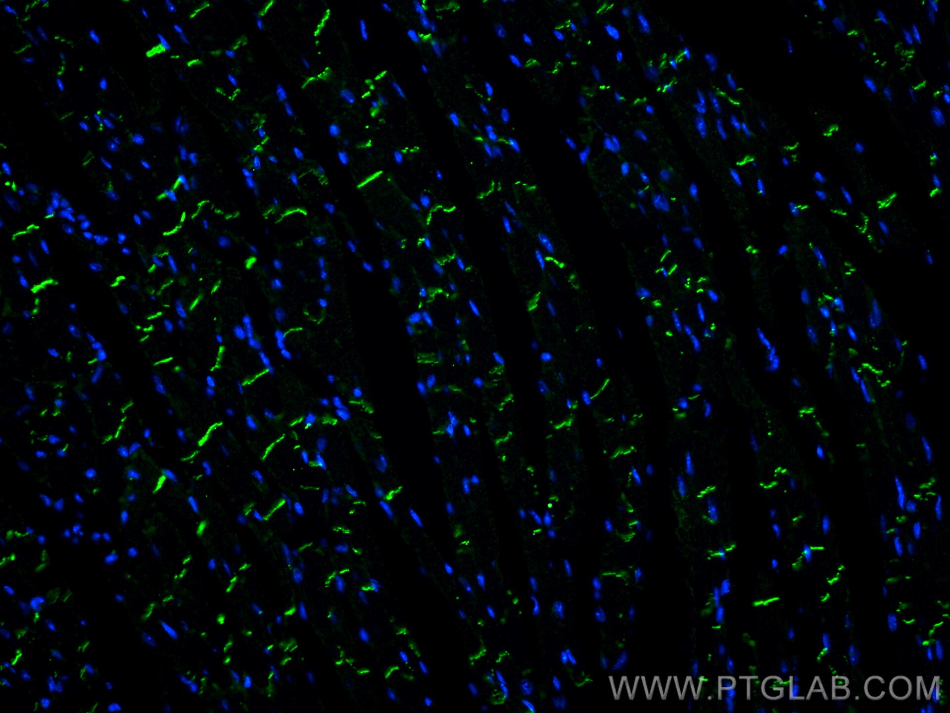
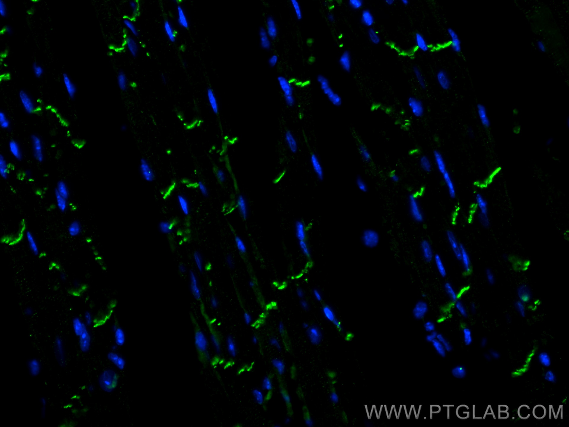
| Positive IF-P detected in | mouse heart tissue |
Recommended dilution
| Application | Dilution |
|---|---|
| Western Blot (WB) | WB : 1:5000-1:50000 |
| Immunohistochemistry (IHC)-AUTOSTAINER | IHC-AUTOSTAINER : 1:3000-1:12000 |
| Immunohistochemistry (IHC) | IHC : 1:7500-1:30000 |
| Immunofluorescence (IF)-P | IF-P : 1:200-1:800 |
| It is recommended that this reagent should be titrated in each testing system to obtain optimal results. | |
| Sample-dependent, Check data in validation data gallery. | |
Product Information
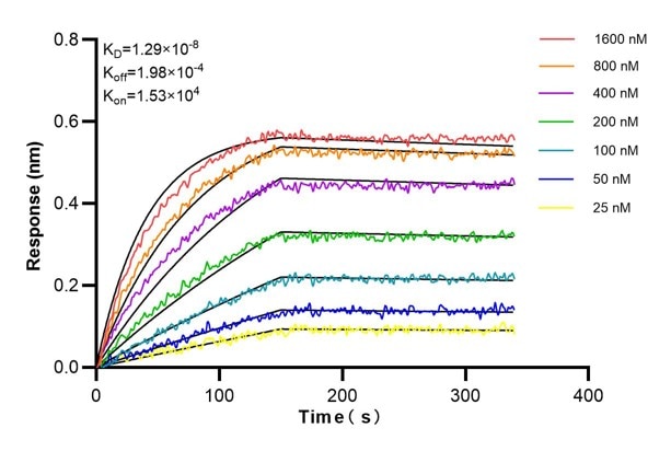
66219-1-Ig targets N-cadherin in WB, IHC, IHC-Autostainer, IF-P, ELISA applications and shows reactivity with human, mouse, rat, pig, rabbit samples.
| Tested Reactivity | human, mouse, rat, pig, rabbit |
| Cited Reactivity | human, mouse, rat, pig, canine, monkey |
| Host / Isotype | Mouse / IgG1 |
| Class | Monoclonal |
| Type | Antibody |
| Immunogen |
CatNo: Ag4996 Product name: Recombinant human N-cadherin protein Source: e coli.-derived, PET28a Tag: 6*His Domain: 33-350 aa of BC036470 Sequence: KTGFPEDVYSAVLSKDVHEGQPLLNVKFSNCNGKRKVQYESSEPADFKVDEDGMVYAVRSFPLSSEHAKFLIYAQDKETQEKWQVAVKLSLKPTLTEESVKESAEVEEIVFPRQFSKHSGHLQRQKRDWVISPINLPENSRGPFPQELVRIRSDRDKNLSLRYSVTGPGADQPPTGIFIINPISGQLSVTKPLDREQIARFHLRAHAVDINGNQVENPIDIVINVIDMNDNRPEFLHQVWNGTVPEGSKPGTYVMTVTAIDADDPNALNGMLRYRIVSQAPSTPSPNMFTINNETGDIITVAAGLDREKVQQYTLIIQ Predict reactive species |
| Full Name | cadherin 2, type 1, N-cadherin (neuronal) |
| Calculated Molecular Weight | 906 aa, 100 kDa |
| Observed Molecular Weight | 130 kDa |
| GenBank Accession Number | BC036470 |
| Gene Symbol | N-cadherin |
| Gene ID (NCBI) | 1000 |
| RRID | AB_2881610 |
| Conjugate | Unconjugated |
| Form | Liquid |
| Purification Method | Protein G purification |
| UNIPROT ID | P19022 |
| Storage Buffer | PBS with 0.02% sodium azide and 50% glycerol, pH 7.3. |
| Storage Conditions | Store at -20°C. Stable for one year after shipment. Aliquoting is unnecessary for -20oC storage. 20ul sizes contain 0.1% BSA. |
Background Information
Cadherins are a family of transmembrane glycoproteins that mediate calcium-dependent cell-cell adhesion and play an important role in the maintenance of normal tissue architecture. N-cadherin (neural cadherin), also known as CDH2 (cadherin 2), is a 130-kDa transmembrane protein and a classical member of the cadherin superfamily which also include E-, P-, R-, and B-cadherins. Expression of N-cadherin has been reported on various cell types including neurons, endothelial cells and cardiac myocytes (PMID: 11282032; 9508779; 8125202). N-cadherin has functions in early brain morphogenesis, synaptogenesis and synaptic plasticity (PMID: 23321619). The N-cadherin ectodomain can be cleaved, leading to the generation of a 90 kD N-terminal fragment (PMID: 16998833; 17028923).
Protocols
| Product Specific Protocols | |
|---|---|
| IF protocol for N-cadherin antibody 66219-1-Ig | Download protocol |
| IHC protocol for N-cadherin antibody 66219-1-Ig | Download protocol |
| WB protocol for N-cadherin antibody 66219-1-Ig | Download protocol |
| Standard Protocols | |
|---|---|
| Click here to view our Standard Protocols |
Publications
What published studies show
The N-cadherin Monoclonal antibody (1D8B3), catalog number 66219-1-Ig, has accumulated 292 citations. These appear in high-impact journals including Nature Communications, Cancer Research, Cell Stem Cell, Cell Death & Disease, Molecular Cancer, Advanced Science, and Science Advances. Research fields span epithelial-mesenchymal transition (EMT), oncology (lung, breast, gastric, liver, colorectal, and other cancers), fibrosis, wound healing, stem cell biology, cardiovascular disease, and reproductive biology, with WB, IHC, and IF as the primary applications.
| Species | Application | Title |
|---|---|---|
Mol Cancer YTHDF2 mediates the mRNA degradation of the tumor suppressors to induce AKT phosphorylation in N6-methyladenosine-dependent way in prostate cancer. | ||
Cell Stem Cell Long-term engraftment and maturation of autologous iPSC-derived cardiomyocytes in two rhesus macaques | ||
Cancer Res Proteomic Profiling of Serum Extracellular Vesicles Identifies Diagnostic Signatures and Therapeutic Targets in Breast Cancer | ||
Cancer Res SRSF9 Forms Phase-separated Condensates to Promote Ovarian Cancer Progression by Inducing RNA Alternative Splicing that is Inhibited by m6A Modification | ||
J Extracell Vesicles Extracellular vesicles derived from Akkermansia muciniphila promote placentation and mitigate preeclampsia in a mouse model | ||
Autophagy Dual roles of CXCR4 (C-X-C motif chemokine receptor 4) in promoting entry of ebolavirus and targeting excessive glycoprotein for reticulophagic degradation to facilitate viral fitness |
Reviews
What customers say
Across three reviews, performance is mixed. For Western Blot, the antibody performed well at 1:2000 dilution, producing a clear 140 kD band in U2OS cells. For Immunofluorescence, results were inconsistent: one user at 1:400 on iPSC-derived cardiomyocytes noted suboptimal membrane localization compared to an alternative pan-cadherin antibody, while another at 1:100 on 3D cholangiocyte organoids reported no detectable signal. Overall, the antibody appears reliable for WB but may require optimization or alternative reagents for IF applications, particularly in 3D or non-standard cell systems.
The reviews below have been submitted by verified Proteintech customers who received an incentive for providing their feedback.
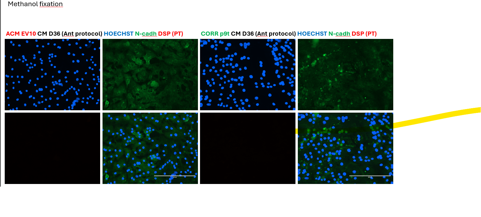
Kiara (Verified Customer) (07-28-2025) | This primary antibody was not internally validated for IF, but there was a paper published that it worked on 4% PFA-fixed H9 ESCs, so I gave it a shot anyway. However, the staining did not show a nice membrane localization that our lab usually sees when using a different rabbit pan-cadherin primary antibody.
 |
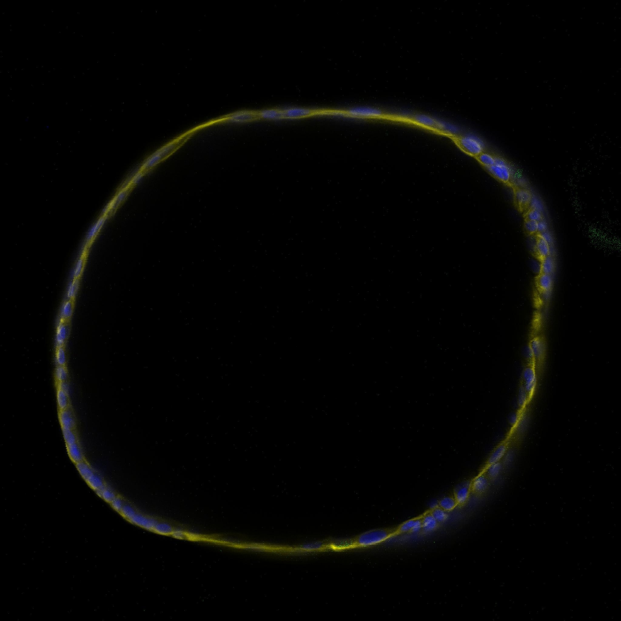
Davide (Verified Customer) (06-13-2023) | The antibody didn't produce any signal in 3d primary human cholangiocyte organoids. I would recommend either to try other antibody concentration or to try another Ab. It was tried on a cell condition which was recently reported to stimulate the CDH2 protein expression. However it should also be present in control condition at low levels.
 |
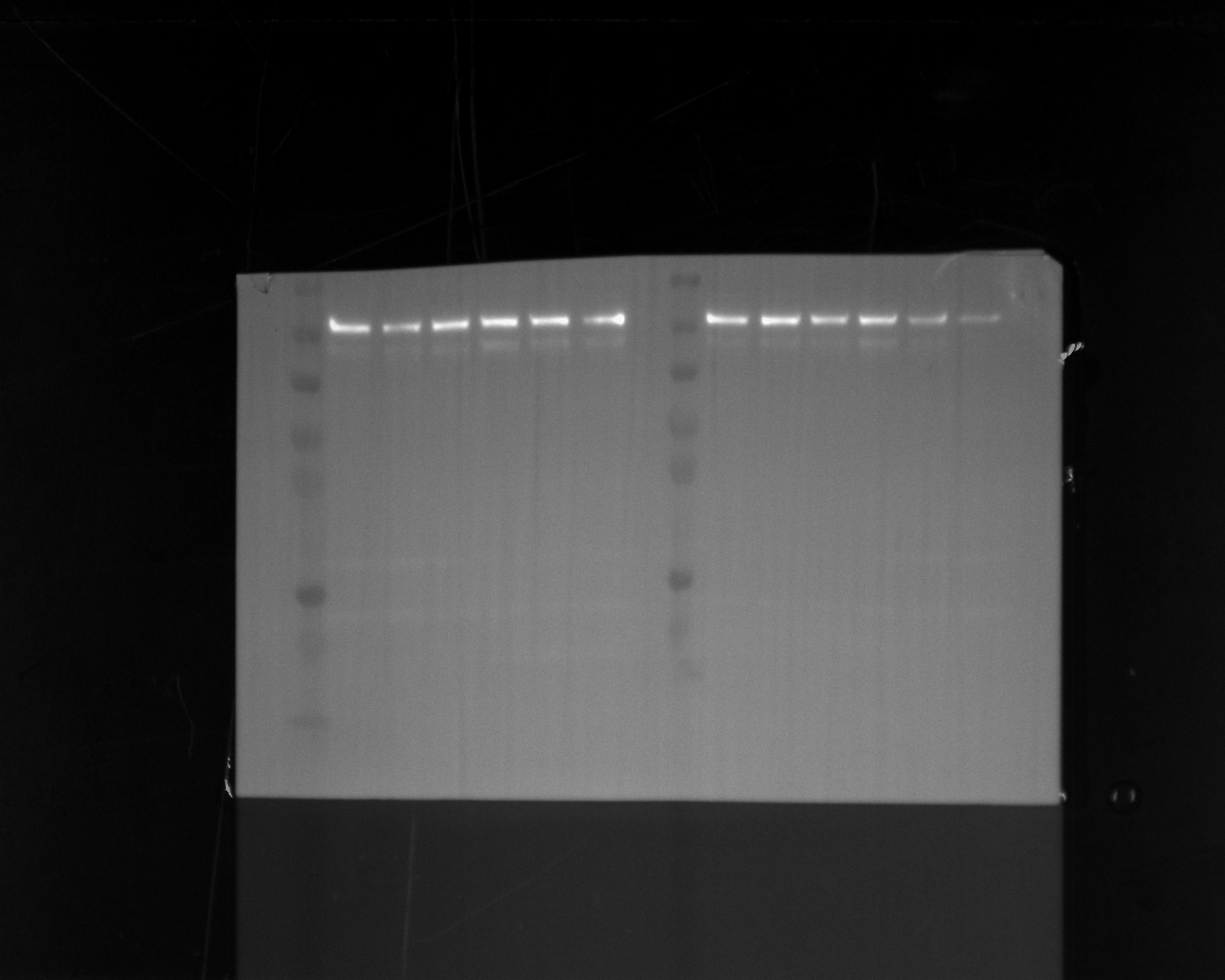
Udesh (Verified Customer) (10-13-2022) | Used the Ab at 1:2000 dilution in 5% NFDM and observed a band at 140 kD. Blocked in 5% NFDM for 1H at RT.
 |

































