





Tested Applications
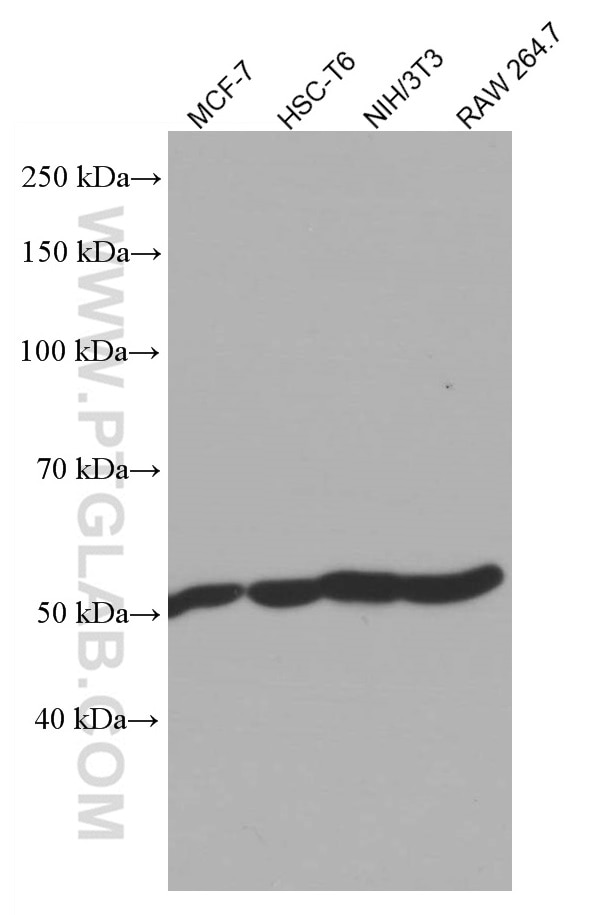
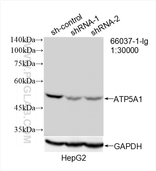
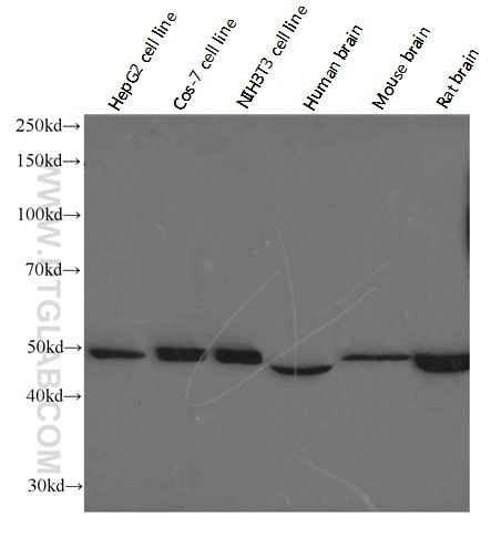
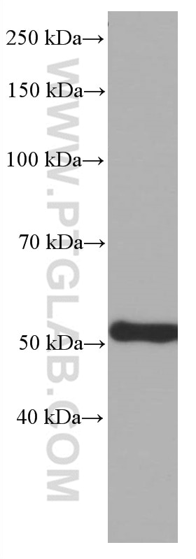
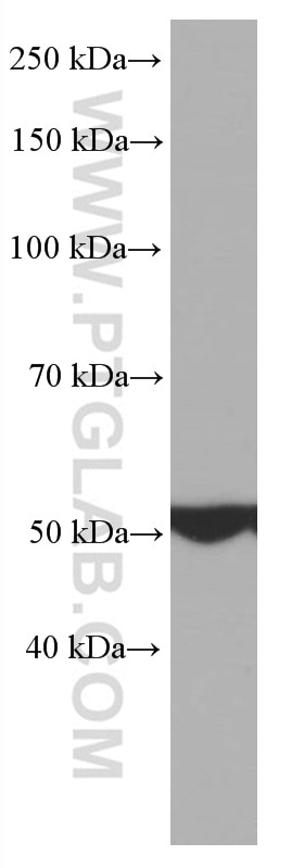
| Positive WB detected in | MCF-7 cells, HeLa cells, HEK-293 cells, HepG2 cells, HSC-T6 cells, NIH/3T3 cells, RAW 264.7 cells |
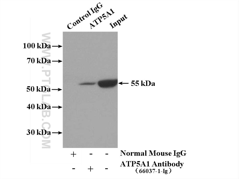
| Positive IP detected in | mouse heart tissue |
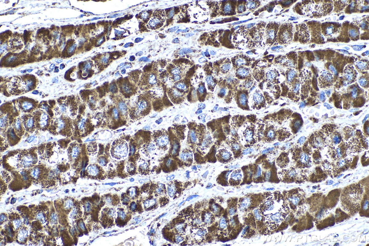
| Positive IHC detected in | human liver cancer tissue, Hepatocellular carcinoma tissue, human colon cancer tissue Note: suggested antigen retrieval with TE buffer pH 9.0; (*) Alternatively, antigen retrieval may be performed with citrate buffer pH 6.0 |
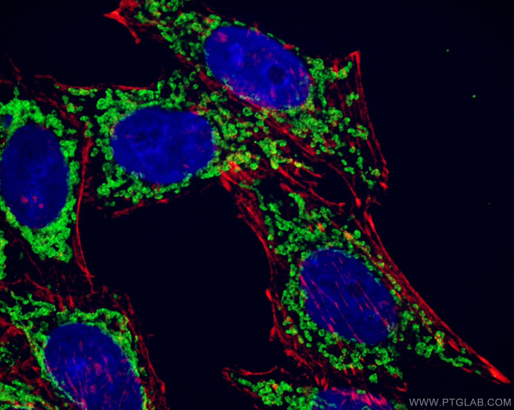
| Positive IF/ICC detected in | HeLa cells, HepG2 cells |
| Positive FC (Intra) detected in | HeLa cells |
Recommended dilution
| Application | Dilution |
|---|---|
| Western Blot (WB) | WB : 1:5000-1:50000 |
| Immunoprecipitation (IP) | IP : 0.5-4.0 ug for 1.0-3.0 mg of total protein lysate |
| Immunohistochemistry (IHC) | IHC : 1:1000-1:4000 |
| Immunofluorescence (IF)/ICC | IF/ICC : 1:400-1:1600 |
| Flow Cytometry (FC) (INTRA) | FC (INTRA) : 0.25 ug per 10^6 cells in a 100 µl suspension |
| It is recommended that this reagent should be titrated in each testing system to obtain optimal results. | |
| Sample-dependent, Check data in validation data gallery. | |
Product Information
66037-1-Ig targets ATP5A1 in WB, IHC, IF/ICC, FC (Intra), IP, CoIP, ELISA applications and shows reactivity with human, mouse, rat, monkey samples.
| Tested Reactivity | human, mouse, rat, monkey |
| Cited Reactivity | human, mouse, rat, hamster |
| Host / Isotype | Mouse / IgG2b |
| Class | Monoclonal |
| Type | Antibody |
| Immunogen |
CatNo: Ag8119 Product name: Recombinant human ATP5A1 protein Source: e coli.-derived, PET28a Tag: 6*His Domain: 203-553 aa of BC064562 Sequence: GRGQRELIIGDRQTGKTSIAIDTIINQKRFNDGSDEKKKLYCIYVAIGQKRSTVAQLVKRLTDADAMKYTIVVSATASDAAPLQYLAPYSGCSMGEYFRDNGKHALIIYDDLSKQAVAYRQMSLLLRRPPGREAYPGDVFYLHSRLLERAAKMNDAFGGGSLTALPVIETQAGDVSAYIPTNVISITDGQIFLETELFYKGIRPAINVGLSVSRVGSAAQTRAMKQVAGTMKLELAQYREVAAFAQFGSDLDAATQQLLSRGVRLTELLKQGQYSPMAIEEQVAVIYAGVRGYLDKLEPSKITKFENAFLSHVVSQHQALLGTIRADGKISEQSDAKLKEIVTNFLAGFEA Predict reactive species |
| Full Name | ATP synthase, H+ transporting, mitochondrial F1 complex, alpha subunit 1, cardiac muscle |
| Calculated Molecular Weight | 60 kDa |
| Observed Molecular Weight | 50 kDa |
| GenBank Accession Number | BC064562 |
| Gene Symbol | ATP5A1 |
| Gene ID (NCBI) | 498 |
| RRID | AB_11044196 |
| Conjugate | Unconjugated |
| Form | Liquid |
| Purification Method | Protein A purification |
| UNIPROT ID | P25705 |
| Storage Buffer | PBS with 0.02% sodium azide and 50% glycerol, pH 7.3. |
| Storage Conditions | Store at -20°C. Stable for one year after shipment. Aliquoting is unnecessary for -20oC storage. 20ul sizes contain 0.1% BSA. |
Background Information
The ATP5A1 gene encodes the α subunit of mitochondrial ATP synthase which produces ATP from ADP in the presence of a proton gradient across the membrane. The mitochondrial ATP synthase, also known as Complex V or F1F0 ATP synthase, is a multi-subunit enzyme complex consisting of two functional domains, the F1-containing the catalytic core and the Fo- containing the membrane proton channel. F0 domain has 10 subunits: a, b, c, d, e, f, g, OSCP, A6L, and F6. F1 is composed of subunits α, β, γ, δ, ε, and a loosely attached inhibitor protein IF1. Recently defect in ATP5A1 has been linked to the fatal neonatal mitochondrial encephalopathy. ATP5A1 is localized in the mitochondria and anti-ATP5A1 can be used as the loading control for mitochondrial or Complex V proteins. This antibody recognizes the endogenous ATP5A1 protein in lysates from various cell lines and tissues. The predicted MW of ATP5A1 is 60 kDa, while it undergoes the transit peptide cleavage to become a mature form around 50-55 kDa. Several isoforms of ATP5A1 exist due to the alternative splicing.
Protocols
| Product Specific Protocols | |
|---|---|
| FC protocol for ATP5A1 antibody 66037-1-Ig | Download protocol |
| IF protocol for ATP5A1 antibody 66037-1-Ig | Download protocol |
| IHC protocol for ATP5A1 antibody 66037-1-Ig | Download protocol |
| IP protocol for ATP5A1 antibody 66037-1-Ig | Download protocol |
| WB protocol for ATP5A1 antibody 66037-1-Ig | Download protocol |
| Standard Protocols | |
|---|---|
| Click here to view our Standard Protocols |
Publications
What published studies show
Anti-ATP5A1 (Cat# 66037-1-Ig) has 56 citations across journals including Nature, Nature Cell Biology, Nature Communications, Cell Reports, Science Advances, Redox Biology, and Cell Death & Disease. Research fields span mitochondrial biology and energy metabolism, neurodegeneration (Alzheimer's, Parkinson's), cardiovascular disease, renal injury, diabetes, thermogenesis, cancer, and oxidative stress. Applications include WB, IF, IHC, and CoIP across human, mouse, rat, and hamster species.
| Species | Application | Title |
|---|---|---|
Signal Transduct Target Ther A two-strata energy flux system driven by a stress hormone prioritizes cardiac energetics | ||
Nat Cell Biol Mitochondrial NADPH fuels mitochondrial fatty acid synthesis and lipoylation to power oxidative metabolism | ||
Nat Cell Biol AIDA directly connects sympathetic innervation to adaptive thermogenesis by UCP1. | ||
Autophagy AKT-mediated phosphorylation of ATG4B impairs mitochondrial activity and enhances the Warburg effect in hepatocellular carcinoma cells. | ||
Nat Commun Defective vascular smooth muscle cell tafazzin impairs mitochondrial function and promotes atherosclerosis in preclinical models. |
Reviews
What customers say
Most reviewers report excellent performance for Western Blot, with strong results in rat heart tissue, human breast cancer cell mitochondrial fractions, and SimpleWestern applications. Immunofluorescence results are mixed: one user found consistent, reliable staining in HULEC cells, while another experienced low signal and high background when using saponin permeabilization in MDA-MB-231 cells. Overall, the antibody is well-regarded for WB and dot blot applications, with good price-quality ratio. Users should note that permeabilization method may affect IF performance.
The reviews below have been submitted by verified Proteintech customers who received an incentive for providing their feedback.
Coralie (Verified Customer) (04-14-2026) | This antibody is working really good for my Dot Blots and Western Blots ! My samples are proteins extracted from rat heart tissue. The price-quality ratio is great, I would recommend without hesitation.
|
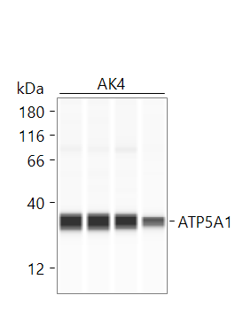
Anis (Verified Customer) (06-26-2025) | Jess - SimpleWestern, 1:40 in antibody diluent 2 Used as a marker for mitochondria isolation purity
 |
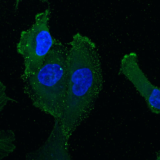
Paulina (Verified Customer) (03-26-2025) | Fixation: 2% PFA for 20min. Permeabilization/antibody dilution solution: 0.05% saponin, 5% horse serum in PBS. Primary antibody diluted 1:100 in saponin solution for 1hr at room temperature, followed by anti-mouse conjugated to AlexaFluor 568 (1:200 in saponin solution) for 1 hr at room temperature. I got very low signal and a lot of background specks, so this antibody does not work well with the saponin permeabilization.
 |
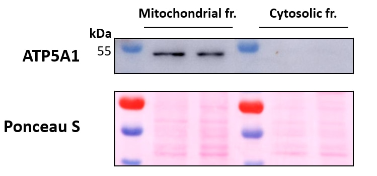
Marina (Verified Customer) (09-12-2022) | Mitochondrial fractions were obtained from whole cell lysates of 2 immortalized human breast cancer cell lines using a commercial kit, leading to pure mitochondrial fractions (ATP5A1 positive) and fractions comprising the cytosols minus the mitochondria (ATP5A1 negative). Primary antibody incubation was performed overnight at 4ºC. Ponceau S is used as total protein loading control.
 |
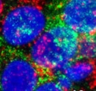
Cassandr (Verified Customer) (02-10-2022) | Consistently works antibody
 |












