
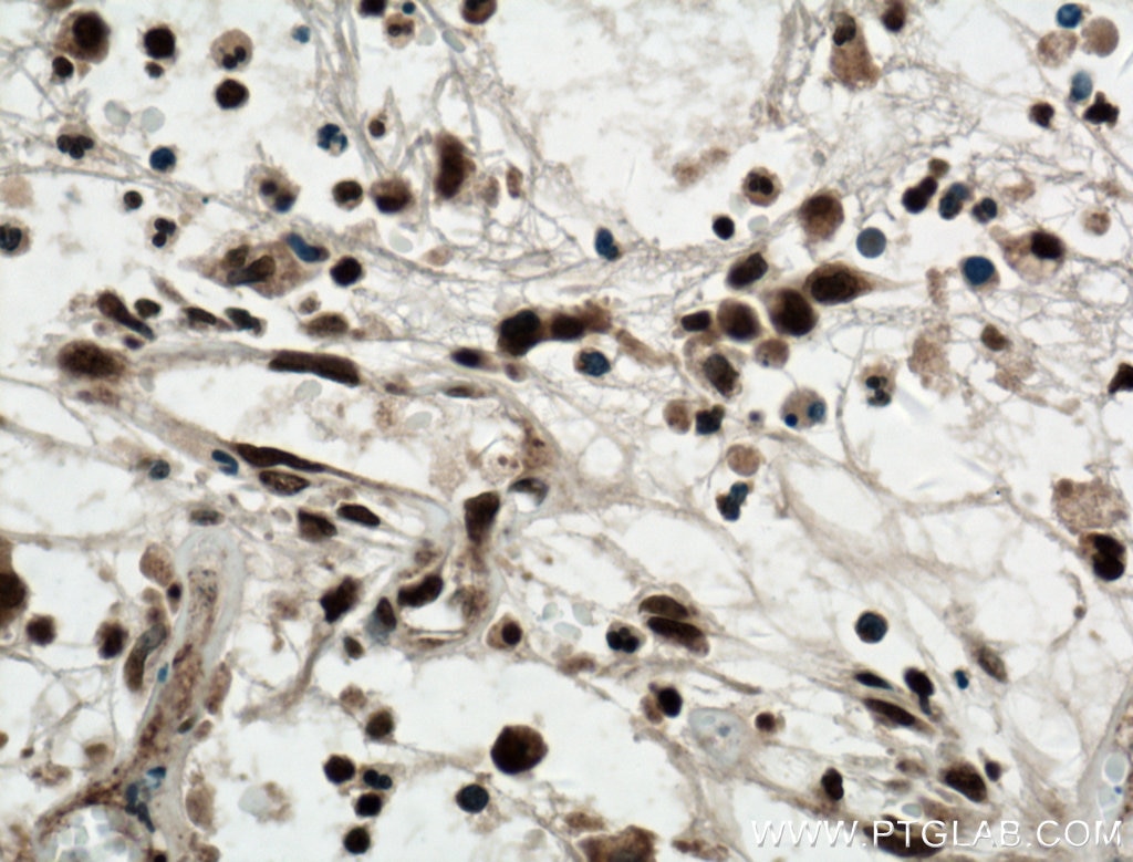
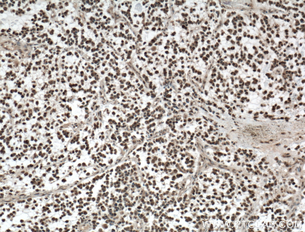
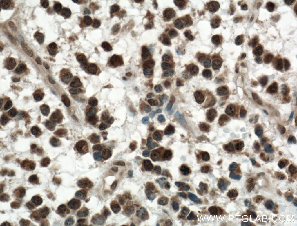
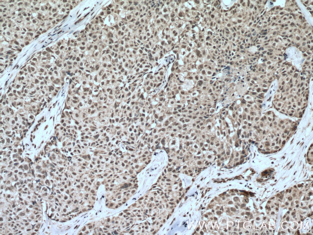
Tested Applications
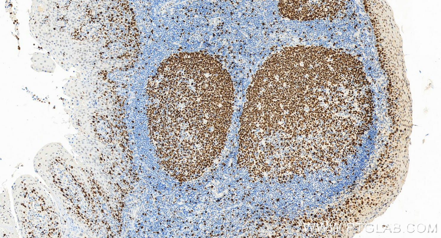
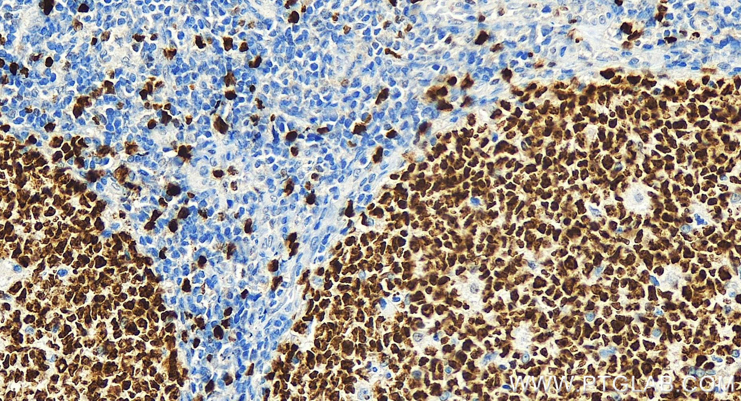
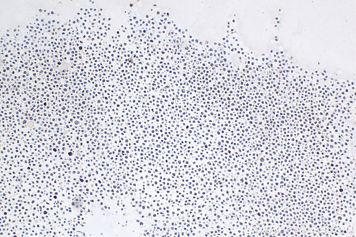
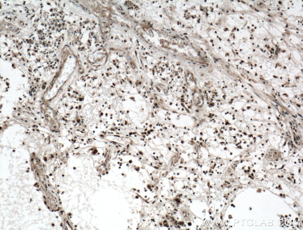
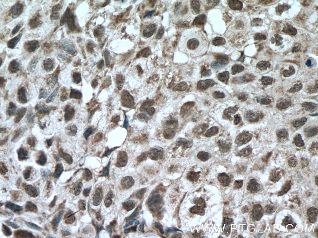
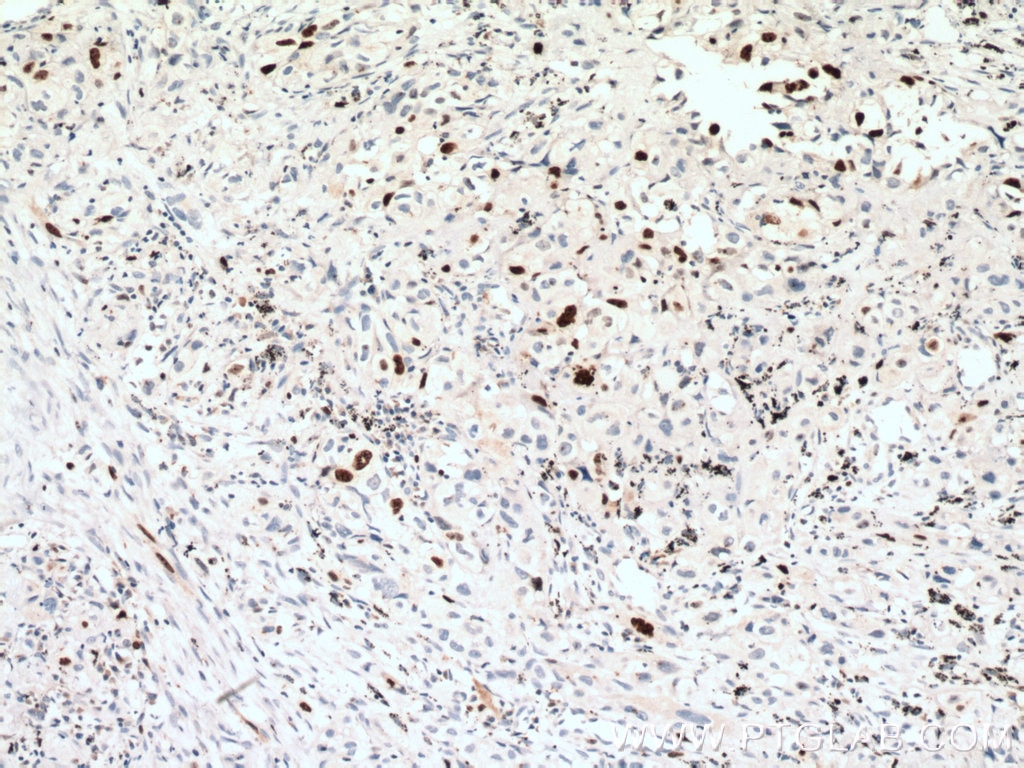
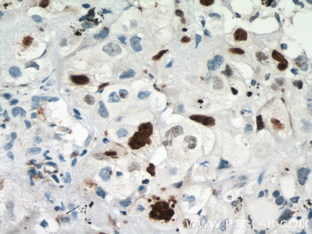
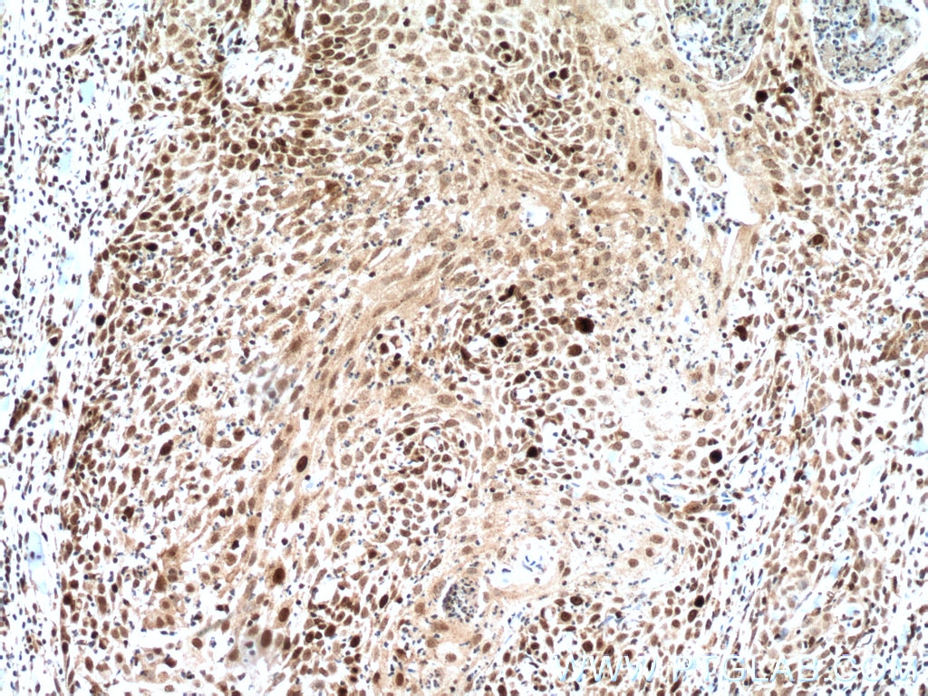
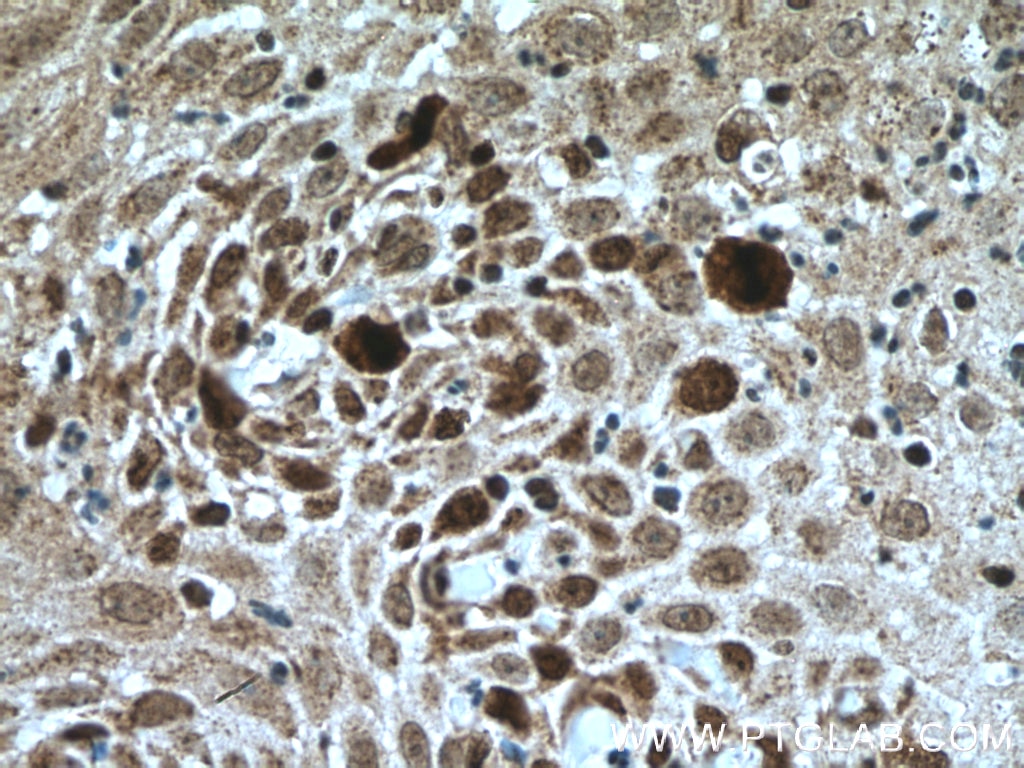
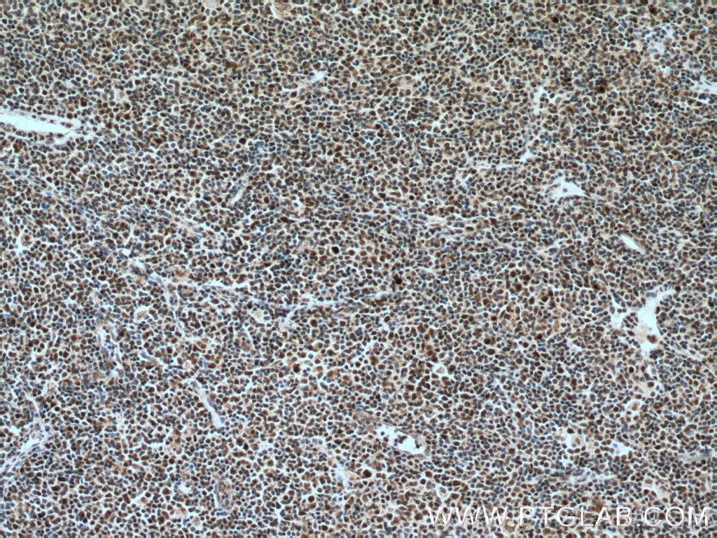
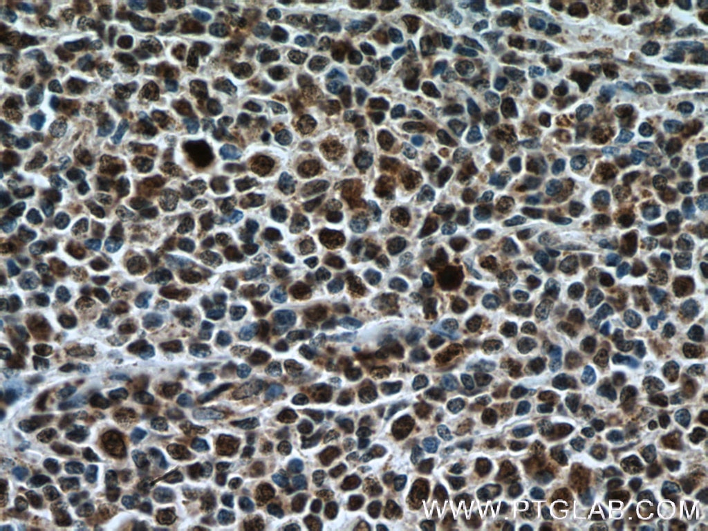
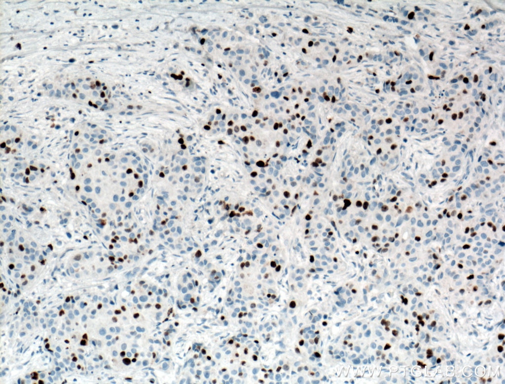
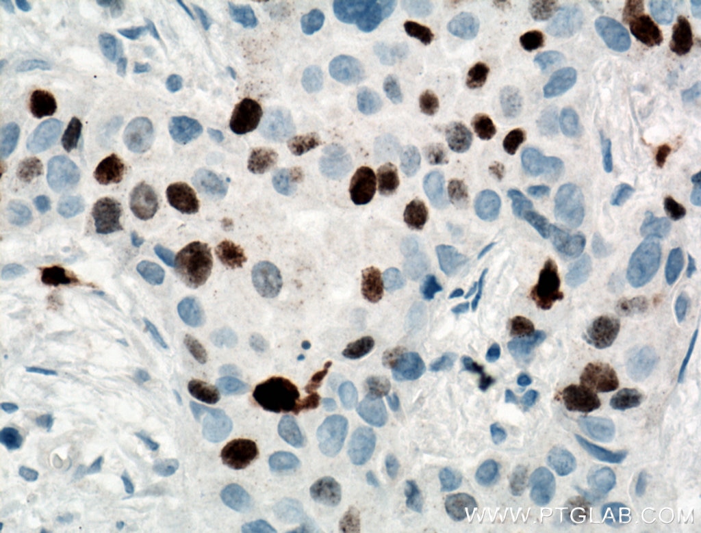
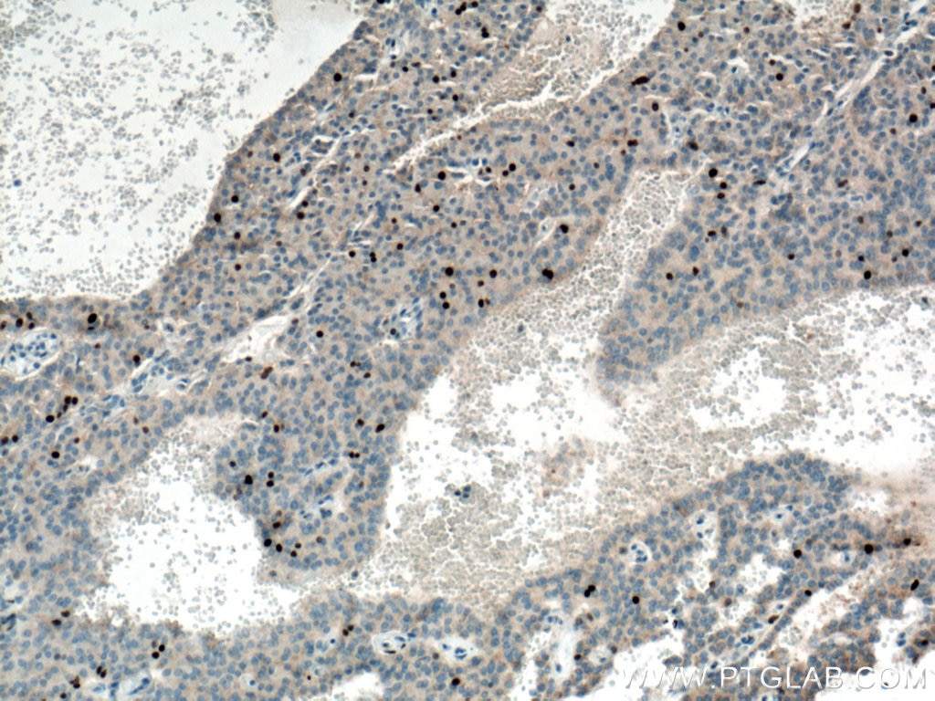
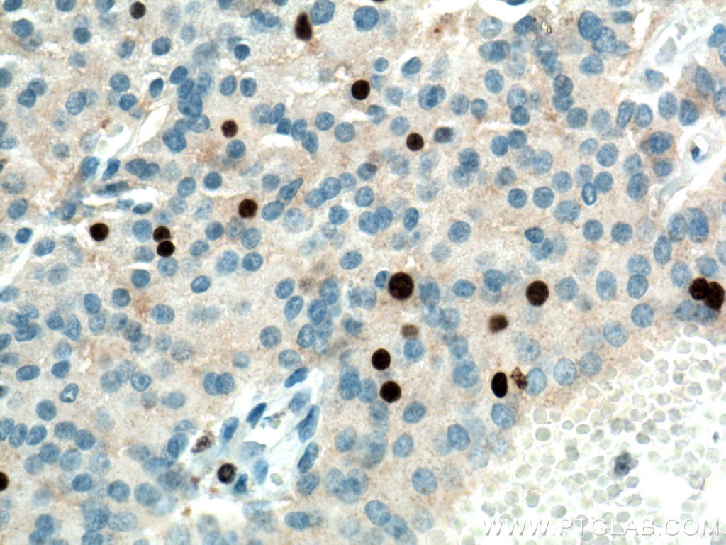
| Positive IHC detected in | human tonsillitis tissue, human colon cancer tissue, human gliomas tissue, human lung cancer tissue, human skin cancer tissue, human lymphoma tissue, human breast cancer tissue, Insulinoma tissue, K-562 cells Note: suggested antigen retrieval with TE buffer pH 9.0; (*) Alternatively, antigen retrieval may be performed with citrate buffer pH 6.0 |
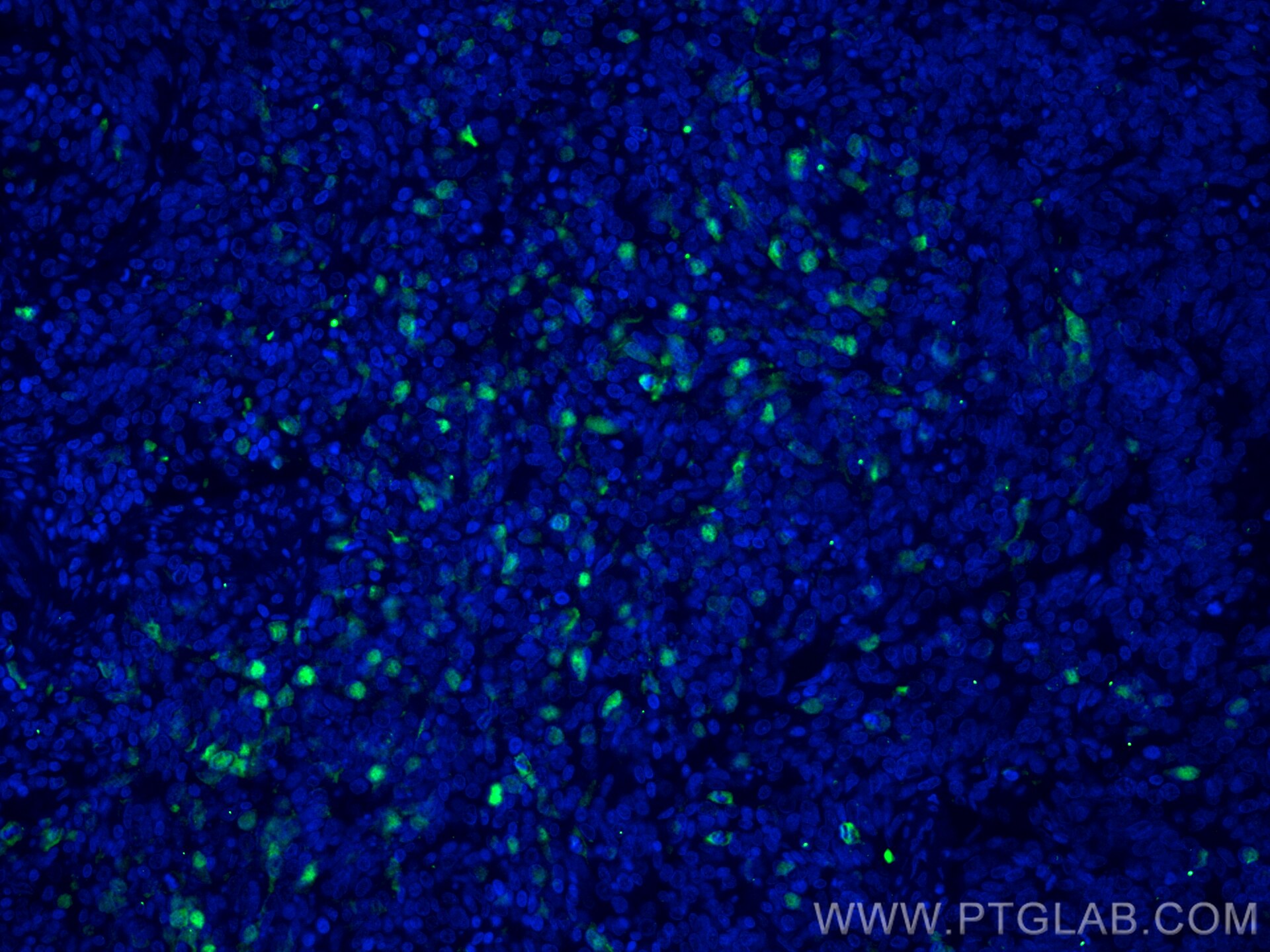
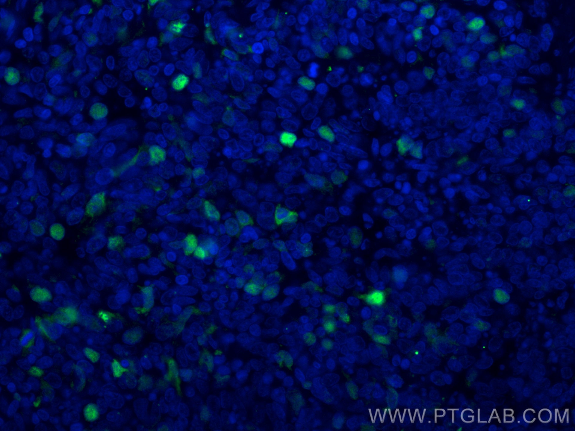
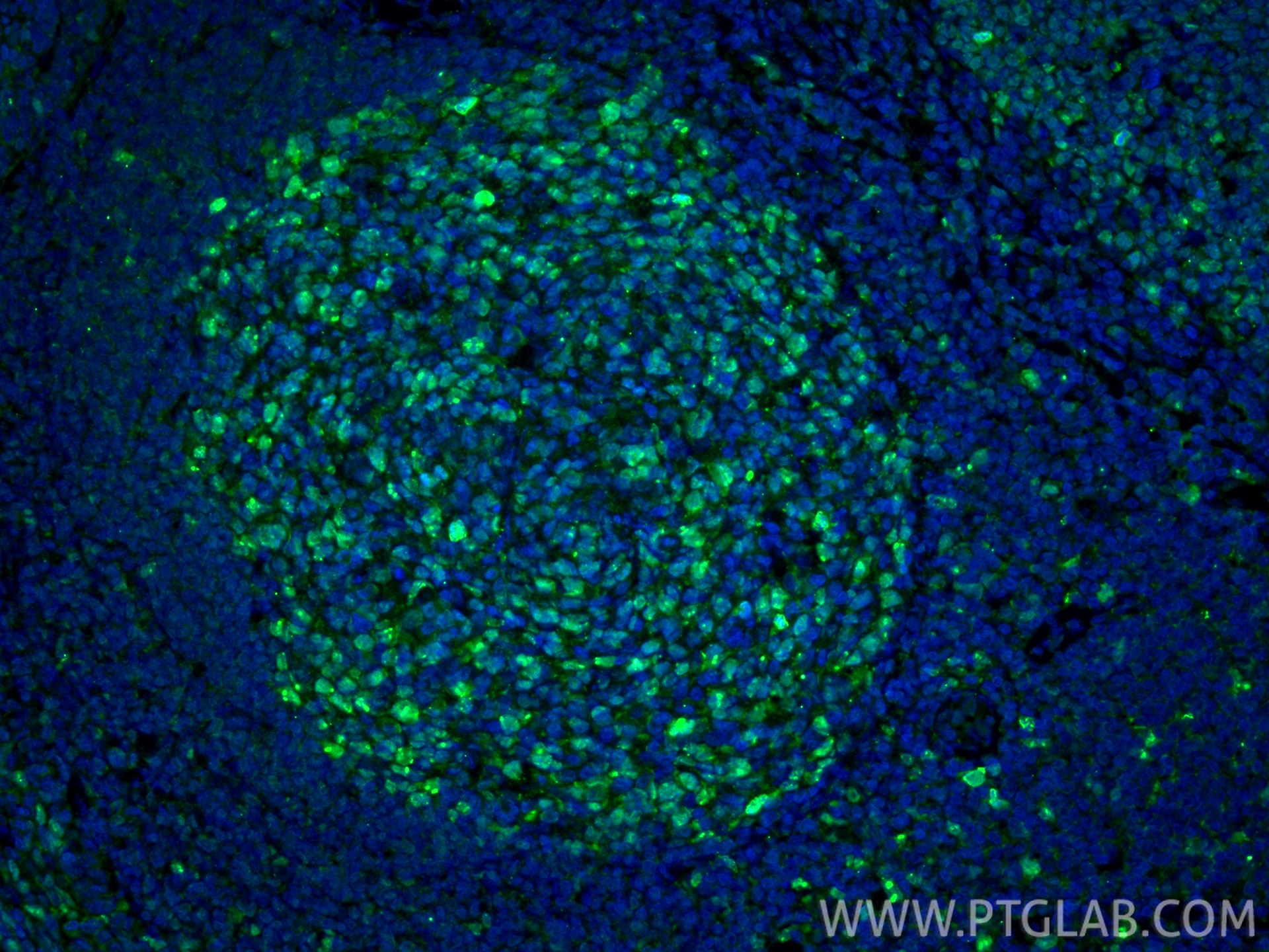
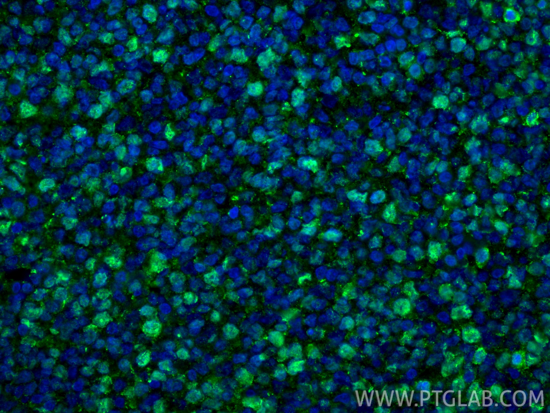
| Positive IF-P detected in | human tonsillitis tissue, human lung cancer tissue |
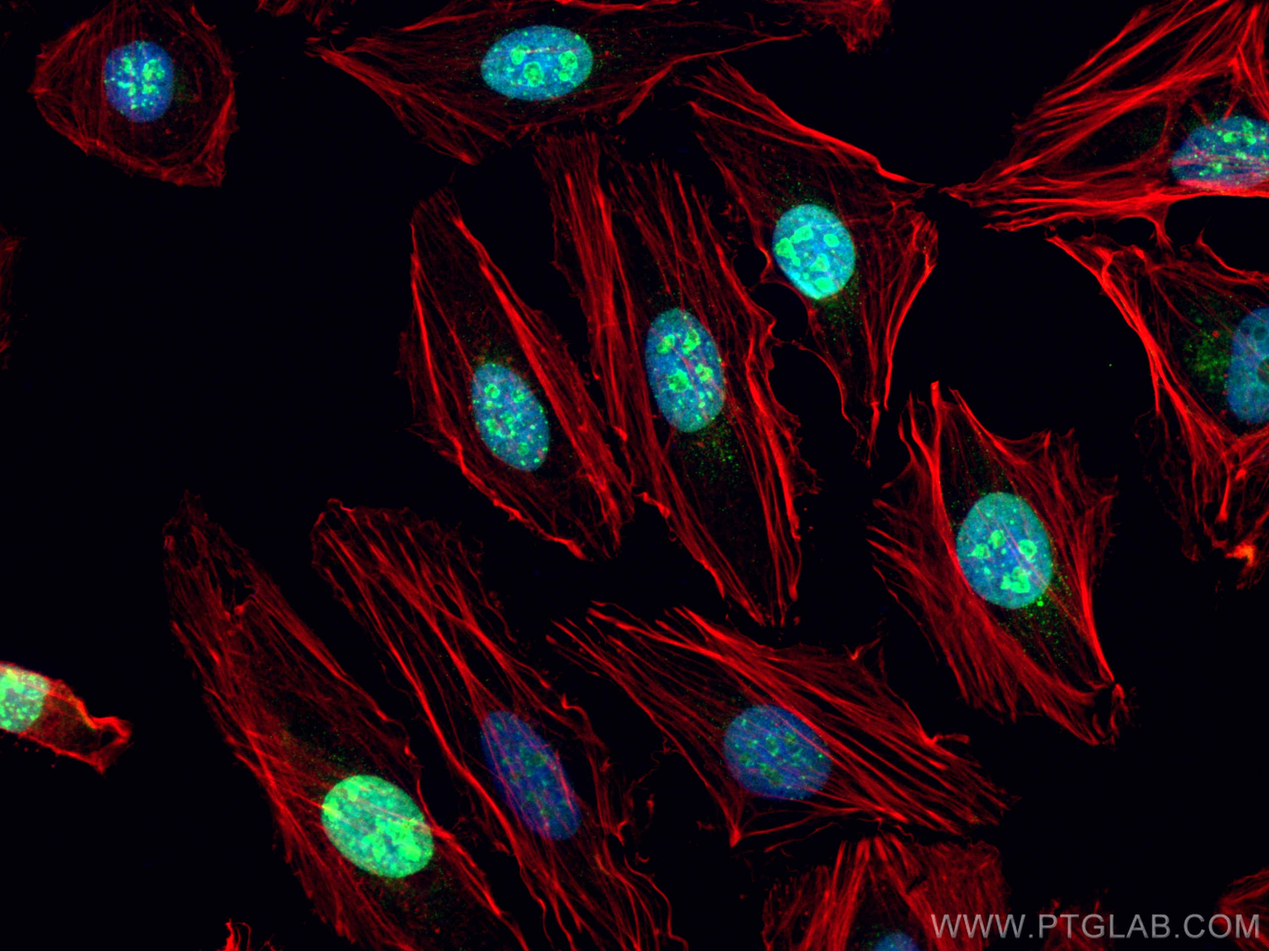
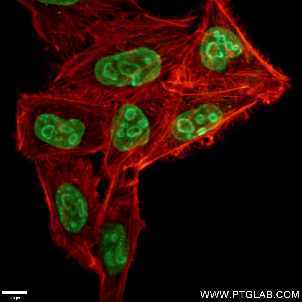
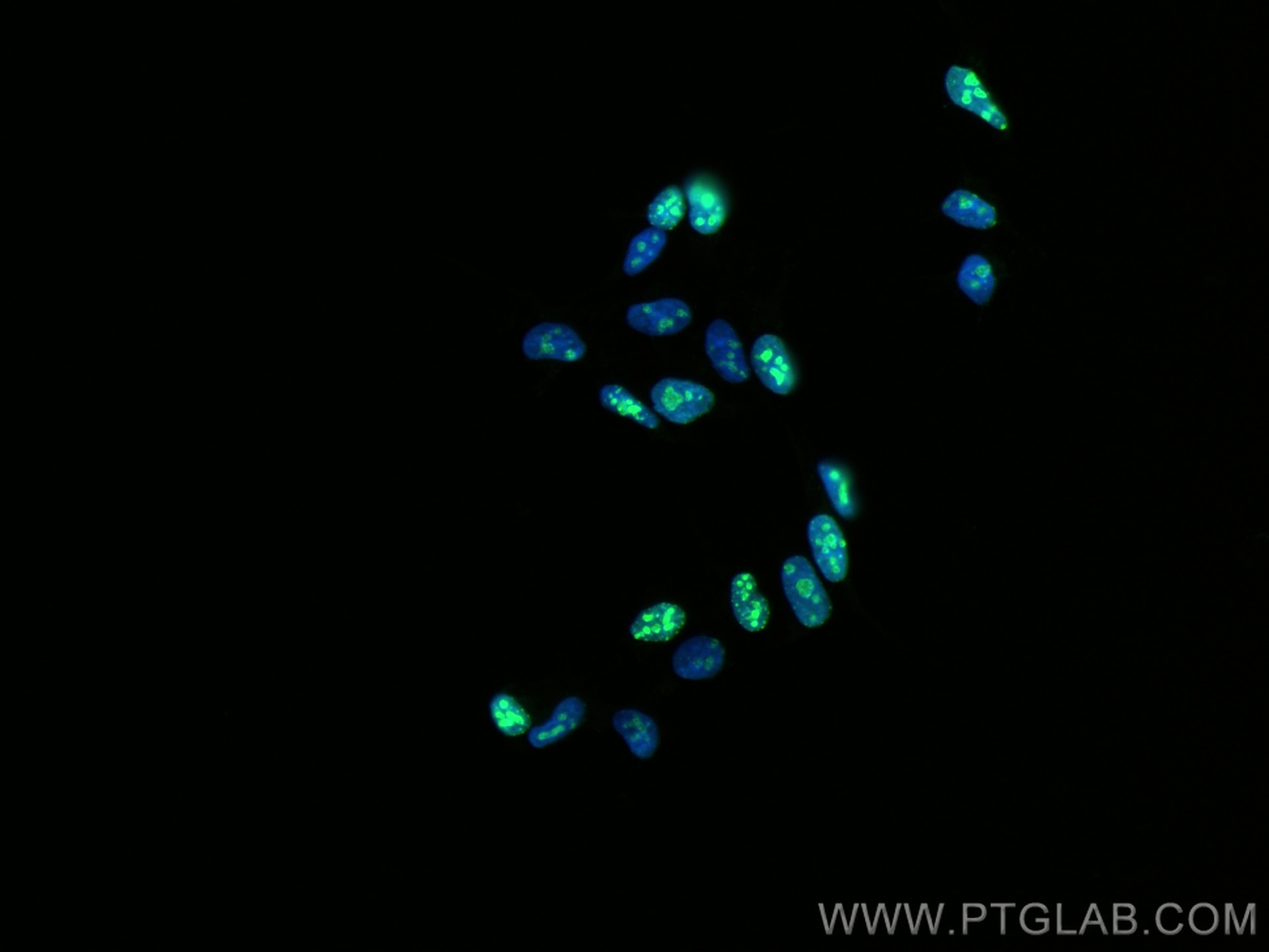
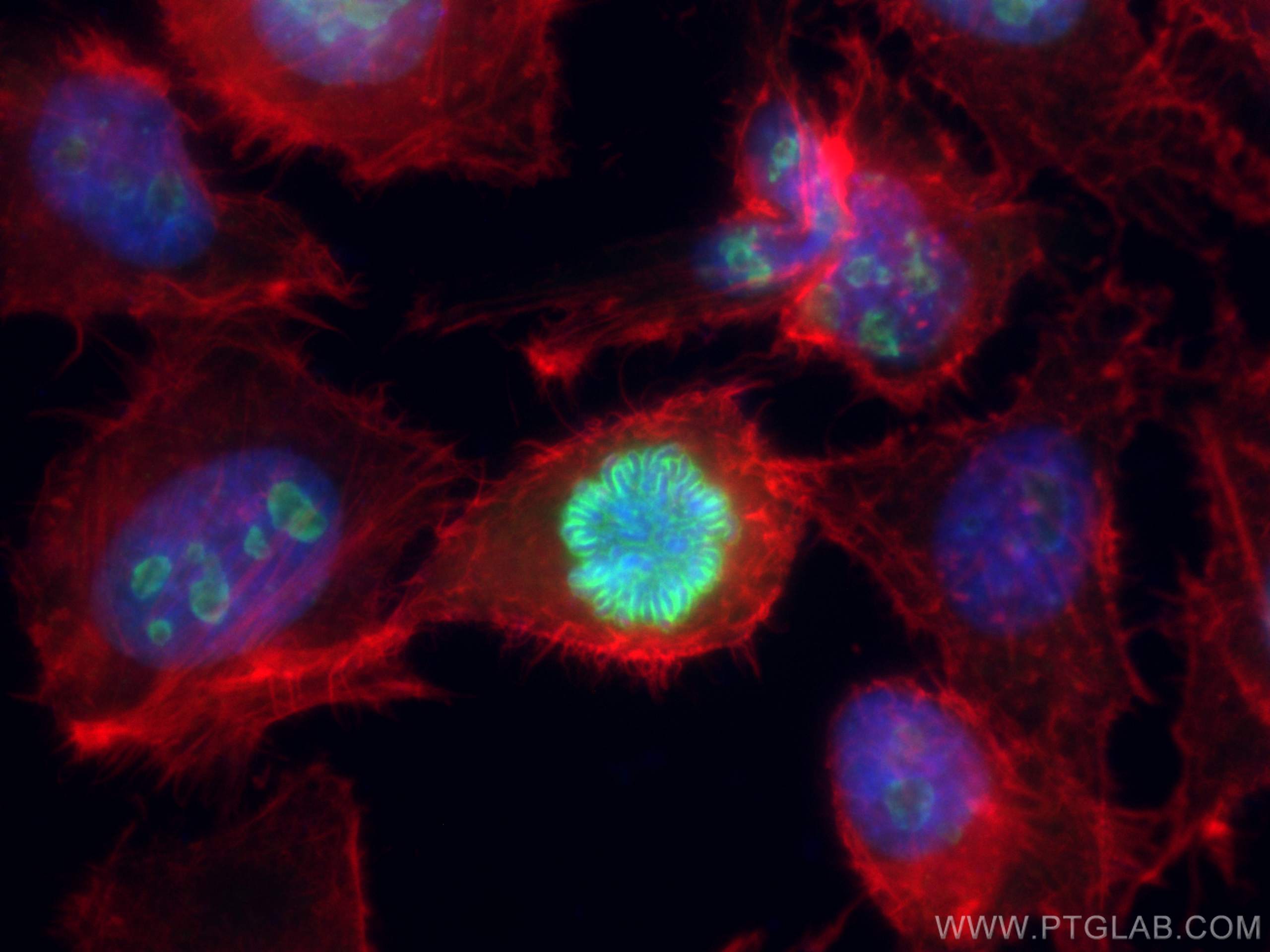
| Positive IF/ICC detected in | HeLa cells, HEK-293 cells |
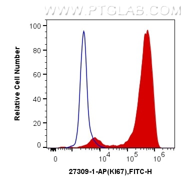
| Positive FC (Intra) detected in | Jurkat cells |
Recommended dilution
| Application | Dilution |
|---|---|
| Immunohistochemistry (IHC) | IHC : 1:4000-1:16000 |
| Immunofluorescence (IF)-P | IF-P : 1:200-1:800 |
| Immunofluorescence (IF)/ICC | IF/ICC : 1:50-1:500 |
| Flow Cytometry (FC) (INTRA) | FC (INTRA) : 0.40 ug per 10^6 cells in a 100 µl suspension |
| It is recommended that this reagent should be titrated in each testing system to obtain optimal results. | |
| Sample-dependent, Check data in validation data gallery. | |
Product Information
27309-1-AP targets Ki-67 in IHC, IF/ICC, IF-P, FC (Intra), ELISA applications and shows reactivity with human samples.
| Tested Reactivity | human |
| Cited Reactivity | human, rabbit, sheep |
| Host / Isotype | Rabbit / IgG |
| Class | Polyclonal |
| Type | Antibody |
| Immunogen |
CatNo: Ag26266 Product name: Recombinant human KI67 protein Source: e coli.-derived, PGEX-4T Tag: GST Domain: 1201-1300 aa of NM_002417 Sequence: AGTLPGSKRQLQTPKEKAQALEDLAGFKELFQTPGHTEELVAAGKTTKIPCDSPQSDPVDTPTSTKQRPKRSIRKADVEGELLACRNLMPSAGKAMHTPK Predict reactive species |
| Full Name | antigen identified by monoclonal antibody Ki-67 |
| Calculated Molecular Weight | 359 kDa |
| GenBank Accession Number | NM_002417 |
| Gene Symbol | KI67 |
| Gene ID (NCBI) | 4288 |
| RRID | AB_2756525 |
| Conjugate | Unconjugated |
| Form | Liquid |
| Purification Method | Antigen affinity purification |
| UNIPROT ID | P46013 |
| Storage Buffer | PBS with 0.02% sodium azide and 50% glycerol, pH 7.3. |
| Storage Conditions | Store at -20°C. Stable for one year after shipment. Aliquoting is unnecessary for -20oC storage. 20ul sizes contain 0.1% BSA. |
Background Information
The Ki-67 protein (also known as MKI67) is a cellular marker for proliferation. Ki67 is present during all active phases of the cell cycle (G1, S, G2 and M), but is absent in resting cells (G0). Cellular content of Ki-67 protein markedly increases during cell progression through S phase of the cell cycle. Therefore, the nuclear expression of Ki67 can be evaluated to assess tumor proliferation by immunohistochemistry. It has been demonstrated to be of prognostic value in breast cancer. In head and neck cancer, several studies have reported an association between high proliferative activity and poorer prognosis.
Protocols
| Product Specific Protocols | |
|---|---|
| FC protocol for Ki-67 antibody 27309-1-AP | Download protocol |
| IF protocol for Ki-67 antibody 27309-1-AP | Download protocol |
| IHC protocol for Ki-67 antibody 27309-1-AP | Download protocol |
| Standard Protocols | |
|---|---|
| Click here to view our Standard Protocols |
Publications
What published studies show
| Species | Application | Title |
|---|---|---|
Signal Transduct Target Ther Exosomes secreted from cardiomyocytes suppress the sensitivity of tumor ferroptosis in ischemic heart failure | ||
Signal Transduct Target Ther FBXW7β loss-of-function enhances FASN-mediated lipogenesis and promotes colorectal cancer growth | ||
Cancer Cell Proteogenomic characterization of non-functional pancreatic neuroendocrine tumors unravels clinically relevant subgroups | ||
Cancer Cell Epithelial cells activate fibroblasts to promote esophageal cancer development | ||
J Hematol Oncol Regulation of Hippo-YAP signaling by insulin-like growth factor-1 receptor in the tumorigenesis of diffuse large B-cell lymphoma. |
Reviews
What customers say
The reviews below have been submitted by verified Proteintech customers who received an incentive for providing their feedback.
Ahmad (Verified Customer) (07-02-2026) | Works wel
|
Udesh (Verified Customer) (08-12-2025) | Worked well in Western, also detected a non specific band at 70 kDa
|
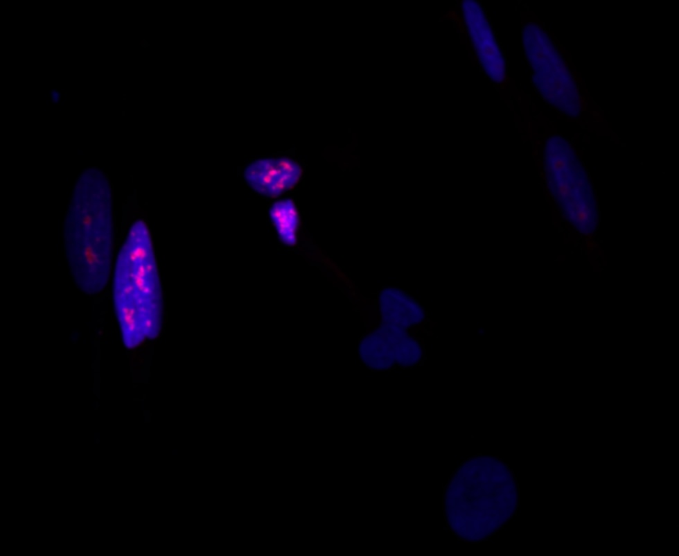
Celine (Verified Customer) (07-09-2025) | Works well in immunoflurescence with fixed cells
 |
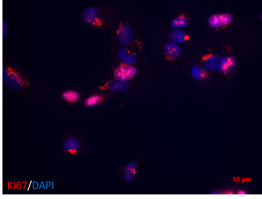
Guo-rong (Verified Customer) (08-30-2022) | Clear staining
 |
Alessandro (Verified Customer) (07-27-2022) | No aspecific staining, great outcome
|
Ana (Verified Customer) (02-15-2022) | Both, 1:50 and 1:100 dilutions worked well
|
kes (Verified Customer) (01-17-2022) | Works at 1:200 dilution for IHC
|
Yan (Verified Customer) (02-18-2020) | 1:100 work for cell.
|





























