

Tested Applications
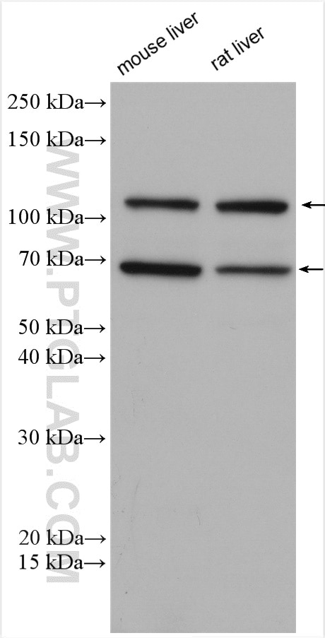
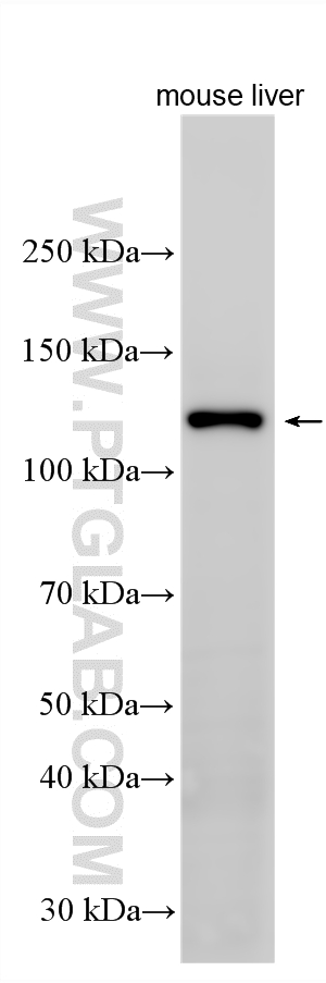
| Positive WB detected in | mouse liver tissue, rat liver tissue |
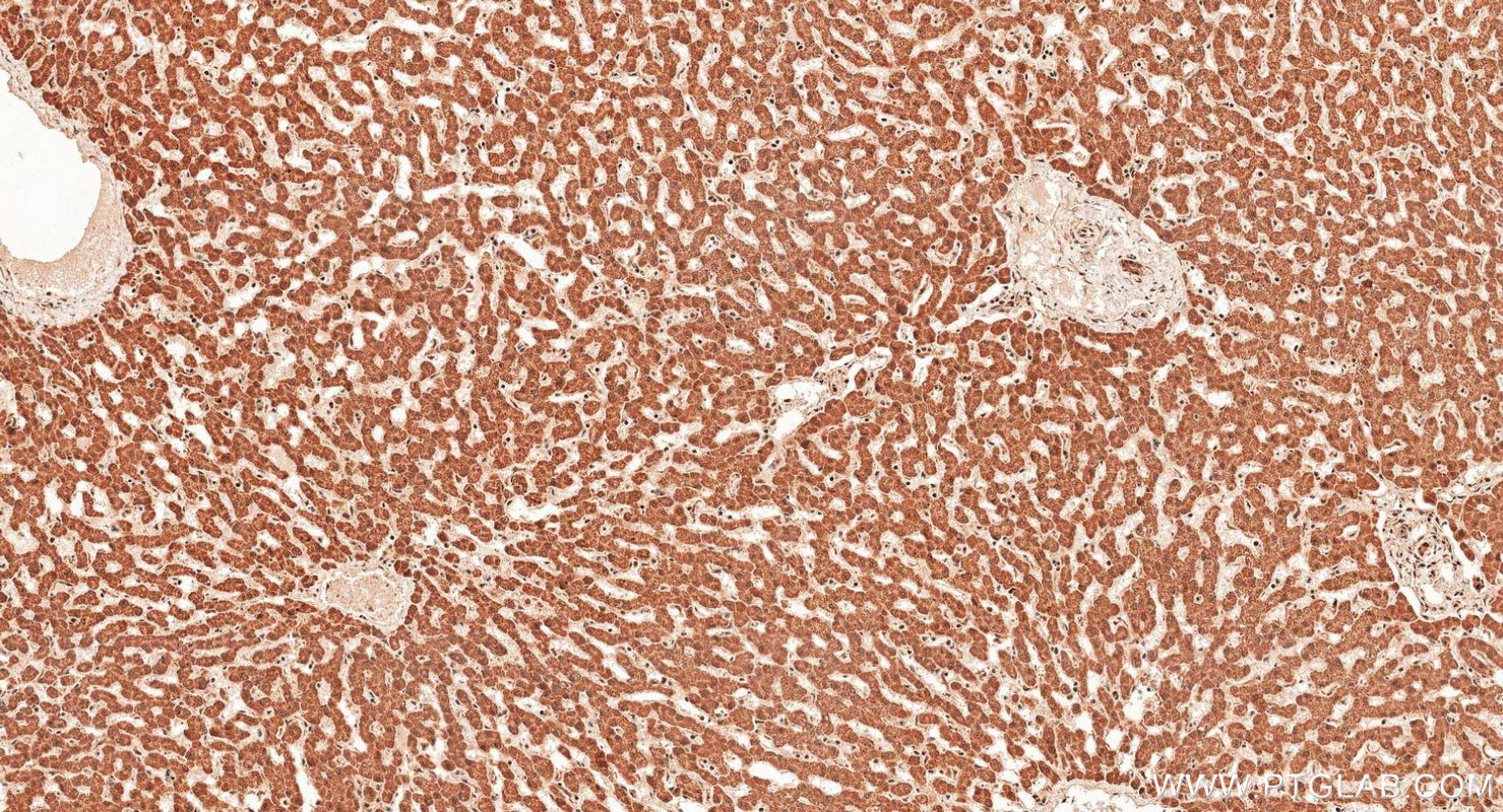
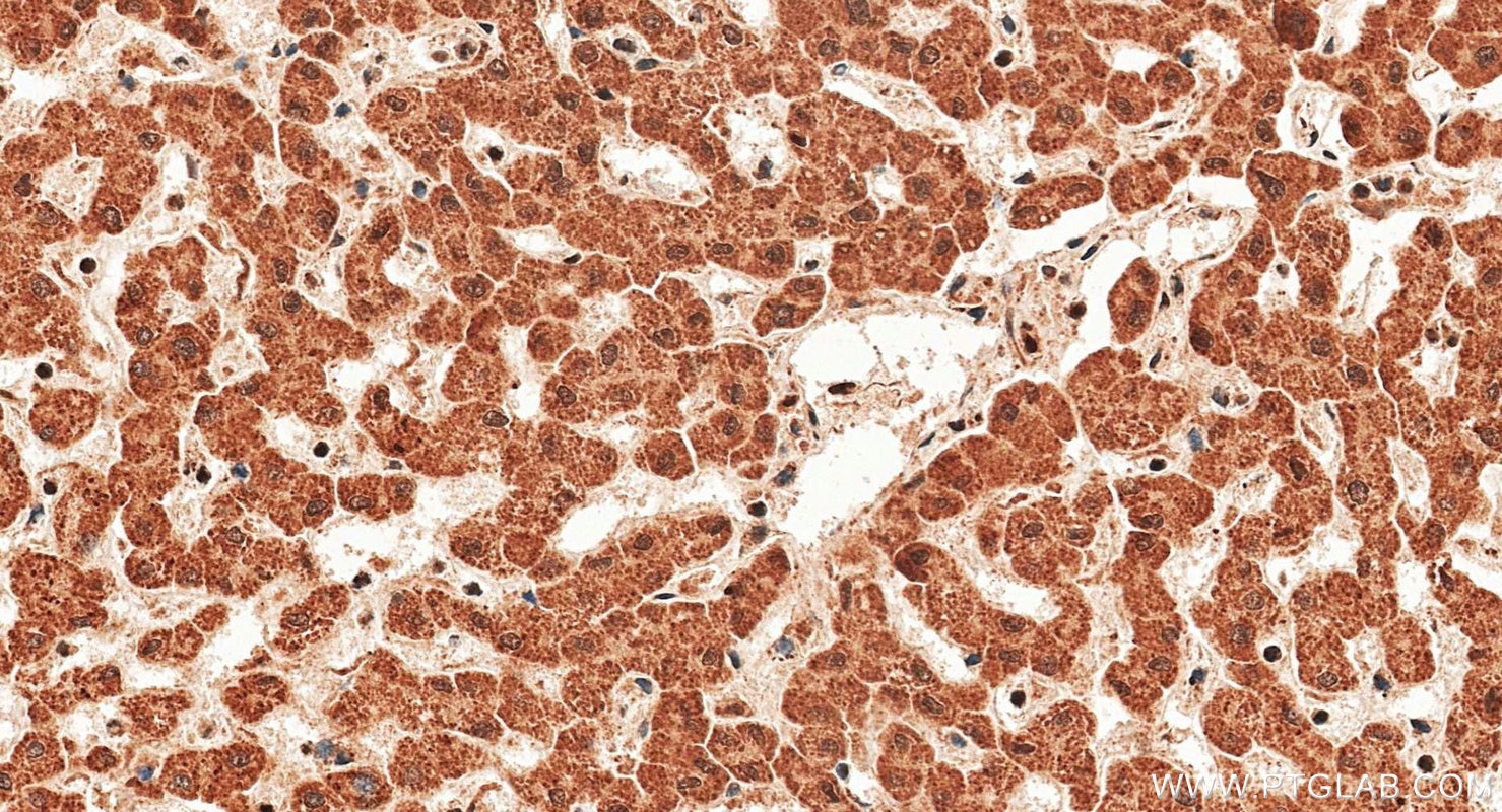
| Positive IHC detected in | human liver tissue Note: suggested antigen retrieval with TE buffer pH 9.0; (*) Alternatively, antigen retrieval may be performed with citrate buffer pH 6.0 |
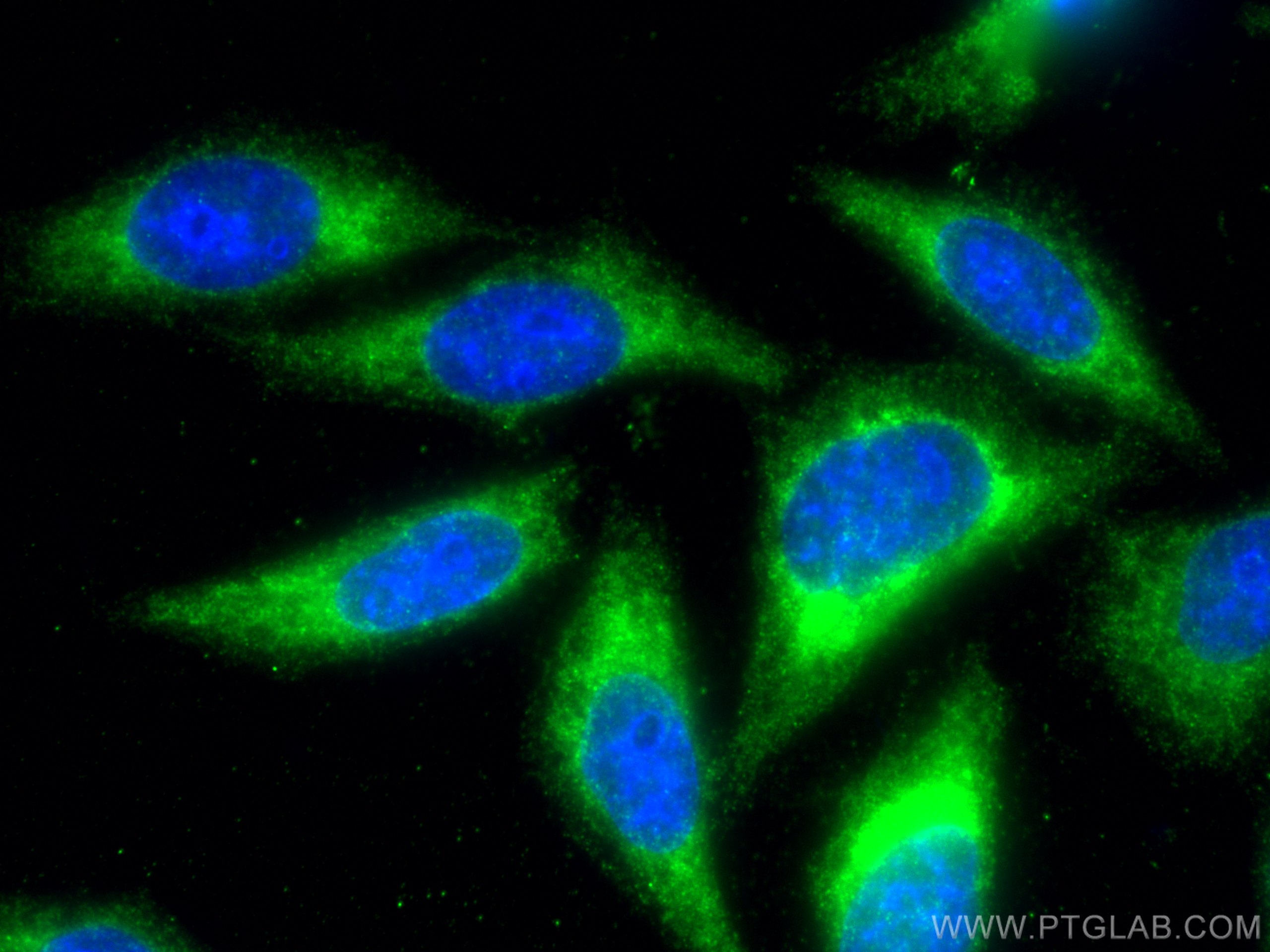
| Positive IF/ICC detected in | HepG2 cells |
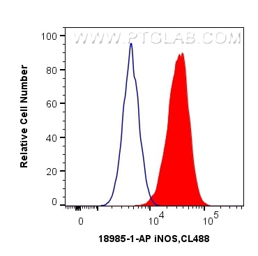
| Positive FC (Intra) detected in | HepG2 cells |
Recommended dilution
| Application | Dilution |
|---|---|
| Western Blot (WB) | WB : 1:2000-1:10000 |
| Immunohistochemistry (IHC) | IHC : 1:250-1:1000 |
| Immunofluorescence (IF)/ICC | IF/ICC : 1:50-1:500 |
| Flow Cytometry (FC) (INTRA) | FC (INTRA) : 0.40 ug per 10^6 cells in a 100 µl suspension |
| It is recommended that this reagent should be titrated in each testing system to obtain optimal results. | |
| Sample-dependent, Check data in validation data gallery. | |
Product Information
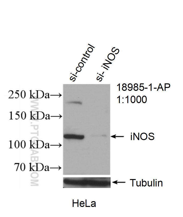
18985-1-AP targets iNOS in WB, IF/ICC, FC (Intra), IP, ELISA applications and shows reactivity with human, mouse, rat samples.
| Tested Reactivity | human, mouse, rat |
| Cited Reactivity | human, mouse, rat, pig, rabbit, chicken, bovine, goat |
| Host / Isotype | Rabbit / IgG |
| Class | Polyclonal |
| Type | Antibody |
| Immunogen |
Peptide Predict reactive species |
| Full Name | nitric oxide synthase 2, inducible |
| Calculated Molecular Weight | 131 kDa |
| Observed Molecular Weight | 110-130 kDa, 65-70 kDa |
| GenBank Accession Number | NM_000625 |
| Gene Symbol | iNOS |
| Gene ID (NCBI) | 4843 |
| RRID | AB_2782960 |
| Conjugate | Unconjugated |
| Form | Liquid |
| Purification Method | Antigen affinity purification |
| UNIPROT ID | P35228 |
| Storage Buffer | PBS with 0.02% sodium azide and 50% glycerol, pH 7.3. |
| Storage Conditions | Store at -20°C. Stable for one year after shipment. Aliquoting is unnecessary for -20oC storage. 20ul sizes contain 0.1% BSA. |
Background Information
NOS2, also named as iNOS and NOS2A, produces nitric oxide (NO) which is a messenger molecule with diverse functions throughout the body. NO is a reactive free radical which acts as a biologic mediator in several processes, including neurotransmission, antimicrobial and antitumoral activities. NOS2 is a nitric oxide synthase which is expressed in liver and is inducible by a combination of lipopolysaccharide and certain cytokines. iNOS has a very short half-life due to rapid degradation by calpain. iNOS monomer is a direct substrate of calpain I and can be cleaved by calpain I at the canonical CaM-binding site(503-532aa) of iNOS, and then a ~70 kDa band can be detected by western (PMID:11786228). This antibody is specific to NOS2.
Protocols
| Product Specific Protocols | |
|---|---|
| FC protocol for iNOS antibody 18985-1-AP | Download protocol |
| IF protocol for iNOS antibody 18985-1-AP | Download protocol |
| IHC protocol for iNOS antibody 18985-1-AP | Download protocol |
| WB protocol for iNOS antibody 18985-1-AP | Download protocol |
| Standard Protocols | |
|---|---|
| Click here to view our Standard Protocols |
Publications
What published studies show
The iNOS (NOS2) Polyclonal Antibody (Cat# 18985-1-AP) has accumulated 1,372 citations in journals including Cell Metabolism, Advanced Materials, Nature Communications, ACS Nano, Biomaterials, Theranostics, Advanced Science, Journal of Nanobiotechnology, Nature Biomedical Engineering, and Cell Death & Disease. Associated research fields encompass macrophage polarization, tumor immunology, neuroinflammation, bone regeneration, wound healing, atherosclerosis, spinal cord injury, stroke, osteoarthritis, periodontitis, ferroptosis, and nanomedicine-based therapeutics.
| Species | Application | Title |
|---|---|---|
Cell Metab Hexokinase 2 senses fructose in tumor-associated macrophages to promote colorectal cancer growth | ||
Cell Metab Macrophage PD-1 regulates energy expenditure and metabolic dysfunction under immune checkpoint blockade. | ||
Cell Metab Hepatic acetyl-CoA metabolism modulates neuroinflammation and depression susceptibility via acetate | ||
Exploration (Beijing) Activating Ferroptosis of M1 Macrophages: A Novel Mechanism of Asiaticoside Encapsuled in GelMA for Anti-Inflammation in Diabetic Wounds. | ||
Adv Mater 3D Bioprinted Human Synovium-Cartilage Models Mimic Rheumatoid Arthritis Microenvironment and Recapitulate In Vivo Therapeutic Responses. | ||
Adv Mater Sequential Targeted Enzyme-Instructed Self-Assembly Supramolecular Nanofibers to Attenuate Intervertebral Disc Degeneration |
Reviews
What customers say
Five out of six reviewers gave this antibody a 5-star rating, praising its performance in Western blot and IHC with clean, specific bands and strong signal at recommended dilutions. One reviewer reported a 1-star experience using it for immunofluorescence on mouse brain tissue, where the antibody appeared to cross-react with nNOS rather than specifically staining iNOS-positive microglia, despite testing multiple dilutions. Overall, the antibody is well-regarded for WB and IHC applications, though caution is advised for IF in tissues expressing related NOS isoforms.
The reviews below have been submitted by verified Proteintech customers who received an incentive for providing their feedback.
MALLIKARJUNA (Verified Customer) (10-24-2025) | WORKED GOOD FOR IF
|
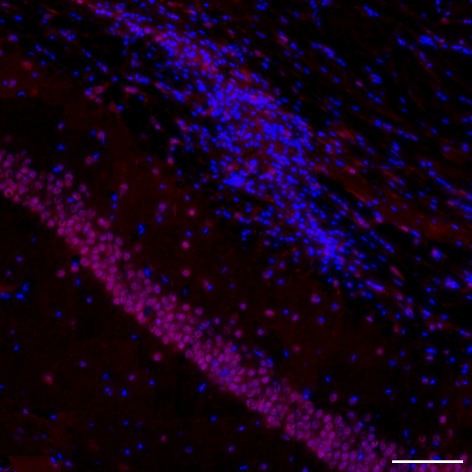
Marion (Verified Customer) (11-24-2024) | DAPI (blue) + iNOS (red) We expected iNOS staining on microglia cells. However, only nuclei of CA1 neurons were stained here. After discussion with the support, we came to the joint conclusion that the antibody must surely cross-link with the nNOS protein. We tested dilutions of 1/100, 1/200, 1/400, 1/600, 1/800 and 1/1000.
 |
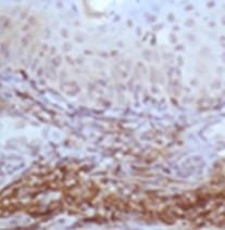
Yasuyo (Verified Customer) (03-01-2022) | This is one of the best IHC signaling I ever got for iNOS. Amazing image. I recommend this for iNOS detection by IHC.
 |
Xie (Verified Customer) (10-06-2021) | Works nice for WB assay.
 |
Prasanth (Verified Customer) (08-19-2019) | This is one of the cleanest and clarity blot I ever got for iNOS. Amazing clarity and no non specific bands. I recommend this for iNOS detection by western blot.
|
Elena (Verified Customer) (08-14-2019) | We tried this antibody with 2 cell lines and it worked just fine. We will use this in the future for other cell lines as well. The antibody produced strong bands at the suggested concentration and we diluted it further.
|








