Immunoprecipitation
Immunoprecipitation (IP) is a technique used to purify and enrich a protein of interest from a complex mixture. It is widely used to investigate protein expression, relative abundance, size, upregulation or downregulation, stability, post-translational modifications (PTMs), and interactions with other proteins.
Reagents to Support Your Immunoprecipitation Workflow
Explore antibodies, VHH bead-based reagents, and supporting tools for immunoprecipitation and post-immunoprecipitation analysis.
Primary antibodies for IP
Explore polyclonal or monoclonal primary antibodies for the IP of untagged proteins.
Anti-IgG VHH Beads for IP as smart Protein A/G alternative New
Anti-IgG VHH Beads use secondary VHHs to selectively bind primary rabbit and/or mouse IgGs, offering consistent affinity and precise targeting with zero leaching, setting a new standard for IP.
Nano-Traps for one-step IP
Nano-Traps use primary VHHs coupled to beads, targeting commonly used tags like GFP, Myc, HA, V5, or endogenous targets like Ubiquitin, SUMO, NEDD8, or ISG15 with high affinity.
Isotype control antibodies
Find the right negative control antibody for your IP antibody to ensure highly specific results.
Antibodies for post-IP Western Blot
Explore antibodies for the detection of precipitated proteins and their interaction partners.
Conformation specific HRP-Goat Anti-Rabbit IgG Recombinant Secondary Antibody
Western Blot supporting reagents
Explore specialized reagents to support and accelerate your post-IP Western Blot.
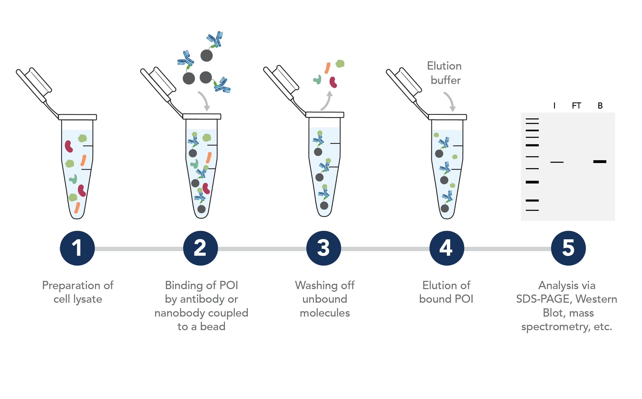
Overview of a Standard Immunoprecipitation Workflow
Are you new to IP or struggling to get reliable results? Discover how to optimize your protocol and troubleshoot common challenges with our practical tips and tricks.
Immunoprecipitation Method Comparison
Compare Protein A/G, anti-IgG VHH Beads, and Nano-Traps to identify the best immunoprecipitation reagent for your workflow.
| Protein A/G | Anti-IgG VHH Beads | Nano-Traps | |
|---|---|---|---|
 |
 |
 |
|
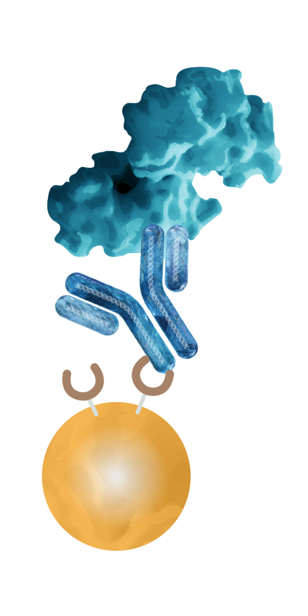
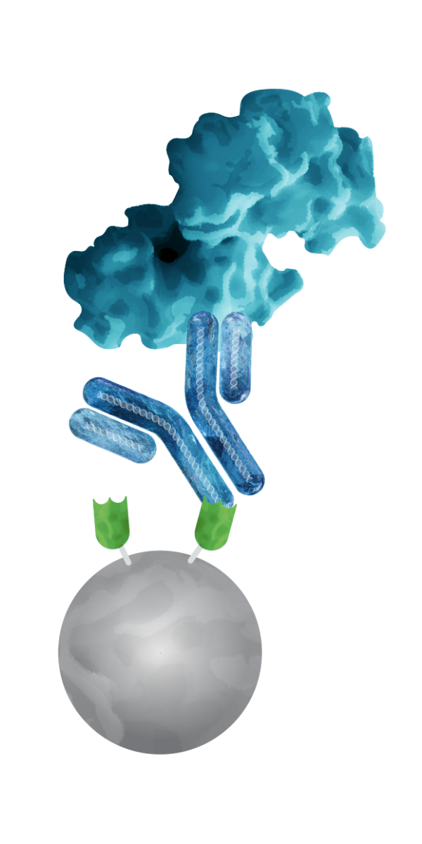
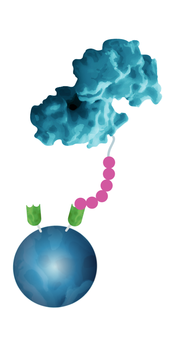
| Binding via | Bacterial Protein A or G | Recombinant Secondary VHH | Recombinant Primary VHH |
| Binding target | IgG | IgG | Protein or peptide tag Endogenous proteins |
| Species-specific IgG recognition? | No | Yes | Binds directly to target |
| Background in SDS-PAGE and Western Blot |
High Protein A/G leaching into IP fraction and unspecific binding of detection antibodies; heavy and light chain antibody bands possible. |
Low Heavy and light chain antibody bands possible. You can use SpeedAb Kits or conformation-specific secondary antibodies to avoid recognition of your pulldown antibody. |
Ultra-low |
| Duration | > 3h | > 3h | < 2h |
| Binding affinity to IgG or tag? |
Very low to high (depending on IgG species) |
High (optimized for the respective IgG subtype) |
High to very high |
| Ready-to-use? | Depends on vendor | Yes | Yes |
| Use for | Standard IPs if background is not critical | Standard IPs where clean, efficient, and consistent performance is required, especially when working with antibody species that have low affinity for Protein A/G. | IPs of tagged proteins, especially for low abundant targets and workflows requiring stringent washing (e.g., IP-MS), or for applications where speed and reproducibility are key. |
| Learn more about anti-IgG VHH Beads | Learn more about Nano-Traps |
Species specificity of anti-rabbit IgG / anti-mouse IgG VHH agarose
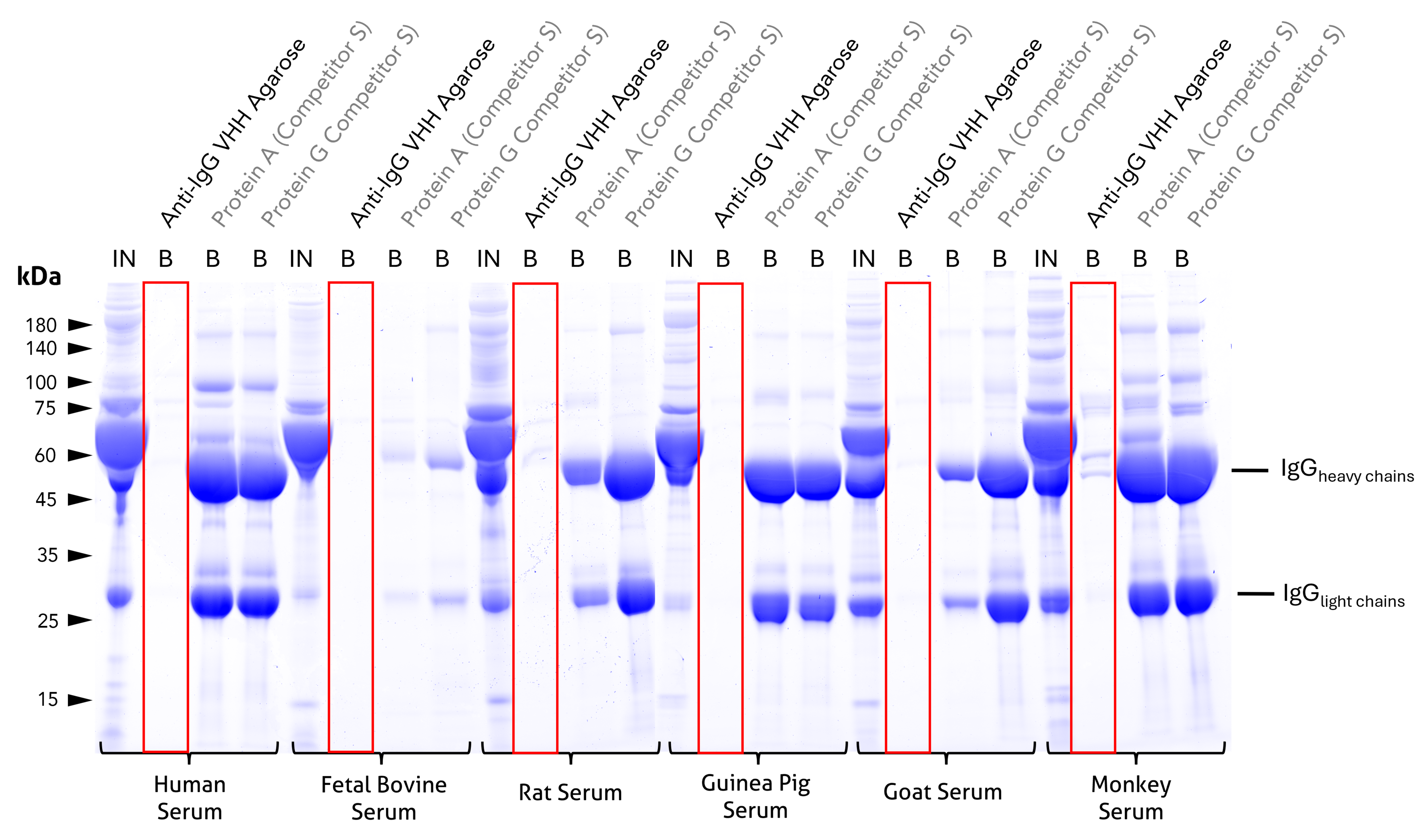
Cross-reactivity test and comparison to competitor Protein A and Protein G beads. Coomassie-stained SDS-PAGE gel showing input (IN) and bound (B) fractions for different beads incubated with 1:10 diluted sera of relevant model species. Unlike the tested protein A or G beads, bound fractions of Proteintech anti-rabbit IgG / anti-mouse IgG VHH agarose reveal no cross-reactivity to human, bovine, rat, guinea pig, or goat IgGs and only minor cross-reactivity to cyno monkey IgGs. For IN and B fractions 1% and 25% were loaded, respectively. Lanes of anti-IgG VHH beads are highlighted red. Results were reproducible for anti-rabbit IgG / anti-mouse IgG Magnetic VHH Agarose.
Clean precipitation and detection of PCNA with anti-IgG VHH Beads
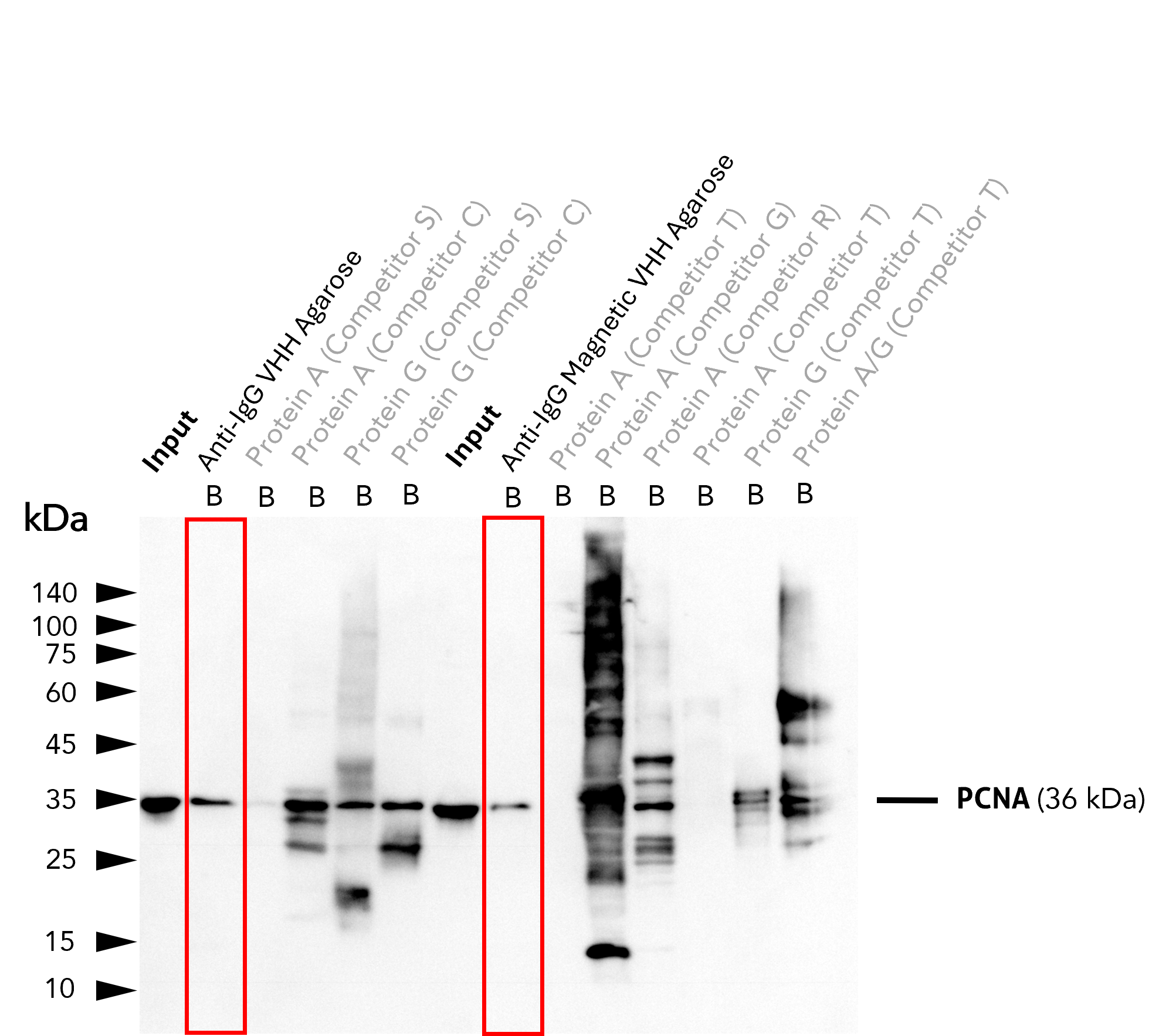
IP of PCNA using 5 µg of anti-PCNA antibody (IgG1, 60097-1-Ig) by anti-rabbit IgG / anti-mouse IgG VHH beads. For Western blot analysis, the PCNA polyclonal antibody (10205-2-AP, 1:2000) was labeled using a conformation-specific HRP-conjugated secondary to avoid staining of heavy and light chain of the IP-antibody. In contrast to many competitors' protein A and G beads, clean pull-down of PCNA by Proteintech anti-rabbit IgG / anti-mouse IgG VHH Beads (Ag. = Agarose; M.Ag. = Magnetic Agarose) facilitates unambiguous identification of the target protein. Other beads show leaching of protein A or protein G fragments into the final IP fractions, which can lead to binding of the detection antibody and unspecific signals, as reported previously (Grant et al., 2019, Biol Proced Online, doi: 10.1186/s12575-019-0095-z). For input (IN) and bound (B) fractions, 1% and 30% were loaded, respectively. Lanes of anti-IgG VHH beads are highlighted in red.
Remarkably low background
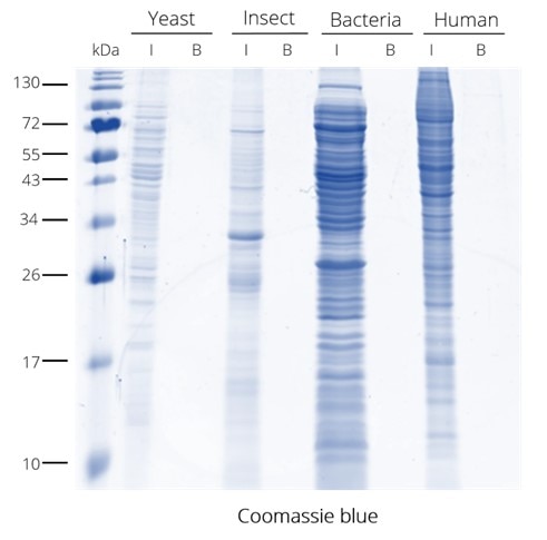
IP from different cell lysates without V5-tagged proteins using V5-Trap. The V5-Trap has remarkably low background with no non-specific binding seen in the bound fraction.
No heavy or light chain bands
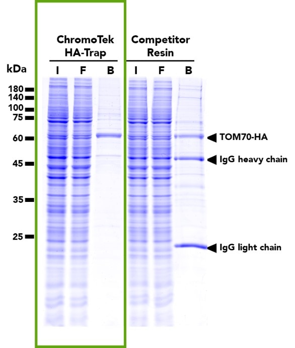
Pulldown of TOM70-HA fusion protein from transfected HEK293T cells using either HA-Trap (left) or a competitor resin (right). Pulldowns with HA-Trap result in far cleaner, single-band pulldowns with significantly less artifact binding compared to the competitor product.
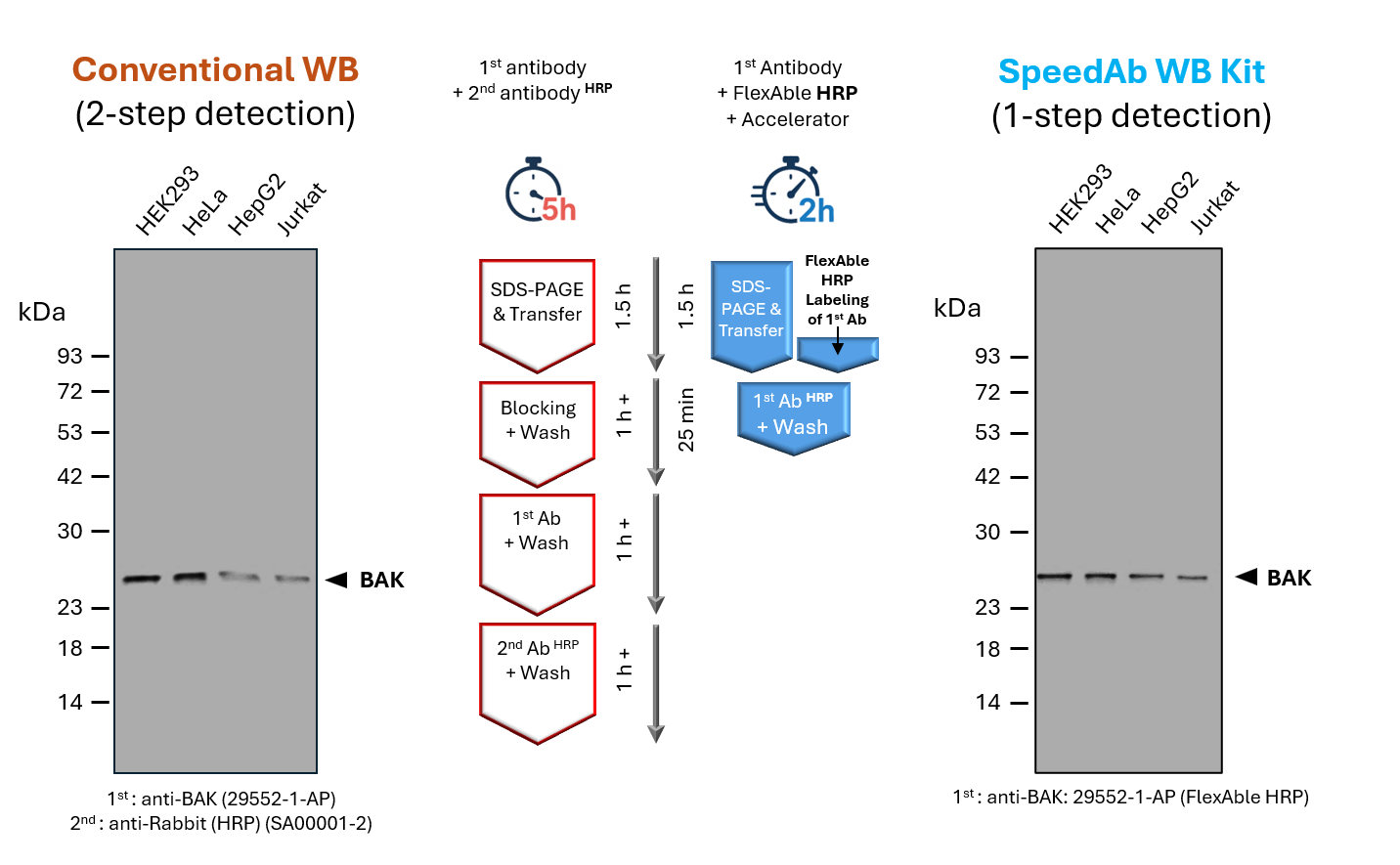
SpeedAb WB Kit for detection of rabbit IgG
(left) Classical Western blot detection of BAK in lysates from different human cell lines. A primary anti-BAK antibody (rabbit) was used in combination with a secondary anti-rabbit antibody (HRP), after blocking and incubation with antibodies for 1 h, respectively. The totaltime of ~ 5 h includes SDS-PAGE, blotting, blocking, primary and secondary antibody incubation, and washing steps.
(right) Detection of BAK in lysates from different human cell lines via SpeedAb WB Kit. The primary anti-BAK antibody was labeled with FlexAble HRP (anti-rabbit: #KFA005) via a simple 15min protocol, enabling1-step detection. The blotting accelerator speeds up binding of the antibody to the target protein, cutting the antibody incubation time to 25minor less and the total time including SDS-PAGE, blotting, antibody incubation and a final washing step to ~2 h.
Watch why GFP-Trap gives the best results for immunoprecipitation (IP)
Low background, no extra bands, and high specificity will improve your pulldown assay significantly. Alpaca Alice shows how it works. Visit www.ptglab.com/nano-trap-free-sample/ to order a free test sample.
Resources
Explore supporting videos and blog articles to help optimize your IP workflow.
Supporting Videos
- How to Optimize Your IP Workflow IP Troubleshooting
- IP-MS Workshop
- Optimizing IP of FP-Tagged Proteins in Plant Cell Extracts
- Save hours on your IP
- View other upcoming webinars
Supporting Blogs
Featured Products
Explore our most cited reagents for immunoprecipitation, including VHH-based Nano-Traps, anti-IgG beads, tag antibodies, and supporting controls.
Anti-Rabbit IgG/anti-Mouse IgG VHH Agarose
An affinity resin for IP of all subtypes of rabbit and mouse IgG. It consists of rabbit and mouse IgG specific VHHs (Nanobodies) coupled to Agarose beads. Also available with Magnetic Agarose.
Anti-Rabbit IgG/anti-Mouse IgG VHH AgaroseGFP-Trap®
An affinity resin for IP of GFP-fusion proteins. It consists of a GFP-specific VHH (Nanobody) coupled to (magnetic) agarose beads or magnetic particles M-270.
GFP-Trap®Myc-Trap®
An affinity resin for IP of Myc-tagged proteins. It consists of an anti-Myc VHH (Nanobody) coupled to (magnetic) agarose beads or magnetic particles M-270.
Myc-Trap®RFP-Trap®
An affinity resin for IP of RFP-fusion proteins. It consists of an RFP-specific VHH (Nanobody) coupled to (magnetic) agarose beads or magnetic particles M-270.
RFP-Trap®V5-Trap®
An affinity resin for IP of V5-tagged proteins. It consists of an anti-V5 VHH (Nanobody) coupled to (magnetic) agarose beads or magnetic particles M-270.
V5-Trap®6*His, His-Tag Monoclonal Antibody (1B7G5)
Monoclonal mouse anti-6×His antibody for detecting and analyzing His-tagged recombinant proteins in applications such as Western blot, IP, IF/ICC, FC, and ELISA.
6*His, His-Tag Monoclonal Antibody (1B7G5)HRP-Goat Anti-Rabbit IgG Conformation Specific Recombinant Secondary Antibody
HRP-conjugated conformation-specific recombinant goat anti-rabbit secondary antibody for WB, IP-WB, and ELISA.
HRP-Goat Anti-Rabbit IgG Conformation Specific Recombinant Secondary AntibodyRabbit IgG control Polyclonal Antibody
Normal rabbit IgG isotype control antibody used to assess non-specific binding and serve as a negative control in applications such as WB, IP, and ELISA.
Rabbit IgG control Polyclonal AntibodyFAQs
Choose the right immunoprecipitation reagent and learn how to optimize your workflow for cleaner results, higher yield, and better downstream detection.
Which immunoprecipitation reagent should I choose for my experiment?
The optimal immunoprecipitation reagent depends on your target protein, for example whether it is tagged or untagged and high- or low-abundance, as well as on the experimental conditions and downstream application.
Nano-Traps are recommended for the IP of tagged proteins (e.g., fluorescent protein tags like GFP-tag and RFP-tag, or peptide tags like Strep, His, V5, Myc, FLAG, etc.) and selected endogenous proteins (e.g., Ubiquitin, SUMO1, NEDD8, p53, PARP1, etc.), especially for low-abundance targets, applications requiring stringent lysis or washing conditions (e.g., ubiquitination assays, plant lysates, or IP-MS or AP-MS), or workflows where speed, sensitivity, and reproducibility are critical.
Anti-IgG VHH Beads offer a cleaner, more efficient alternative to conventional Protein A/G. Unlike Protein A/G, which binds IgGs from multiple species with varying affinities, anti-IgG VHH Beads are optimized for their respective species or isotype, resulting in higher specificity and reduced background. This is especially beneficial when working with serum samples or samples containing BSA, FCS, or HSA, such as commercial antibody formulations, since this can lead to substantial background when using Protein A/G (bovine or human IgG contamination).
When are anti-IgG VHH Beads a better choice than Protein A/G for immunoprecipitation?
While Protein A and G remain the most common choices for antibody capture, their broad and unspecific Fc binding and potential for leaching from beads can compromise specificity and lead to interfering background in sensitive assays. Our anti-IgG VHH Beads use secondary VHHs coupled to agarose or magnetic agarose beads and are optimized for targeted precision and clean results.
When are Nano-Traps a better choice than Protein A/G for immunoprecipitation?
Nano-Traps use primary VHHs coupled to magnetic or agarose beads and are the preferred choice for immunoprecipitation of proteins that carry an affinity tag such as GFP, Myc, HA, or V5. They are also available for a range of endogenous targets, including Ubiquitin, SUMO, NEDD8, and ISG15.
Compared to Protein A/G-based IPs, Nano-Trap-based IPs are cleaner and more efficient. Because Nano-Traps do not rely on antibodies for target capture, they eliminate heavy and light chain antibody contamination in downstream analysis such as Western blot or mass spectrometry. They also enable faster, one-step pulldowns that can be completed in under 2 hours, whereas Protein A/G protocols typically take at least 3 hours since they require an additional incubation step with the IgG or a pre-assembly of beads.
How can I reduce background in immunoprecipitation?
Background in immunoprecipitation experiments can occur due to degradation of the protein of interest, non-specific binding to the beads, or Protein A/G leaching from the beads and interfering with downstream Western blot detection.
To reduce background, we recommend:
- Optimize lysate preparation for your specific cell type and use an appropriate amount of starting material. Too little material will result in low signals, while too much input material will increase the risk of background binding. Add sufficient protease inhibitors to minimize degradation of proteins. Depending on your research question, you may want to add specific inhibitors to counter, for example, dephosphorylation.
- Optimize binding conditions by incubating at 4 °C for a maximum of 1 hour. Longer incubation times or higher temperatures will increase the risk of degrading your protein of interest or of precipitate formation, which will result in background binding.
- Optimize washing conditions by increasing salt or detergent concentrations or frequency of washing. Increasing NaCl or KCl concentrations may help to reduce background in the case of DNA-binding proteins, for example. However, a careful approach is necessary, as overly stringent wash conditions may also inadvertently eliminate real interaction partners of your protein of interest. Use a fresh tube for the final wash step to reduce background from proteins sticking to plastic surfaces.
- Include a pre-clearing step: Pre-clearing is not routinely needed for Nano-Traps but may be relevant for Protein A/G-based approaches. For example, samples such as serum may contain antibodies, which may compete with your primary antibody for Protein A/G binding. Reduce endogenous antibodies with Protein A/G-bead pre-clearing or use the appropriate anti-IgG VHH Beads to circumvent this problem. Samples may also contain antibody-binding proteins, which will lead to background binding, but can be eliminated using pre-clearing with an isotype control.
Tip: Unlike conventional Protein A/G-based approaches, nanobody-based reagents (Nano-Traps, anti-IgG VHH Beads) are designed for highly specific target binding, minimizing background and increasing consistency of your workflows.
Which immunoprecipitation reagent is best for low-abundance targets or stringent washing?
ChromoTek nanobodies exhibit high binding affinities in the picomolar to nanomolar range and are highly target-specific. Their small size and compact structure contribute to their thermostability and robustness under stringent washing and lysis buffer conditions, including high salt, detergents, reducing agents, and chaotropic agents such as urea.
Since Nano-Traps rely exclusively on immobilized nanobodies and do not require a capture antibody, this format offers particular benefits for the pulldown of low-abundance targets, and for IPs under demanding conditions, where high affinity, specificity, and stability are key.
How do I avoid antibody heavy and light chain bands masking my target protein in my Western blot after immunoprecipitation?
To avoid antibody heavy and light chain bands masking your protein of interest, we recommend the following strategies:
- Use different antibody species for capture and detection, so your Western blot secondary antibody does not bind to both.
- Use SpeedAb Kits: If switching species is not feasible, we recommend our SpeedAb Kits. They include FlexAble HRP, a solution to label primary antibodies with HRP. This enables 1-step detection without a secondary antibody and helps circumvent this problem. Note: SpeedAb kits additionally contain our Blotting Accelerator, a solution with special organic compounds that lets you skip the blocking step and speed up antibody incubation when antibodies are diluted in it.
- Use conformation-specific secondaries: Use a secondary antibody that only binds native, but not denatured IgG, such as the HRP-Goat anti-rabbit IgG conformation-specific recombinant secondary antibody.
- Cross-link your antibody: Covalently cross-link your IP antibody to the bead, so it does not elute with your sample.
- Avoid antibody contamination altogether: Our Nano-Traps rely on primary nanobodies that bind directly to their target without requiring a traditional capture antibody, entirely eliminating antibody contamination of your eluate.
How many cells do I need to get enough yield for an IP or Co-IP?
As a starting point, we recommend using lysate from about 0.5–1 × 10⁶ cells per IP reaction for many mammalian cell lines, but the exact number strongly depends on the cell type, cell size, and the expression level of your protein of interest. For highly overexpressed tagged proteins, you can often use fewer cells, whereas for endogenous, low-abundance, or complex-associated targets, especially in Co-IPs, you may need substantially more material, up to the 10⁷-cell range. The optimal cell number should therefore always be determined empirically by a small titration series.
What are the best practices for pulldown of membrane proteins?
For the pulldown of membrane proteins, we recommend using mild nonionic or zwitterionic detergents for cell lysis. Optimize both the detergent concentration and salt conditions to ensure that your membrane protein is fully solubilized while preserving co-precipitation of known interaction partners. Our Lys-Buf provides a mild starting point for testing, whereas our RIPA buffer offers strong lysis but can disrupt weak or transient interactions can be stabilized before or during lysis using gentle crosslinkers or nanodisc/detergent‑free approaches.
Why am I getting a weak signal or no band in Western blot after immunoprecipitation?
A weak or missing signal after an IP typically indicates that the protein degraded, failed to bind the matrix, or is physically obscured on your blot. Troubleshoot your protocol using these tips:
- Include proper experimental controls: Always run an input control and a positive control to verify your target protein is present and your Western blot detection works.
- Ensure your capture antibody is validated for IP. The target epitope must be accessible on the 3D protein surface since the protein is in a native state.
- Check lysis buffer restrictions: Antibodies may lose function in lysates containing reducing agents, extreme pH, or high detergent concentrations.
- Optimize sample preparation: Prevent protein degradation by keeping lysates on ice and using fresh samples with protease inhibitors.
- Optimize wash stringency: If your target-protein interaction is fragile, reduce the number of washes or decrease the salt and detergent concentrations.
- Verify bead-antibody compatibility: Traditional Protein A or G beads have a weak affinity for certain antibody species and subclasses, risking that you lose your antibody during IP.
Where can I find an immunoprecipitation protocol for my application?
Protocols are available in the following places:
- Standard immunoprecipitation protocol.
- Nano-Trap and anti-IgG VHH Bead protocols are available on the product information section of each product page.
- On-bead digestion protocol for IP-MS and AP-MS workflows.