
Industry First: Proteintech Launches IHC Whole Slide Image (WSI) Viewer
Written by: Abel Zhang at Proteintech
-
Whole Slide Imaging (WSI) provides a complete picture of an entire tissue slide, thereby reducing selection bias from single-field, fixed magnification images
-
It can help researchers evaluate for antibody staining consistency, background signal, and tissue reference regions
-
It improves antibody data transparency and traceability by showing the complete original slide scan
-
Proteintech has implemented whole slide images for hundreds of IHC antibodies, an industry first, in an effort to provide reliable validation data
An antibody product page may display an IHC image with a clean, well-defined signal and a clear background, creating a strong impression of product performance. After ordering the product and running the experiment, however, the observed signal may differ substantially from the image shown online.
Does this indicate an antibody quality issue? Not necessarily.
The discrepancy may instead originate from the image itself.
The Issue with Single Field Images
When antibody companies present IHC validation data, the traditional approach is to capture one or more JPG images at fixed magnifications. These images are typically the “most representative” regions chosen by lab personnel and can be prone to selection bias.
If a particular clone stains only marginally across most of the tissue but happens to look striking in one small corner, that corner is likely to be selected for the validation image presented on the antibody product page.
Whole Slide Imaging (WSI): Handing You the Entire Slide
WSI (Whole Slide Imaging) is a technology that digitizes an entire tissue slide into a high-resolution image. A slide scanner takes a series of images, field by field, at 20x or 40x magnification and stitches them together into a complete digital slide. The digital slide can then be browsed and viewed at any magnification.
WSI provides an authentic, full picture of the entire slide, removed from selection bias. It can help to answer several questions, including:
-
Is the staining consistent across the entire slide?
-
Are the negative regions truly clean?
-
Does the normal tissue adjacent to the tumor express the target?
WSI Provides a Complete Staining Overview
Different antibodies against the same target may look similar on a single field image because the field with the best antibody staining was likely chosen. However, when the full slide images of these antibodies are compared side by side, the differences become immediately obvious.
We compared three KRT19 clones: some showed uniform signal across most of the tissue, some only stained well in localized areas, and some had background staining. Viewed at the whole-slide level, it’s more evidently clear which clone has broader applicability.
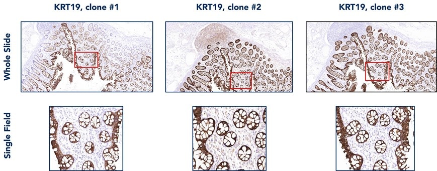
Figure 1: Comparison of the whole slide staining patterns of three different KRT19 antibody clones in colon tissue. Clone 1 shows incomplete staining of all colon glands, clone 2 shows non-specific staining of the lymph node area, and clone 3 shows uniform staining across all colon glands.
WSI can Establish Tissue Reference Points
Browsing through a whole slide image allows for an independent assessment of the relationship between positive and negative staining regions throughout the tissue.
Below, we used CD138 staining in tonsil tissue as an example. Besides the positive expression in plasma cells, the staining of the squamous epithelium (green arrows) itself serves as a reference marker for positive expression, while the mantle zone provides a natural negative control area (red arrow). However, in a localized high-magnification image like the one on the left, these critical reference cues are lost.
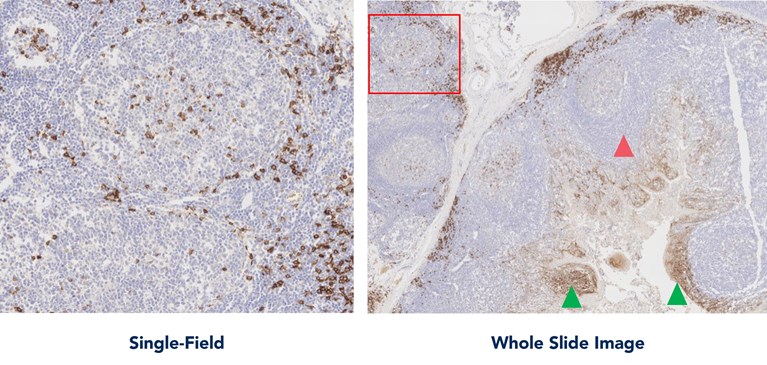
Figure 2: CD138 antibody staining in the tonsil tissue. The whole slide image on the right allows for the establishment of positive and negative staining control regions. The single-field image on the left was taken from the area within the red box of the whole slide image.
WSI Improves Data Transparency and Traceability
Ultimately, the distinction between WSI and single-field JPGs is not defined by resolution alone, but by the extent to which the available information is controlled or fully disclosed. A single-field JPG presents a selected view of the tissue, whereas WSI provides access to the complete slide data and supports independent evaluation.
For diagnostic pathology and antibody screening, this difference is critical. Pathologists need to see the complete tissue architecture to make accurate judgments, while researchers screening antibodies need to confirm that an antibody performs consistently across different regions. Any manually selected partial image inherently carries bias.
Whole slide data has another important value: traceability. The image the viewer sees is the original scan file with no selective cropping or color adjustment. All results can be traced back to the complete slide.
Proteintech’s Whole Slide Image Data and Viewing Platform
Proteintech has already published thousands of WSI images on its product pages, covering nearly a thousand targets, with ongoing expansion underway. Through these whole slide images, we hope to deliver more comprehensive detection information to customers.
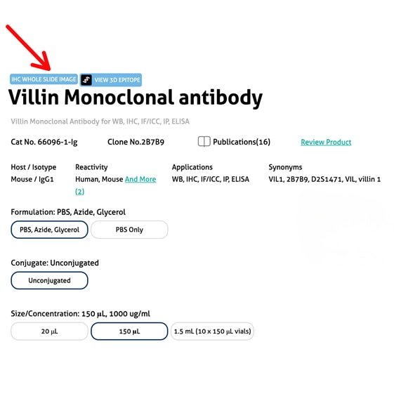
On the Proteintech product pages, antibodies tagged with “IHC Whole Slide Image” indicate that WSI images are available. These images can be accessed through three different methods:
1. Clicking the “IHC Whole Slide Image” tag directly
2. Clicking the “IHC Whole Slide Image” link below the IHC QC data

3. Clicking the “IHC Whole Slide Image” link that appears below the IHC data pop-out images
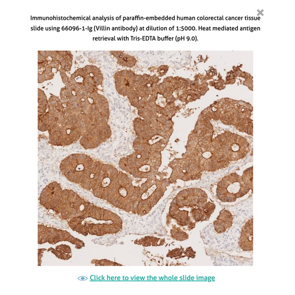
Once the WSI viewing platform has been opened, the controls at the top right of the image can be used to zoom in, zoom out, reset to default size, and view as a full-screen image. The mouse wheel can also be used to zoom in and out of the image, while the mouse itself can be dragged to pan across the entire tissue area. The viewing platform is supported on all standard computer screens as well as mobile phones, tablets, and larger conference displays.
View all antibodies with whole slide images: Results for "IHC whole slide image" | Proteintech Group
FAQs:
Why can single-field IHC images be misleading?
-
Single-field images may only show the "best-looking" region of the tissue, which can introduce selection bias and may not accurately reflect antibody staining across the full tissue slide.
Does a difference between an online IHC validation image and experimental data mean that an antibody is of poor quality?
-
Not necessarily. The discrepancy may come from how the validation image was selected or presented by the manufacturer, rather than from actual antibody performance alone.
Why is WSI useful for comparing different antibodies against the same target?
-
Whole slide images make it easier to determine whether an antibody stains uniformly, in localized areas only, or non-specifically, enabling better selection of the best antibody for a user's application.
Where can Proteintech WSI data be found?
- Whole slide images can be found on select antibody pages tagged with "IHC Whole Slide Image" which links to available WSI data.
Are whole slide images available for all Proteintech antibodies?
-
Not yet. More images are being added on a rolling basis. Contact us if you are looking for whole slide images for a specific antibody, and we will do our best to provide it.

Related Content
Mouse-on-mouse for Immunohistochemistry: tips and tricks
How do I know if the antibody will cross-react?
A beginner's guide to Immunohistochemistry
Protein or peptide antigen? Advantages and disadvantages
Support
Newsletter Signup
Stay up-to-date with our latest news and events. New to Proteintech? Get 10% off your first order when you sign up.
Able™