
Mastering the Transition: A Protocol for Differentiating SH-SY5Y Cells into Neurons
Written by Ceanne Elliott, PhD Candidate at Indiana University School of Medicine
Summary
-
Differentiation transitions proliferative SH-SY5Y cells into post-mitotic, neuron-like cells with greater biological relevance for neuroscience research.
-
Retinoic acid initiates the differentiation process, while subsequent treatment with brain-derived neurotrophic factor (BDNF) promotes neurite outgrowth, cellular branching, and the development of mature neuronal signaling networks.
-
The 10-day protocol involves seeding cells on Day −1, introducing retinoic acid on Day 0, and adding retinoic acid and BDNF through sequential medium changes on Days 3, 6, and 9.
-
Careful reagent preparation and cell handling are essential for successful differentiation, particularly when preparing light-sensitive retinoic acid and performing medium changes without disturbing the cells.
-
Successful differentiation is demonstrated by extensive neurite outgrowth, formation of interconnected neuron-like networks, and increased expression of the neuronal markers βIII-tubulin and synaptophysin.
Introduction
The SH-SY5Y human neuroblastoma cell line is widely used in neuroscience research and is considered a foundational in vitro model for investigating the molecular pathogenesis of neurodegenerative disorders. These cells exhibit intrinsic catecholaminergic properties, making them particularly valuable for modeling neuronal biology1. However, in their basal culture state, SH-SY5Y cells continuously proliferate and exhibit an immature neuroblast phenotype, lacking the functional synaptic structures and specialized signaling networks of adult human neurons. To achieve true biological relevance in neurological studies, it is necessary to transition these cells out of the cell cycle. Differentiation forces SH-SY5Y cells into a post-mitotic state, promotes extensive neurite outgrowth, and allows for the establishment of transcriptomic and proteomic signatures of mature neurons—ultimately providing a reproducible system for examining complex neuronal pathways1.
Understanding Neuronal Differentiation Reagents
In their undifferentiated state, SH-SY5Y cells exhibit an immature catecholaminergic profile characterized by proliferative proteins like proliferating cell nuclear antigen (PCNA) and early neuronal markers like nestin2 . To transition these cells into a post-mitotic state, Retinoic Acid (RA) is introduced to initiate the differentiation program. As a powerful growth inhibitor and cellular differentiation-promoter, RA drives the cells toward a cholinergic or dopaminergic phenotype, marked by increased choline acetyl transferase (ChAT) activity and vesicular monamine transporter (VMAT) expression3. RA treatment can also establish a mature dopaminergic phenotype; however, achieving this usually requires the co-administration of supplementary agents like phorbol esters2.
While RA begins the differentiation process, terminal functional maturation requires the subsequent addition of Brain-Derived Neurotrophic Factor (BDNF). Following RA pre-treatment, BDNF promotes extensive neurite outgrowth, axonal branching, and the maturation of regulatory networks essential for synaptic neurotransmission4. This sequence yields fully differentiated cells that express mature neuronal markers like grown-associated protein (GAP-43), neuronal nuclei (NeuN), synaptophysin (SYN), beta-3-tubulin (TUBB3), and microtubule associated protein 2 (MAP2), while remaining free of glial markers such as glial fibrillary acidic protein (GFAP)5.
Maintaining this advanced phenotype in vitro demands a highly specialized approach to the culture environment. Neurobasal medium provides crucial metabolic support while specifically excluding excitatory amino acids, such as glutamate and aspartate, to prevent excitotoxicity in mature cells2. This is paired with B-27, an optimized, serum-free supplement necessary for the long-term survival and viability of post-mitotic neurons6.
This step-by-step guide, based on the protocol described by Targett et al.3, produces a reliable population of differentiated neuron-like cells. The resulting culture displays extensive branching and expresses key pre- and post-synaptic markers, providing an accurate cholinergic and glutamatergic model for in vitro experiments3.
Tips Before Starting Differentiation Protocol
-
Passage numbers less than 10 are ideal for performing the differentiation protocol.
-
Make Differentiation 1 (Table 2) and Differentiation 2 (Table 3) Mediums without retinoic acid (RA). RA should be added to the culture medium immediately before adding to cells.
-
Medium changes should be performed carefully to not disturb the cells. Remove the medium by tilting the plate and placing the pipette tip on the side of the well. New medium should be added by tilting the plate and gently pipetting the medium on the slide of the plate.
-
Medium and DPBS should be warmed to 37°C before adding to the cells.
-
For a 6-well plate, use ~1mL of DPBS for wash steps and ~1mL of trypsin (1X) to split cells. For a 10cm dish, use ~3-4mL of DPBS for wash steps and ~3-4mL of trypsin (1X) to split cells.
-
Prepare differentiation reagents, such as retinoic acid (RA), brain-derived neurotrophic factor (BDNF), and B-27 (see “Preparation of Differentiation Reagents” section below).
-
If samples will be collected at multiple time points, plate a separate well for each collection time point on Day −1. Fixing, lysing, scraping, or pelleting cells is destructive, so the same well cannot be used for subsequent time points.
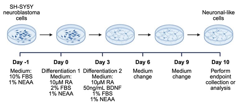
Figure 1. Overview of SH-SY5Y differentiation protocol using RA and BDNF. Image adapted from Targett et al3.
Preparation of Differentiation Reagents
B-27: Aliquot 50X B-27 into single-use aliquots (e.g. 21µL) and store in -20°C.
BDNF: Resuspend 10µg of HumanKine® BDNF protein (HZ-1335) in 1mL neurobasal medium with 1X B-27 supplement for stock concentration of 10µg/mL BDNF.
-
For example, if you need 50X B-27 to be diluted to 1X B-27 in 1mL neurobasal medium, add 20µL of 50X B-27 to 980µL neurobasal medium and mix thoroughly.
-
Aliquot the resuspended BDNF in media into single-use aliquots (e.g. 250µL) and store in -20°C.
RA: Resuspend 50mg in 33.3mL of 95% ethanol for a final concentration of 5mM RA. Note: RA is sensitive to light, temperature, and air!! Perform the following steps in the dark in a cell culture hood:
-
Wrap aluminum foil around a 50mL tube containing 33.3mL of 95% ethanol. Place the tube on ice.
-
Spin down the tube containing 50mg RA and add 1mL of ice cold 95% ethanol and mix thoroughly
-
Add the resuspended 1mL of RA to remaining 33.3mL 95% ethanol in the 50mL tube.
-
Vortex and/or pipette with a serological pipette to mix.
-
Make single-use aliquots (e.g. 51µL or 100µL), ideally in light sensitive tubes, and store at -20°C.
-
Longer term aliquots should be stored at -80°C.
Use the following recipes to prepare complete growth and differentiation medium:
|
Complete Growth Medium |
Quantity for 50 mL |
|
1:1 ratio of EMEM to F-12K |
22mL EMEM and 22mL F-12K |
|
10% FBS |
5mL FBS |
|
1% NEAA |
500µL 100X NEAA stock |
|
1% pen/strep |
500µL 100X pen/strep stock |
Table 1. EMEM/F-12K Medium.
|
Differentiation 1 Medium |
Quantity for 50 mL |
|
1:1 ratio of EMEM to F-12K |
24mL EMEM and 24mL F-12K |
|
2% FBS |
1mL FBS |
|
1% NEAA |
500µL 100X NEAA stock |
|
1% pen/strep |
500µL 100X pen/strep stock |
|
Add RA immediately before use to a final concentration of 10µM. Protect from light. |
|
Table 2. Differentiation 1 Medium.
|
Differentiation 2 Medium |
Quantity for 50 mL |
|
1:1 ratio of EMEM to F-12K |
24.125mL EMEM and 24.125mL F-12K |
|
1% FBS |
500µL FBS |
|
1% NEAA |
500µL 100X NEAA stock |
|
1% pen/strep |
500µL 100X pen/strep stock |
|
50ng/mL BDNF |
250µL 10µg/mL BDNF |
|
Add RA immediately before use to a final concentration of 10µM. Protect from light. |
|
Table 3. Differentiation 2 Medium.
Preparation of Cells for Differentiation
-
Thaw SH-SY5Y cells by incubating at 37°C. Add thawed cells to 4mLs of warmed Complete Growth Medium and mix by pipetting. Centrifuge cells for 5-10 minutes at 300 × g. Remove the supernatant and resuspend 450,000-500,000 cells in 3mL of Complete Growth Medium to plate in a 6-well plate.
Tip: If more cells are needed, seed approximately 2-3 million cells in 10 mL of Complete Growth Medium in a 10 cm tissue-culture dish. This corresponds to a seeding density of approximately 5 × 10^4 cells/cm² which has been optimized previously7 . Under these conditions, the cells should reach 70–90% confluency within approximately one week, although growth rate may vary with passage number and culture conditions. Replace the medium every 3–4 days until the cells reach 70–80% confluency by washing cells with warm DPBS and placing new media on the side of the dish.
-
After 3-4 days, check the confluency of cells. If cells are not 70-80% confluent, perform a medium change by removing the medium and adding fresh medium. Repeat this step every 3-4 days until cells are 70-80% confluent. If cells are 70-80% confluent, proceed to Step 1 of Differentiation Protocol.
Differentiation Protocol
-
Day -1: Split and seed cells.
Split cells by first washing wells with warmed DPBS. Pipette DPBS on the side of the well and tilt gently, then remove. Add 1mL of trypsin (1X) to detach the cells, incubate the cells for 3-5 minutes, and count cells using counting method of choice. Cells should be plated at roughly 10,000 cells/cm². For a well in a 6-well plate, the surface area is 9.6cm, so seed anywhere between 80,000-100,000 cells per well. Calculate the amount needed for seeding and add to 3mL of Complete Growth Medium in a new well within the 6-well plate.
Tip: If you plan to collect cells at various time points throughout the differentiation protocol, plate at the concentration which should be 80-90% confluent by the timepoint you need.
-
Day 0: Change Complete Growth Medium to Differentiation 1 Medium.
Before starting, calculate the amount of RA needed to be added to Differentiation 1 Medium before placing on cells. Use C1V1=C2V2 to calculate and make sure concentration/volume units are the same.
-
For example, if you need to make 5mL of Differentiation 1 medium:
Concentration 1 = 5mM = 5,000µM
Concentration 2= 10µM
Volume 1 = unknown
Volume 2 = 5mL
-
(5,000µM)*(V1) = (10µM)*(5mL)
(5,000)*(V1) = 50
V1 = 0.01 mL = 10µL
-
Add 10µL of 5mM stock RA to 4,990µL of Differentiation 1 medium in the dark and mix thoroughly. Keep RA on ice!
Perform the following steps in the dark: Remove media from well, wash cells by adding warmed DPBS, and add 3mL of Differentiation 1 medium containing RA to cells by pipetting on the side of the well.
-
Day 3: Change media to Differentiation 2 medium.
Before starting, calculate the amount of RA needed to be added to medium before placing on cells. Use the same steps used on Day 1 of the protocol for calculations.
Perform the following steps in the dark: Remove media from well, wash cells by adding warmed DPBS, and add 3mL of Differentiation 2 medium containing RA to cells by pipetting on the side of the well.
-
Day 6: Add fresh Differentiation 2 medium.
Before starting, calculate the amount of RA needed to be added to medium before placing on cells. Use the same steps used on Day 1 of the protocol for calculations.
Perform the following steps in the dark: Remove media from well, wash cells by adding warmed DPBS, and add 3mL of Differentiation 2 medium containing RA to cells by pipetting on the side of the well.
-
Day 9: Add fresh Differentiation 2 medium.
Before starting, calculate the amount of RA needed to be added to medium before placing on cells. Use the same steps used on Day 1 of the protocol for calculations.
Perform the following steps in the dark: Remove media from well, wash cells by adding warmed DPBS, and add 3mL of Differentiation 2 medium containing RA to cells by pipetting on the side of the well.
-
Day 10: Perform endpoint collection or analysis.
At Day 10, the differentiated cells can be processed according to the downstream application. Cells can be analyzed immediately in the culture plate, fixed for immunocytochemistry or imaging⁸, lysed for protein or nucleic acid analysis, or harvested and pelleted for later analysis. If samples will not be processed immediately, use storage conditions appropriate for the planned assay.
A frozen cell pellet should be used for downstream molecular analysis and should not be returned to culture. If live differentiated cells are needed after Day 10, maintain a separate culture under appropriate differentiation conditions or begin a new differentiation experiment using cryopreserved, undifferentiated SH-SY5Y cells.
To pellet cells for downstream protein analysis, cool the centrifuge to 4°C before beginning. Scrape cells with a cell scraper in the well. Remove medium and place in a 5mL or 15mL conical tube and spin for 5-10 min at 300-400 × g (at 4°C). After centrifugation, wash cells with 3mL of cold DPBS by performing another 5 min spin at 400g. Remove the supernatant and store the cell pellet at -80°C.
Phenotypic Changes Across Differentiation
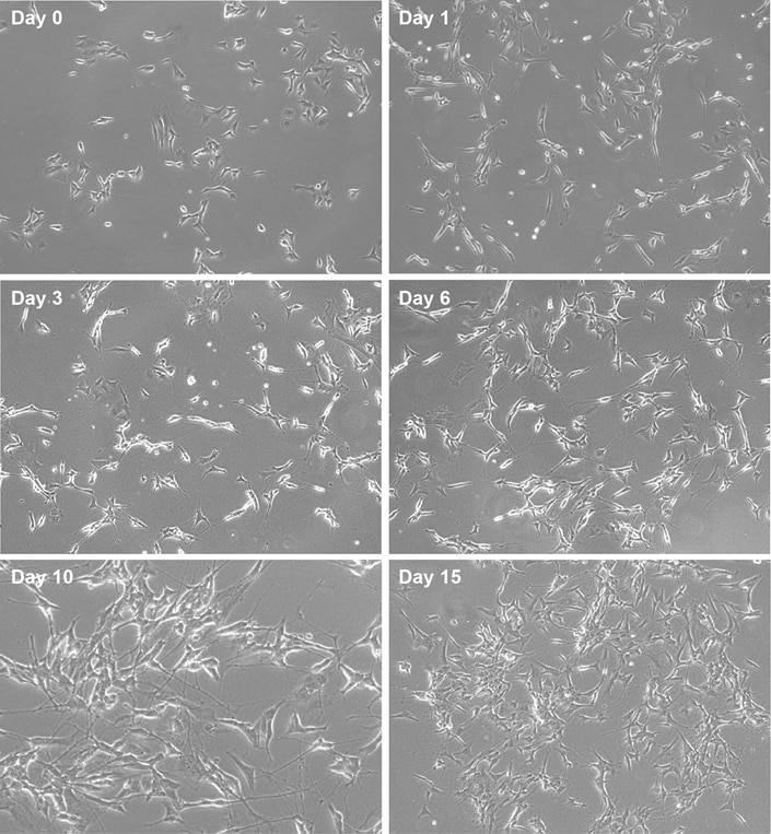
Figure 2. Phenotypic changes in SH-SY5Y cells during neuronal differentiation. Phase-contrast images of SH-SY5Y cells collected throughout the differentiation protocol. Day 0 shows undifferentiated cells before treatment. Day 1 shows early changes in cell morphology and neurite extension. Day 3 shows increased cell elongation and early neurite outgrowth before the medium change. Day 6 shows greater neurite outgrowth, increased cellular connectivity, and the formation of neuron-like networks before the medium change. By Day 10, the cells display extensive neurite outgrowth and branching, forming an interconnected neuron-like network; these phenotypes continue through Day 15.
Confirmation of Differentiation Markers
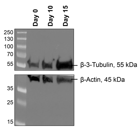
Figure 3. Confirmation of neuronal differentiation by βIII-tubulin (TUBB3) expression. Western blot showing βIII-tubulin (TUBB3, ~55 kDa; 1:25,000) and β-actin (45 kDa; 1:100,000) protein expression in SH-SY5Y cells collected at Day 0, Day 10, and Day 15 during the differentiation protocol. β-Actin was used as a loading control to confirm equal protein loading across samples. βIII-tubulin is a neuron-specific microtubule protein that is highly expressed in mature and differentiating neurons, where it contributes to microtubule assembly, neurite outgrowth, and maintenance of neuronal morphology9. Therefore, increased βIII-tubulin expression over the course of differentiation confirms successful acquisition of a mature neuronal phenotype in SH-SY5Y cells. (Note: Bands are cut closely on the right due to using mutant SH-SY5Y cell lines next to wild-type SH-SY5Y, which are present in lanes 1-3).
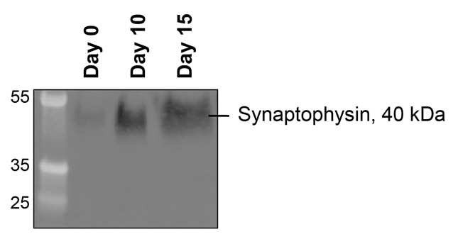
Figure 4. Confirmation of neuronal differentiation by synaptophysin (SYN) expression. Western blot showing synaptophysin (~40 kDa; 1:50,000) protein expression in SH-SY5Y cells collected at Day 0, Day 10, and Day 15 of a neuronal differentiation protocol. β-Actin, shown in Figure 3, was used as the loading control for these samples. Synaptophysin is a synaptic vesicle membrane protein and a marker of mature neurons that increases as neurons develop synaptic vesicles and acquire the machinery required for synaptic transmission10. As a result, increased synaptophysin expression during differentiation is consistent with successful maturation of SH-SY5Y cells toward a neuronal phenotype. (Note: Bands are cut closely on the right due to using mutant SH-SY5Y cell lines next to wild-type SH-SY5Y, which are present in lanes 1-3).
|
Proteintech Reagents Used for Differentiation and Characterization |
Catalog Number |
|
HumanKine® Recombinant Human BDNF Protein |
|
|
TUBB3-specific/TUJ1 Monoclonal Antibody |
|
|
Synaptophysin Monoclonal Antibody |
|
|
Multi-rAb® HRP-Goat Anti-Mouse Recombinant Secondary Antibody (H+L) |
Table 4. Proteintech reagents used for SH-SY5Y neuronal differentiation and Western blot analysis.
FAQs
Can differentiated cells be maintained beyond Day 10?
Yes, provided that the cells remain healthy and receive suitable medium and neurotrophic support. Shipley et al. reported that differentiated SH-SY5Y cultures could be maintained for up to two weeks after terminal differentiation and used for subsequent experiments5. However, culture duration can affect cell survival, morphology, and protein expression. Experiments extending beyond Day 10 should therefore include consistent medium changes, matched time-point controls, and confirmation that the differentiated phenotype is maintained.
Do SH-SY5Y cells have to be pelleted at Day 10?
No. Day 10 marks the endpoint of the differentiation protocol, but pelleting is only necessary for experiments that require a harvested cell pellet. Depending on the downstream application, the cells can be analyzed while still attached to the plate, treated with an experimental compound, fixed for imaging, lysed for protein or nucleic acid analysis, or harvested for later processing. Targett et al. lysed cells at Day 10 for Western blot analysis, demonstrating that pelleting is not a required step3.
Can the cells be fixed for staining instead of lysed or pelleted?
Yes. Differentiated SH-SY5Y cells can be fixed directly in the culture plate or on coverslips for immunocytochemistry. Fixing the cells while they remain attached is generally preferred for imaging because it preserves neurite outgrowth, cellular branching, and neuron-like networks. Riegerová et al. fixed RA/BDNF-differentiated SH-SY5Y cells with a paraformaldehyde-based fixation solution before immunofluorescence staining8. The fixation and permeabilization conditions should be optimized for the target antigen and antibody.
Can the cells be used immediately after differentiation?
Yes. The cells do not need to be stored before use. Experimental treatments, live-cell imaging, viability assays, functional measurements, fixation, or cell lysis can be performed at the end of differentiation. If the experiment requires treatment of differentiated cells, it is often best to begin the treatment while the cells remain attached to the original culture plate, since detachment can damage the neurites and disrupt the established network.
Can a frozen Day-10 cell pellet be thawed and returned to culture?
A cell pellet that has simply been stored at -80°C should not be expected to contain viable cells and should only be used for downstream molecular analysis. Viable cryopreservation requires the cells to be suspended in an appropriate cryopreservation medium containing a cryoprotectant, frozen under controlled conditions, and stored below -130°C or in liquid nitrogen. The ATCC protocol for SH-SY5Y cells recommends complete growth medium containing 5% DMSO and storage in the vapor phase of liquid nitrogen11.
Although undifferentiated SH-SY5Y cells can be cryopreserved using this approach, freezing and recovering terminally differentiated cells is not established by the differentiation protocol described here. RA/BDNF-treated cells are post-mitotic and neurotrophic factor-dependent, and their survival and phenotype after detachment and cryopreservation cannot be assumed12. For reproducible experiments, cryopreserve low-passage undifferentiated cells and perform a new differentiation experiment after thawing.
Can the same culture be collected at multiple time points?
No. Prepare separate wells or plates for each destructive collection time point. Once cells are fixed, lysed, scraped, or pelleted, that culture cannot be used at a later time point. For example, if samples will be collected on Days 0, 5, and 10, plate at least one well for each time point, along with any necessary controls and replicates. Seeding density should also be adjusted and optimized for each time point collection. Targett et al. prepared cultures for collection at several points during differentiation, allowing comparisons of changes in morphology and protein expression across the time course3.
Why should retinoic acid be protected from light?
All-trans retinoic acid is sensitive to light, oxygen, and heat. Light exposure can cause isomerization or degradation, potentially changing the effective concentration added to the cells13. Retinoic acid stocks should therefore be prepared under reduced-light conditions, divided into single-use aliquots, protected from light, and added to the differentiation medium immediately before use14.
Why are retinoic acid and BDNF used sequentially?
Retinoic acid initiates differentiation by reducing proliferation and promoting cell-cycle exit and neurite formation. Subsequent BDNF treatment supports survival, neurite extension, and the development of a more mature neuron-like phenotype. Sequential RA and BDNF treatment produce greater neuronal differentiation than either stage alone and leads to cells with extensive neurites and increased expression of neuronal and synaptic proteins3,12.
How can successful differentiation be confirmed?
Differentiation should be assessed using both cell morphology and neuronal marker expression. Successful cultures show reduced proliferation, elongated cell bodies, extensive neurite outgrowth, increased branching, and formation of interconnected networks. These morphological changes can be supported by increased expression of neuronal and synaptic markers, including βIII-tubulin, MAP2, GAP-43, NeuN, and synaptophysin. Targett et al. demonstrated time-dependent neurite formation and changes in neuronal and synaptic markers during the 10-day RA/BDNF protocol3.
References:
-
Pezzini F, Bettinetti L, Di Leva F, et al. Transcriptomic Profiling Discloses Molecular and Cellular Events Related to Neuronal Differentiation in SH-SY5Y Neuroblastoma Cells. Cell Mol Neurobiol. 2017;37(4):665-682.
-
Kovalevich J, Santerre M, Langford D. Considerations for the Use of SH-SY5Y Neuroblastoma Cells in Neurobiology. Methods Mol Biol. 2021;2311:9-23.
-
Targett IL, Crompton LA, Conway ME, Craig TJ. Differentiation of SH-SY5Y neuroblastoma cells using retinoic acid and BDNF: a model for neuronal and synaptic differentiation in neurodegeneration. In Vitro Cell Dev Biol Anim. 2024;60(9):1058-1067.
-
Goldie BJ, Barnett MM, Cairns MJ. BDNF and the maturation of posttranscriptional regulatory networks in human SH-SY5Y neuroblast differentiation. Front Cell Neurosci. 2014;8:325.
-
Shipley MM, Mangold CA, Szpara ML. Differentiation of the SH-SY5Y Human Neuroblastoma Cell Line. J Vis Exp. 2016(108):53193.
-
Martin ER, Gandawijaya J, Oguro-Ando A. A novel method for generating glutamatergic SH-SY5Y neuron-like cells utilizing B-27 supplement. Front Pharmacol. 2022;13:943627.
-
Feles S, Overath C, Reichardt S, et al. Streamlining Culture Conditions for the Neuroblastoma Cell Line SH-SY5Y: A Prerequisite for Functional Studies. Methods Protoc. 2022;5(4).
-
Riegerova P, Brejcha J, Bezdekova D, et al. Expression and Localization of AbetaPP in SH-SY5Y Cells Depends on Differentiation State. J Alzheimers Dis. 2021;82(2):485-491.
-
Mariani M, Karki R, Spennato M, et al. Class III beta-tubulin in normal and cancer tissues. Gene. 2015;563(2):109-114.
-
Yuan X, Li W, Yan Q, Ou Y, Long Q, Zhang P. Biomarkers of mature neuronal differentiation and related diseases. Future Sci OA. 2024;10(1):2410146.
-
ATCC. SH-SY5Y, CRL-2266: culture and cryopreservation information; 2026.
-
Encinas M, Iglesias M, Liu Y, et al. Sequential treatment of SH-SY5Y cells with retinoic acid and brain-derived neurotrophic factor gives rise to fully differentiated, neurotrophic factor-dependent, human neuron-like cells. J Neurochem. 2000;75(3):991-1003.
-
Sharow KA, Temkin B, Asson-Batres MA. Retinoic acid stability in stem cell cultures. Int J Dev Biol. 2012;56(4):273-278.
-
Sigma Aldrich. trans-Retinoic Acid; 2026.

Related Content
Midbrain organoid generation: Practical tips from the bench | Proteintech Group
A Guide to Markers for Oligodendrocyte Identification | Proteintech Group
Markers for Astrocyte Identification | Proteintech Group
Identifying Cortical Cell Types Through Immunofluorescence and Electrophysiology | Proteintech Group
Support
Newsletter Signup
Stay up-to-date with our latest news and events. New to Proteintech? Get 10% off your first order when you sign up.
Able™