
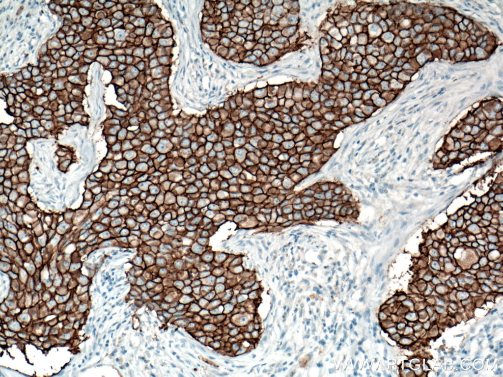
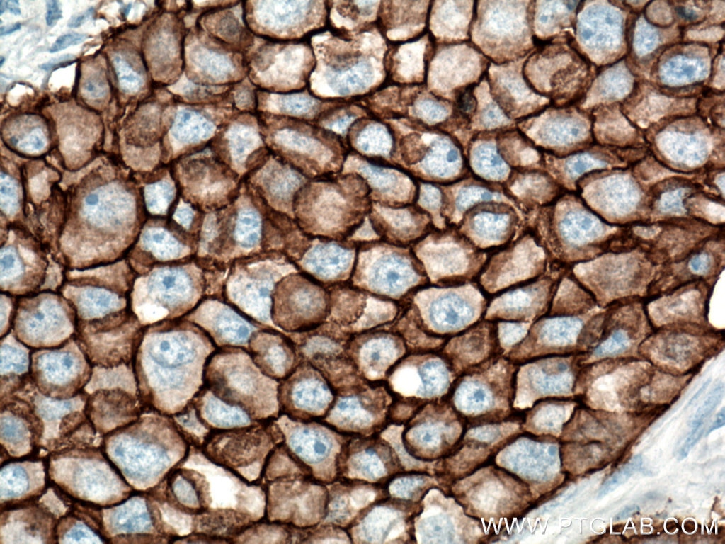
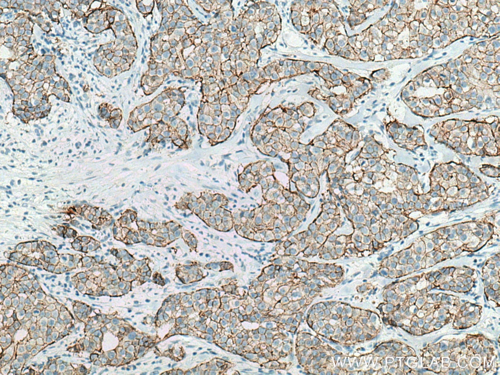
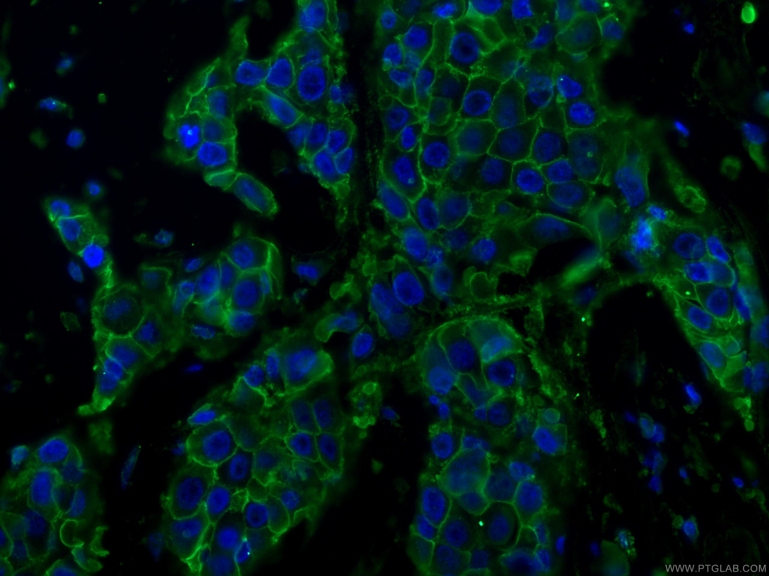

Tested Applications
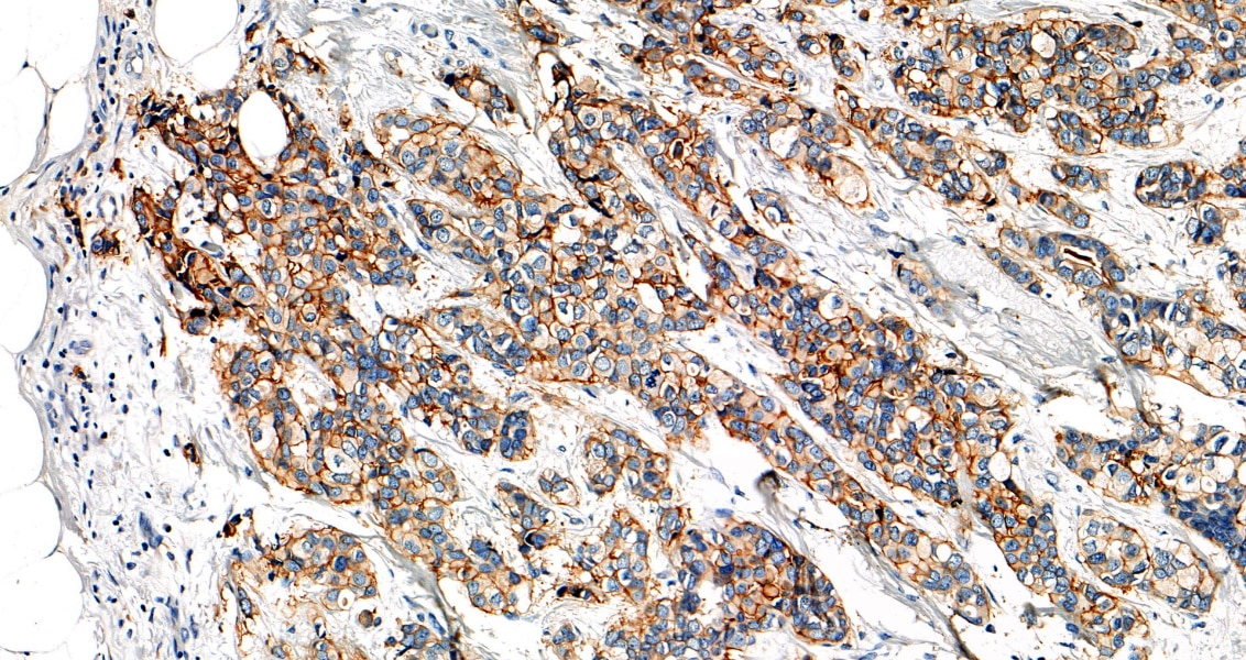
| Positive IHC detected in | human breast cancer tissue Note: suggested antigen retrieval with TE buffer pH 9.0; (*) Alternatively, antigen retrieval may be performed with citrate buffer pH 6.0 |
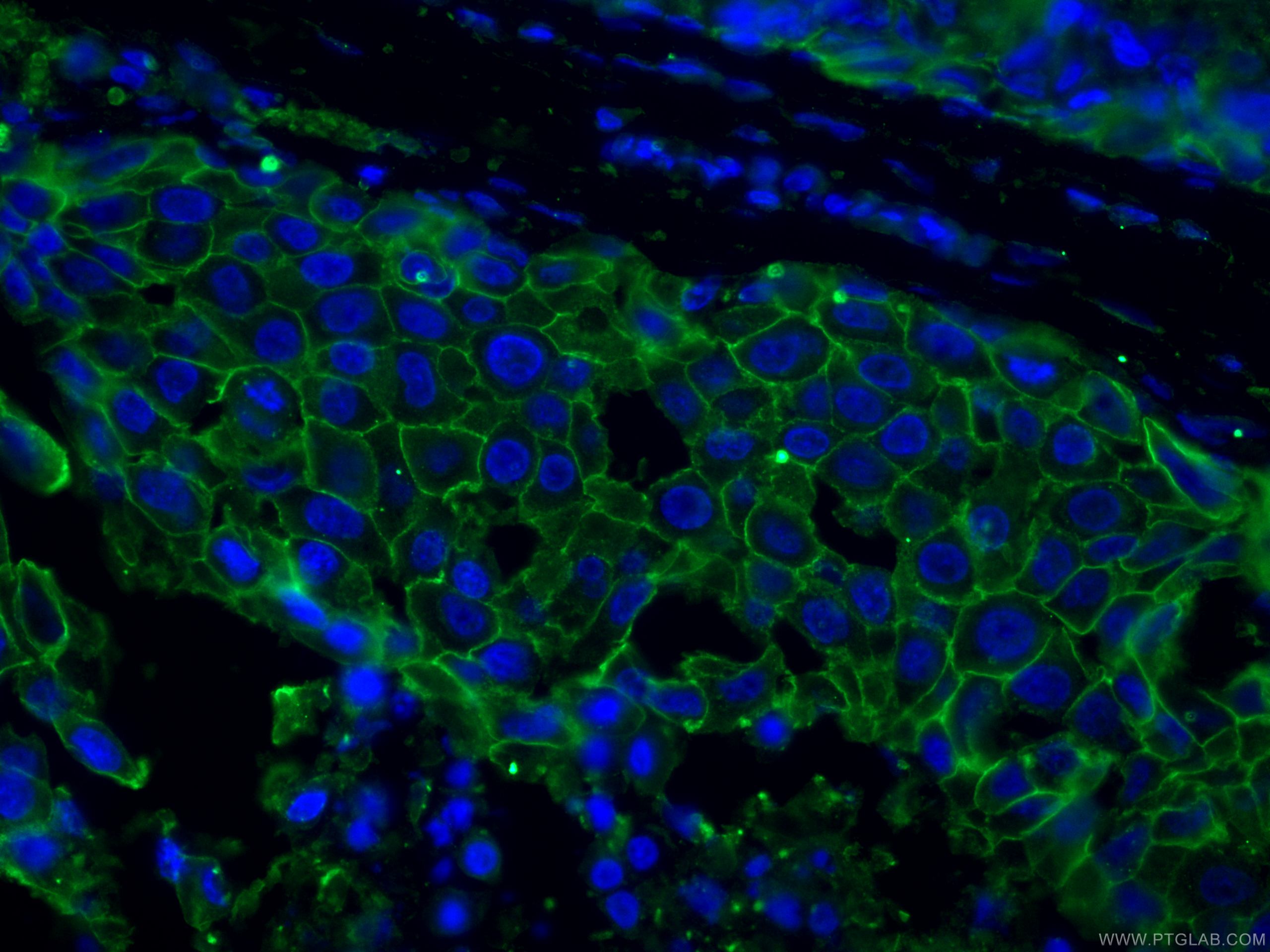
| Positive IF-P detected in | human breast cancer tissue |
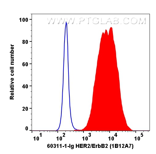
| Positive FC detected in | SK-BR-3 cells |
Recommended dilution
| Application | Dilution |
|---|---|
| Immunohistochemistry (IHC) | IHC : 1:800-1:3200 |
| Immunofluorescence (IF)-P | IF-P : 1:50-1:500 |
| Flow Cytometry (FC) | FC : 0.20 ug per 10^6 cells in a 100 µl suspension |
| It is recommended that this reagent should be titrated in each testing system to obtain optimal results. | |
| Sample-dependent, Check data in validation data gallery. | |
Product Information
60311-1-Ig targets HER2/ErbB2 in WB, IHC, IF-P, ELISA applications and shows reactivity with human samples.
| Tested Reactivity | human |
| Cited Reactivity | human, mouse |
| Host / Isotype | Mouse / IgG2b |
| Class | Monoclonal |
| Type | Antibody |
| Immunogen |
CatNo: Ag16463 Product name: Recombinant human ERBB2 protein Source: e coli.-derived, PGEX-4T Tag: GST Domain: 23-372 aa of BC156755 Sequence: TQVCTGTDMKLRLPASPETHLDMLRHLYQGCQVVQGNLELTYLPTNASLSFLQDIQEVQGYVLIAHNQVRQVPLQRLRIVRGTQLFEDNYALAVLDNGDPLNNTTPVTGASPGGLRELQLRSLTEILKGGVLIQRNPQLCYQDTILWKDIFHKNNQLALTLIDTNRSRACHPCSPMCKGSRCWGESSEDCQSLTRTVCAGGCARCKGPLPTDCCHEQCAAGCTGPKHSDCLACLHFNHSGICELHCPALVTYNTDTFESMPNPEGRYTFGASCVTACPYNYLSTDVGSCTLVCPLHNQEVTAEDGTQRCEKCSKPCARVCYGLGMEHLREVRAVTSANIQEFAGCKKIFG Predict reactive species |
| Full Name | v-erb-b2 erythroblastic leukemia viral oncogene homolog 2, neuro/glioblastoma derived oncogene homolog (avian) |
| Calculated Molecular Weight | 1255 aa, 138 kDa |
| GenBank Accession Number | BC156755 |
| Gene Symbol | HER2/ErbB2 |
| Gene ID (NCBI) | 2064 |
| RRID | AB_2881423 |
| Conjugate | Unconjugated |
| Form | Liquid |
| Purification Method | Protein A purification |
| UNIPROT ID | P04626 |
| Storage Buffer | PBS with 0.02% sodium azide and 50% glycerol, pH 7.3. |
| Storage Conditions | Store at -20°C. Stable for one year after shipment. Aliquoting is unnecessary for -20oC storage. 20ul sizes contain 0.1% BSA. |
Background Information
HER2, ErbB2, and Neu is a 185-kDa transmembrane glycoprotein member of the epidermal growth factor (EGF) receptor family of receptor tyrosine kinases. It has no ligand-binding domain and therefore cannot bind growth factors. However, it does bind tightly to other ligand-bound EGF receptor family members to form a heterodimer, stabilizing ligand binding and enhancing kinase-mediated activation of downstream signalling pathways, such as those involving mitogen-activated protein kinase and phosphatidylinositol-3 kinase. Amplification and/or overexpression of HER2 have been reported in numerous cancers, including breast and ovarian tumors. HER2 is a therapeutic target for the treatment of breast cancer and other carcinomas.
Protocols
| Product Specific Protocols | |
|---|---|
| FC protocol for HER2/ErbB2 antibody 60311-1-Ig | Download protocol |
| IF protocol for HER2/ErbB2 antibody 60311-1-Ig | Download protocol |
| IHC protocol for HER2/ErbB2 antibody 60311-1-Ig | Download protocol |
| Standard Protocols | |
|---|---|
| Click here to view our Standard Protocols |
Publications
What published studies show
The HER2/ErbB2 Monoclonal antibody (Cat# 60311-1-Ig) has 14 citations published in journals including Molecular Cancer, Advanced Materials, ACS Nano, Genome Medicine, Journal of Biological Chemistry, International Journal of Cancer, and Frontiers in Oncology. Research fields span breast cancer (trastuzumab resistance, organoid-based therapy), ovarian cancer (clear cell carcinoma, PET imaging), gastric cancer, urothelial carcinoma, photoimmunotherapy, SERS imaging, and CDK4 inhibitor sensitivity, with applications in IF, IHC, WB, and FC.
| Species | Application | Title |
|---|---|---|
Mol Cancer CMTM6 overexpression confers trastuzumab resistance in HER2-positive breast cancer | ||
Adv Mater Targeted SERS Imaging and Intraoperative Real-Time Elimination of Microscopic Tumors for Improved Breast-Conserving Surgery | ||
ACS Nano Photoinduced Actin Aggregation Involves Cell Death: A Mechanism of Cancer Cell Cytotoxicity after Near-Infrared Photoimmunotherapy | ||
Genome Med Comprehensive molecular characterization identifies therapeutic vulnerabilities in ovarian clear cell carcinoma. | ||
Int Immunopharmacol Development and In vitro antitumor evaluation of a novel anti-p16 antibody fragment-drug conjugate. | ||
Mol Med Rep HPK1 positive expression associated with longer overall survival in patients with estrogen receptor-positive invasive ductal carcinoma‑not otherwise specified. |
Reviews
What customers say
The single review for this product reports a 5-star rating from an immunohistochemistry application on human breast cancer tissue. The user performed staining on FFPE slides using an automated staining, covering, and scanning system, with a 1:5000 dilution and 30-minute incubation. Heat-mediated antigen retrieval was done with Tris-EDTA buffer (pH 9.0), followed by an HRP-conjugated one-step development system, and results were scanned under a 40x lens. Overall, the reviewer was satisfied with the staining outcome.
The reviews below have been submitted by verified Proteintech customers who received an incentive for providing their feedback.
Felletár (Verified Customer) (06-25-2025) | Immunostaining was performed using an automated staining, covering, and scanning system. FFPE slides were incubated for 30 minutes. HRP conjugated one step development system, scanning under 40x lens. Heat mediated antigen retrieval with Tris-EDTA buffer (pH 9.0).
 |







