Use Able AI chat for product recommendations

Microscopy Image Competition

Competition details

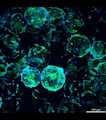
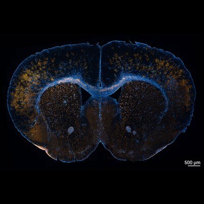
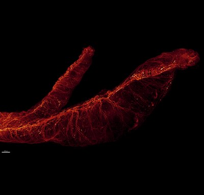
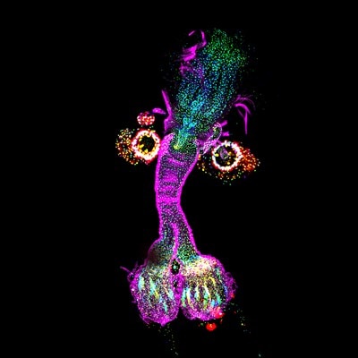
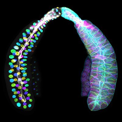
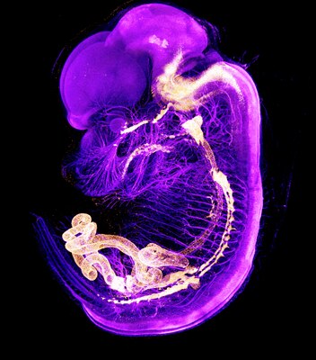
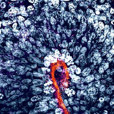
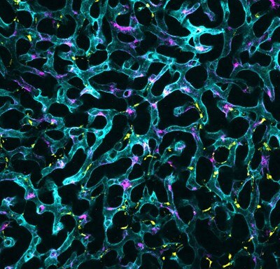
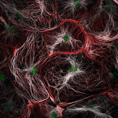
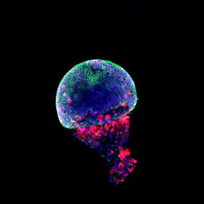
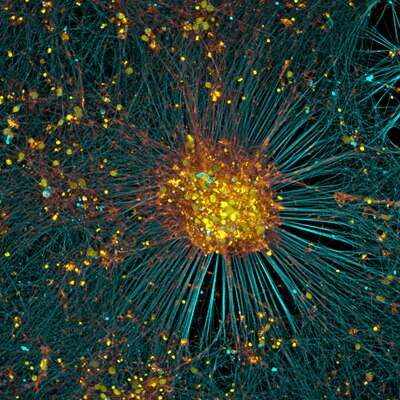
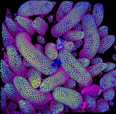
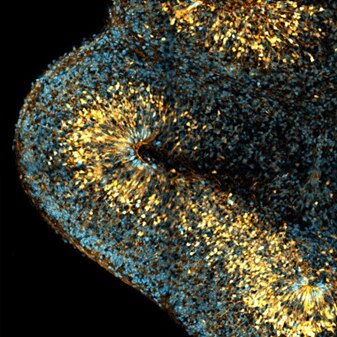
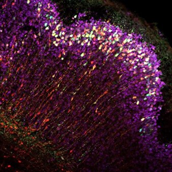
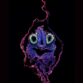
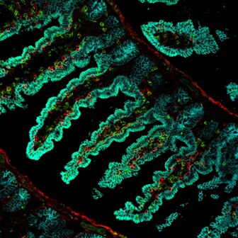
The Proteintech Microscopy Image Competition is back for 2026. Whether you're imaging cells, tissues, organoids or whole organisms, we want to celebrate the incredible images driving scientific discovery.

Submit your best immunofluorescence microscopy image for the chance to win:

  • $200 Amazon Gift Card
  • $1,000 Proteintech Product Grant
  • Professional framed print of your winning image
  • Global recognition across Proteintech’s website, email and social media

No Proteintech products in your image? No problem!

Entries do not have to feature Proteintech products. Two winners will be chosen via public vote: one winner with Proteintech products and one winner without.

Category 1 - Images featuring Proteintech products

Category 2 - Images featuring any products

Scroll down for the full competition rules and terms and conditions. Entries may also be showcased on the Proteintech website, email newsletter, social media channels and other marketing materials.

Submit your entry using the form on the right.


Judging Panel

Headshot of Dr Joe McKellar
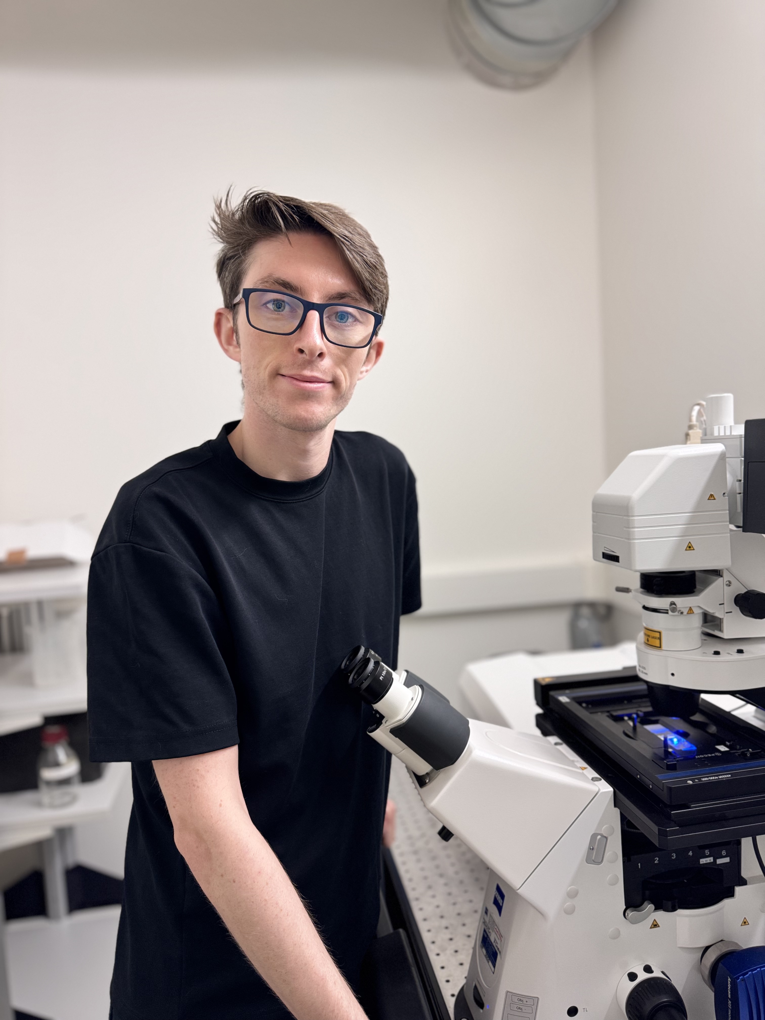
Dr Joe McKellar Guest judge

This year's competition will be judged by a panel of Proteintech scientists, joined by guest judge Dr. Joe McKellar, an internationally recognised microscopy specialist whose award-winning images have been recognised in competitions including Nikon Small World, Evident's Image of the Year Award and was the 2026 winner of CiteAb's Image of the Year Award.

Eight finalists, four images using Proteintech products and four images using any products. Finalists will be selected based on overall aesthetics, technical difficulty, research relevance, originality and suitability for the competition. The overall winner will then be determined by a public vote.

Finalist announcement: 19th October 2026

Entry guidelines

Please include a figure legend containing:

  • Antibody catalogue numbers (Proteintech and/or other suppliers).
  • Sample type, for example mouse brain or 4% PFA-fixed tissue.
  • A colour key for each fluorophore.
  • Imaging technique used, such as confocal, STED, STORM or lightsheet.
  • Objective magnification.
  • Scale bar (recommended).

Accepted image formats: JPEG or PNG (maximum 4 MB).

Voting process

On 20th October 2026, the entry form on this page will be replaced by a voting form. Only one vote per day, per person will be counted. The overall winner will be the entry that receives the most public votes.

  • Public voting period: 19th-25th October 2026
  • Winner announcement: 2nd November 2026

Frequently asked questions

Do I need to use Proteintech antibodies?

No. The competition is open to images generated using antibodies from any supplier.

Can I submit more than one image?

You may submit one image per category.

Can published images be entered?

Yes, provided the publication is Open Access and you have permission from all relevant authors and funders.

Who can enter?

Researchers, students and imaging specialists worldwide.

Key dates

Key dates for the Proteintech Microscopy Image Competition
Event Date
Competition opens 3 August 2026
Entry deadline 5 October 2026
Finalists announced 19 October 2026
Public voting 19–25 October 2026
Winners announced 2 November 2026

Submit your entry

Use the form below to upload your microscopy image and supporting information.

CoraLite fluorescent dye-conjugated antibodies

Proteintech's CoraLite antibodies are directly conjugated with fluorescent dyes: CoraLite488, CoraLite594 and CoraLite647. They offer bright and long-lasting fluorescence and are ideal for immunofluorescence studies requiring multiplex co-labeling without the need for secondary antibodies.

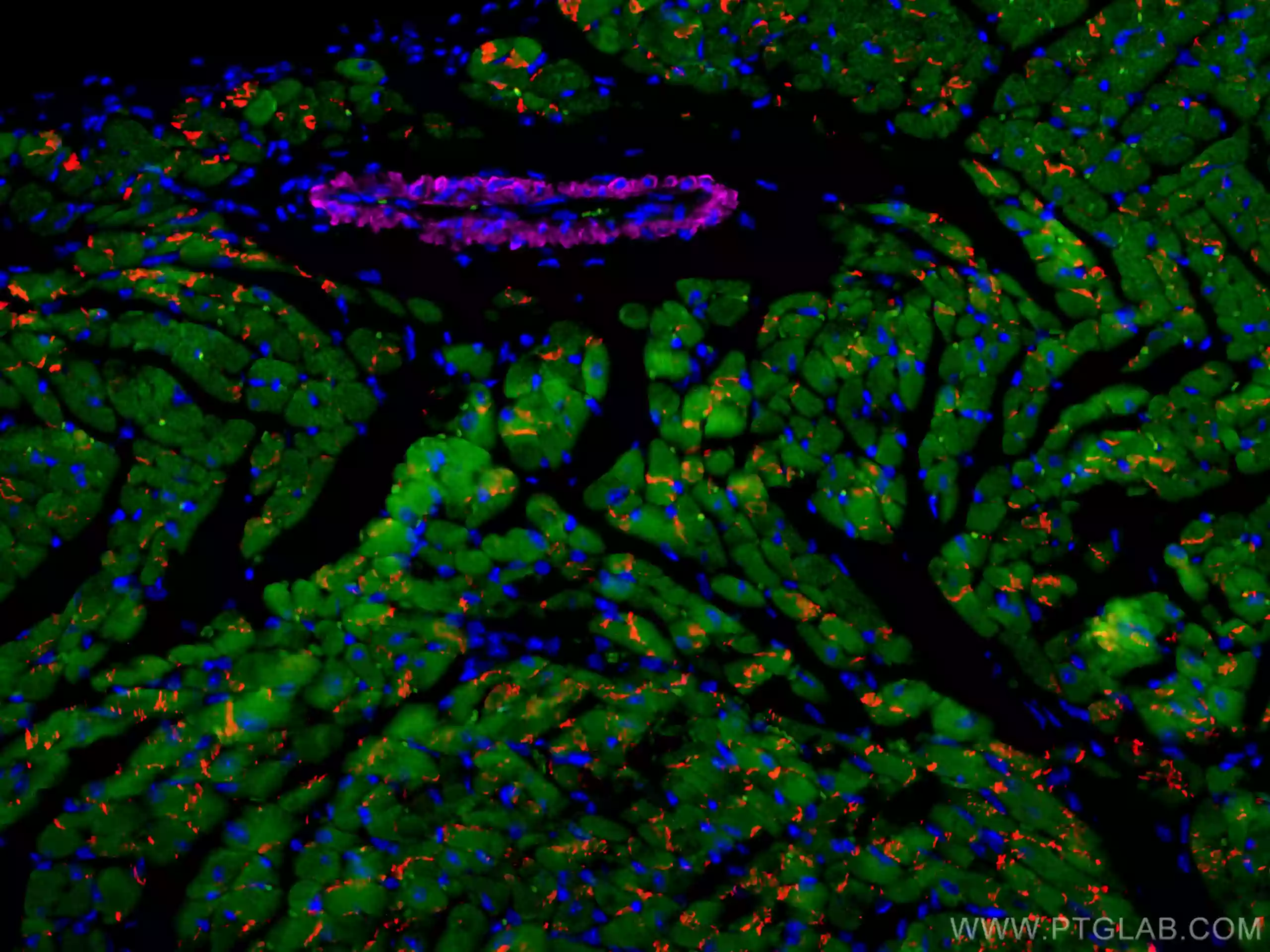
Multiplex immunofluorescence image of mouse heart showing Troponin I, N-cadherin and smooth muscle actin staining.

Antibodies: CL488-66376 (Troponin I), CL594-22018 (N cadherin), CL647-67735 (Smooth muscle actin).

Mouse heart. Expression of Troponin I (green) in cardiac muscle, N cadherin (red) in intercalated discs, and smooth muscle actin (pink) in smooth muscle cells of the cardiac vasculature.
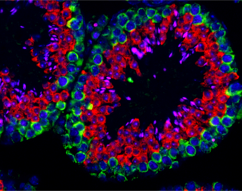
Multiplex immunofluorescence image of mouse testis showing DAZL, BOULE and TNP1 staining.

Antibodies: CL488-12633 (DAZL), CL594-13720 (BOULE), CL647-17178 (TNP1).

Mouse testis. Expression of DAZL (green) in spermatogonia cells, BOULE (red) in spermatocytes, and TNP1 (pink) in spermatids. This image shows differential protein expression during spermatogenesis.

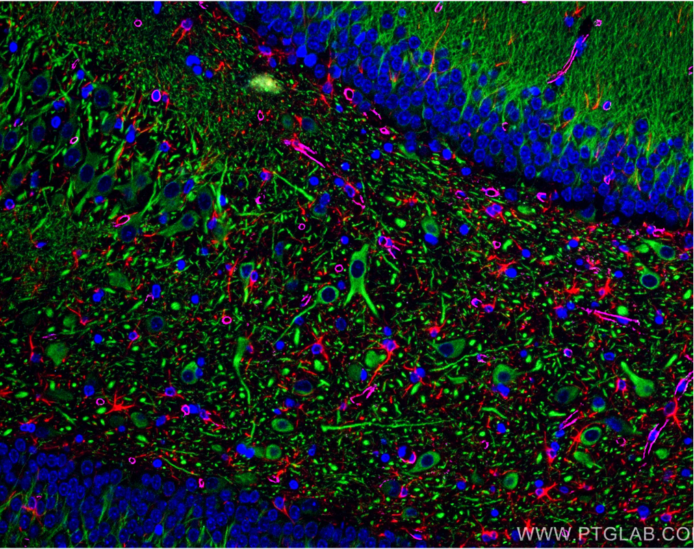
Multiplex immunofluorescence image of mouse brain showing MAP2, GFAP and AQP4 staining.

Antibodies: CL488-17490 (MAP2), CL594-16825 (GFAP), CL647-16473 (AQP4).

Mouse brain. Expression of MAP2 (green) in neurons, GFAP (red) in astrocytes, and AQP4 (pink) in astroglial endfeet.

ChromoTek Nano-secondaries

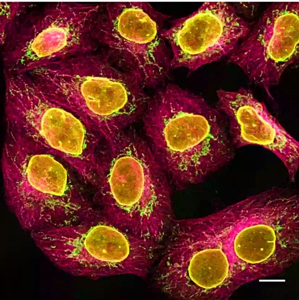
Nano-Secondaries are the next level of secondary antibodies. They enable cleaner images with higher resolution. They consist of VHHs, also named nanobodies, conjugated to Alexa Fluor dyes. Nano-Secondaries have high affinities and bind to primary antibodies in a subclass- and species-specific manner.

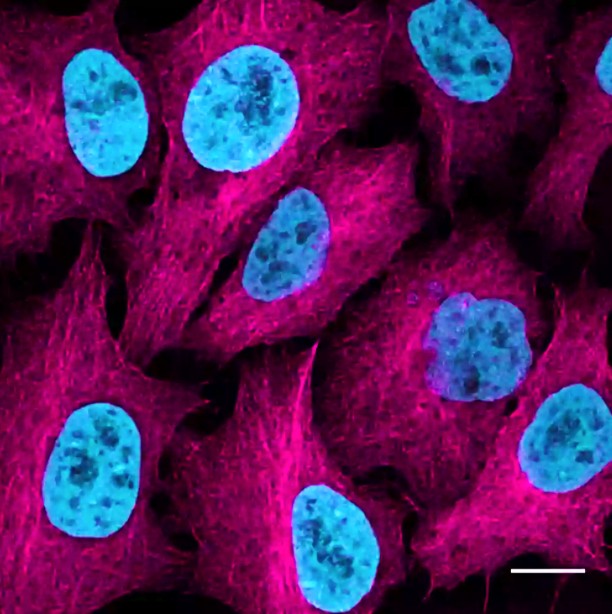
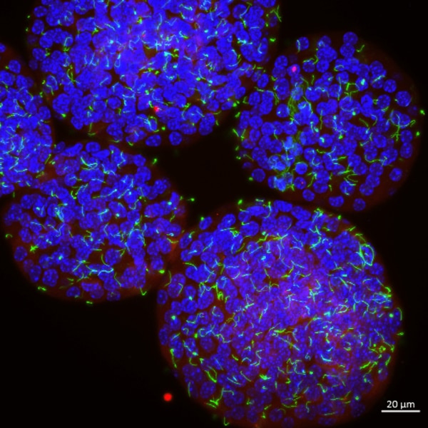
HeLa cells stained with alpaca anti-rabbit IgG VHH Alexa Fluor 647 and a nuclear marker.

HeLa cells stably expressing Tubulin-GFP at near-endogenous level were immunostained with rabbit anti-GFP PABG1 antibody and alpaca anti-rabbit IgG VHH Alexa Fluor 647 (magenta). Nuclei were detected with H2B-RFP and RFP-Booster Atto594 (cyan). Scale bar, 10 μm.

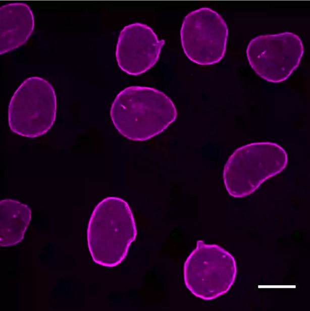
Specific immunofluorescence example demonstrating anti-mouse IgG2b Nano-Secondary performance.

The anti-mouse IgG2b Nano-Secondary is subclass-specific and does not cross-react with IgGs from other commonly used species, including rabbit, or with mouse IgG1 and IgG3 subclasses.

Terms and conditions

  • The competition is open from 3 August to 5 October 2026.
  • Entries must be submitted via the online entry form.
  • A maximum of one entry per person, per category is permitted.
  • Images do not need to feature Proteintech products. Separate awards will be given for images using Proteintech products and images using products from any supplier.
  • Images must be submitted as JPEG or PNG files and be no larger than 4 MB.
  • Entries must be the entrant's original work and must not infringe any third-party copyright.
  • Previously published images may be entered only if the publication is Open Access and permission has been obtained from all relevant authors and funders.
  • Eight finalists, four per category, will be selected by a panel of Proteintech scientists and microscopy experts. Two overall winners will then be chosen by public vote.
  • Public voting is limited to one vote per person, per day. Proteintech reserves the right to remove fraudulent or duplicate votes.
  • Each finalist will receive one CoraLite® antibody. Each winner will receive a $200 Amazon Gift Card, a $1,000 Proteintech Product Grant, and a framed print of their winning image.
  • By entering, participants grant Proteintech permission to use their submitted image, name, institution and image description for promotional purposes related to the competition.
  • Proteintech reserves the right to amend, suspend or cancel the competition and to disqualify entries that do not comply with these Terms and Conditions.

||
New chat

Able

正在加载,请稍候...