Western Blot
Proteintech offer solutions in helping you to achieve publication worthy results in a fast and reliable manner.
Fast Workflow Solutions for Western Blot New
Proteintech offers reagents and Kits that are specialized for a fast Western blot workflow.
Removes Blocking Step and cuts your incubation times to ~15 min
FlexAble HRP Antibody Labeling Kit
Enables 1-step detection by labeling primary antibody with HRP
SpeedAb WB Kit New
Combines both advantages in a Kit to save you time and money
Antibodies for Western Blot
Explore WB products for detection of precipitated proteins and its interaction partners (CoIP).
- Over 13,000 polyclonal and monoclonal primary antibodies
- Tag antibodies against protein- or peptide-tags
- Secondary antibodies
- Nano-Secondary® reagents (VHHs)
- FlexAble HRP for 1 step detection
Control Antibodies
Proteintech offers a wide range of control antibodies for Western Blot
- Provide a consistent reference to normalize target protein expression levels
- Ensure observed changes in target protein levels result from experimental conditions, not variations in sample loading or transfer efficiency
More Supporting Reagents
Explore all our supporting reagents around Western blotting
Achieve optimal detection of your protein of interest
Estimate protein sizes with prestained ladder
Find the buffer best suited for your assay
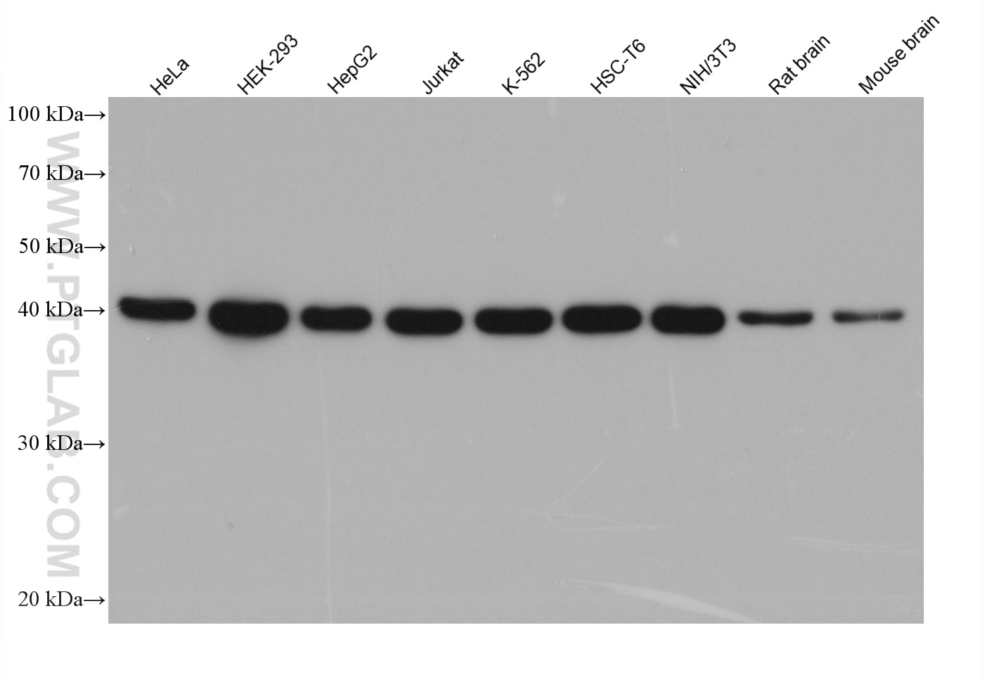
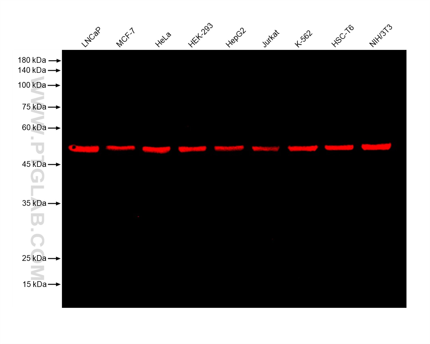
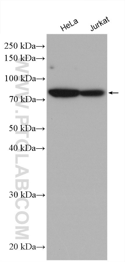
Various lysates were subjected to SDS-PAGE followed by Western blot with rabbit anti-TDP43 recombinant antibody (80001-1-RR) at dilution of 1:20,000. Multi-rAb™ HRP-Goat Anti-Rabbit Recombinant Secondary Antibody (H+L) (RGAR001) was used at 1:20000 for detection.
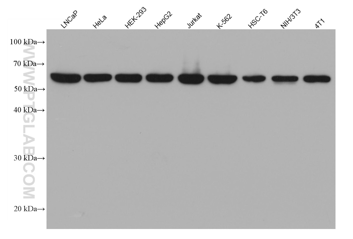
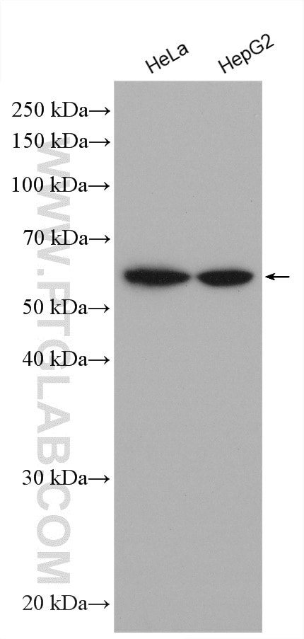
Various lysates were subjected to SDS-PAGE followed by Western blot with U2AF2 mouse monoclonal antibody (68166-1-Ig) at 1:20,000. Multi-rAb™ HRP-Goat Anti-Mouse Recombinant Secondary Antibody (H+L) RGAM001 were used at 1:20000 for detection.
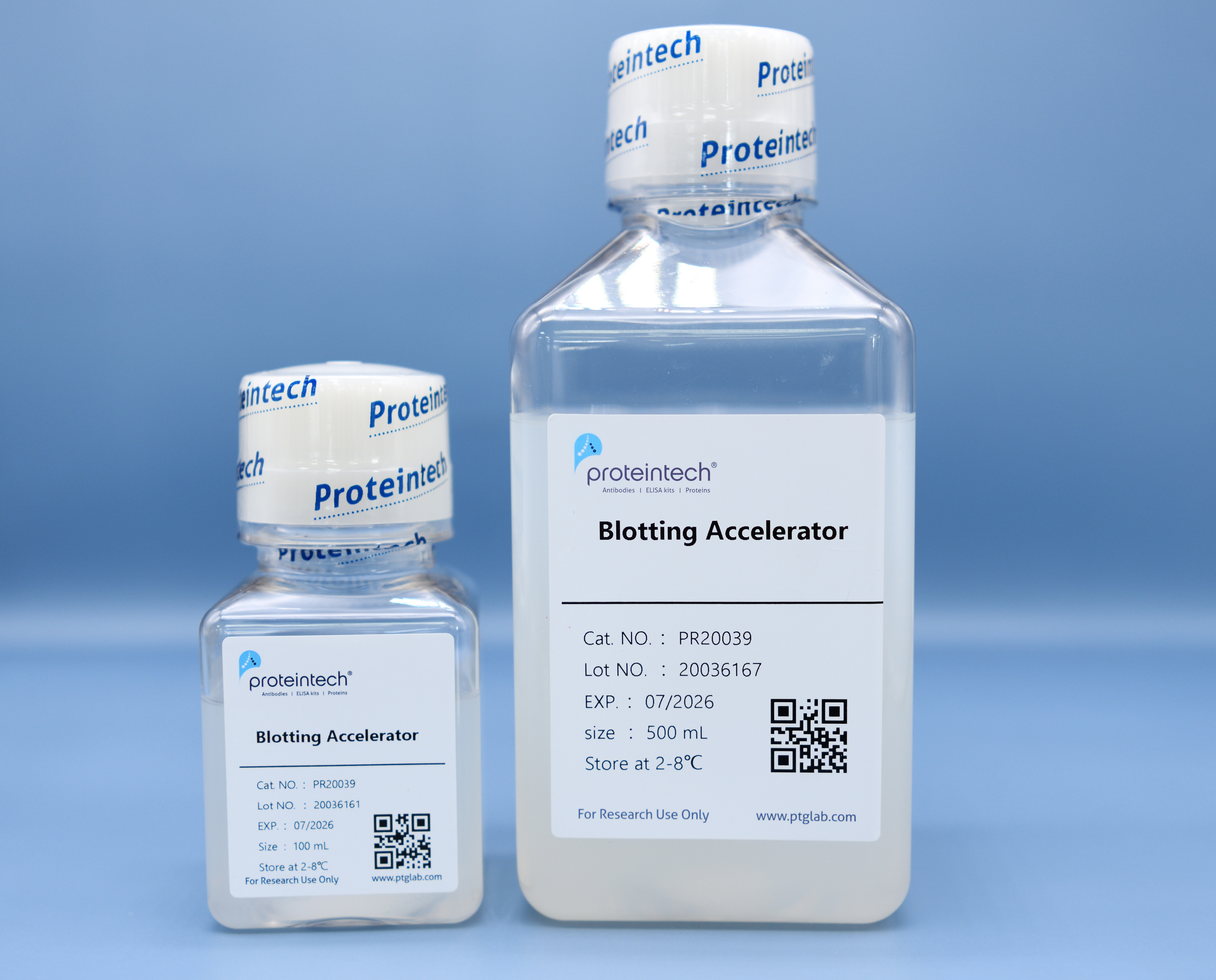
The product is available in two packaging specifications: 100mL and 500mL. Its appearance is in the form of a dilute colloid.
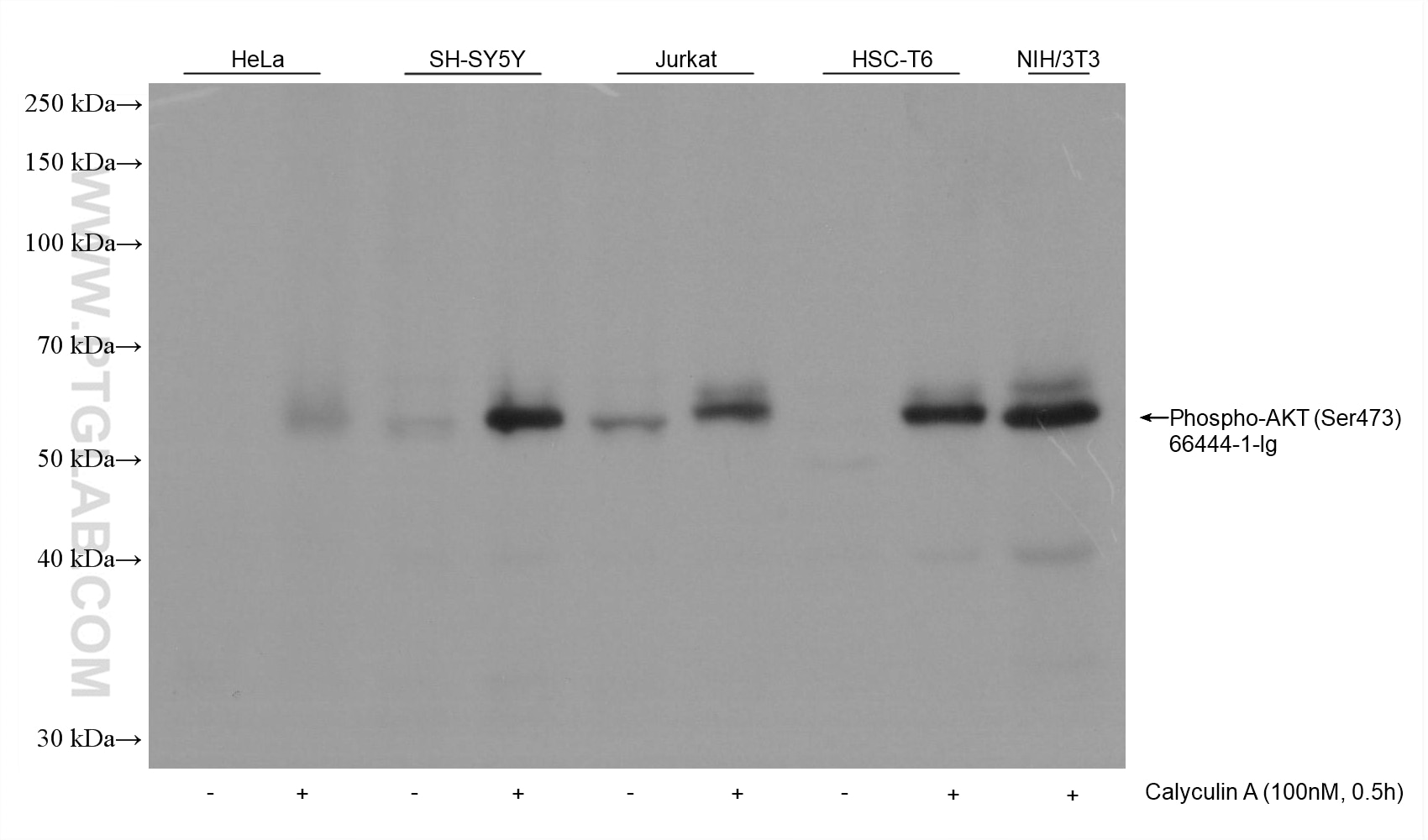
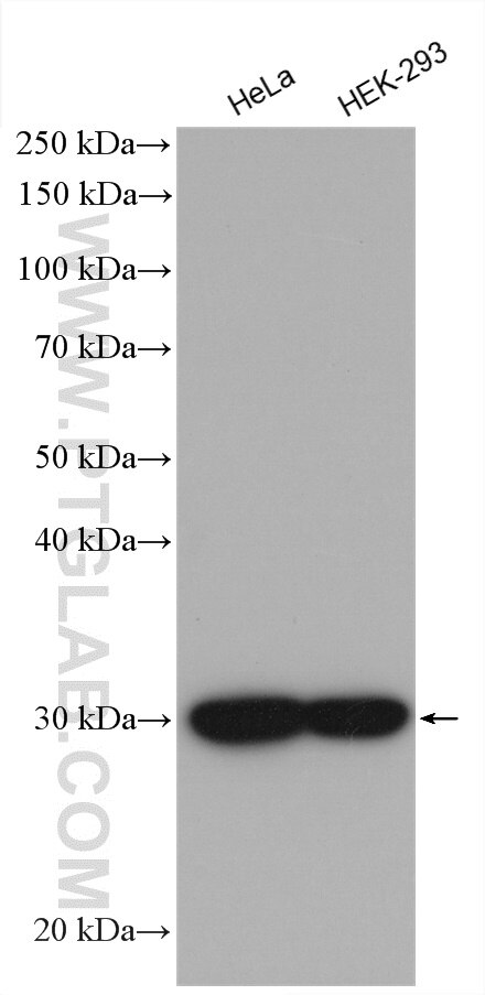
Various samples were loaded at 30 ug per well and subjected to Western blot, unblocked. Primary antibody 66444-1-Ig (Phospho-AKT (Ser473) Mouse Monoclonal Antibody) was diluted with PR20039 at 1:10,000 and incubated at room temperature for 15 minutes. The secondary antibody RGAM001 was diluted with PR20039 at 1:20,000 and incubated at room temperature for 10 minutes.
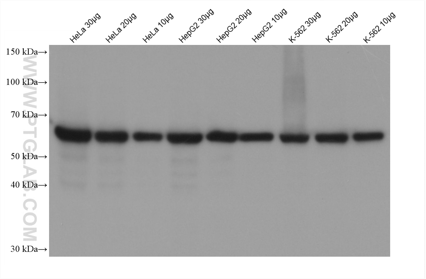
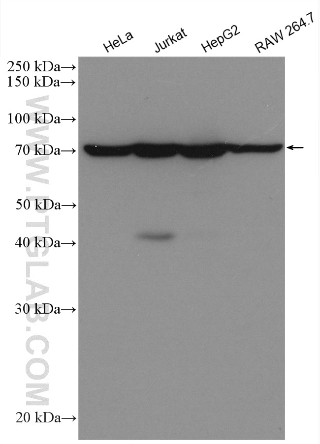
Various samples were loaded at different volumes per well and subjected to Western blot, unblocked. Primary antibody 15282-1-AP (HSPD1 Rabbit Polyclonal Antibody) was diluted with PR20039 at 1:50,000 and incubated at room temperature for 18 minutes. The secondary antibody RGAR001 was diluted with PR20039 at 1:20,000 and incubated at room temperature for 8 minutes.
Various samples were loaded at 30 ug per well and subjected to Western blot, unblocked. Primary antibody 80713-1-RR (Beta Tubulin Rabbit Recombinant Antibody) was diluted with PR20039 at 1:20,000 and incubated at room temperature for 15 minutes. The secondary antibody RGAR006 was diluted with PR20039 at 1:10,000 and incubated at room temperature for 10 minutes.
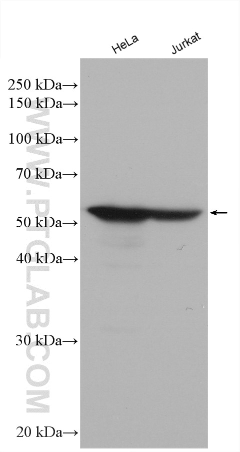
Various cell lysates were subjected to SDS PAGE followed by Western blot with anti-Vimentin antibody (10366-1-AP) labeled with FlexAble HRP Antibody Labeling Kit for Rabbit IgG (KFA005).
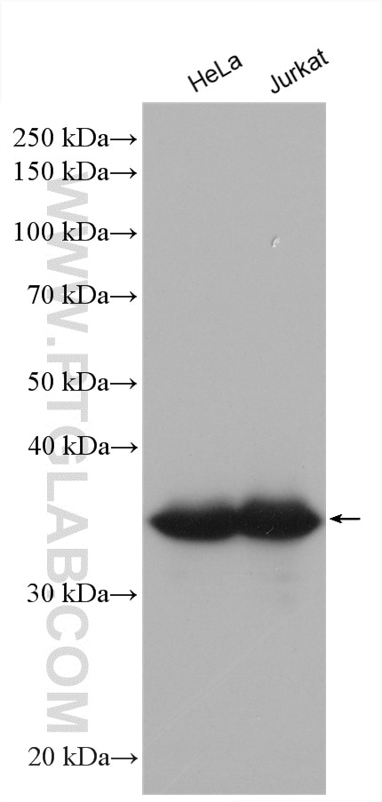
Various cell lysates were subjected to SDS PAGE followed by Western blot with anti-GAPDH antibody (10494-1-AP) labeled with FlexAble HRP Antibody Labeling Kit for Rabbit IgG (KFA005).
Various cell lysates were subject to SDS PAGE followed by Western blot with anti-HSPA5 antibody (66574-1-Ig) labeled with FlexAble HRP Antibody Labeling Kit for Mouse IgG1 (KFA025).
Various cell lysates were subjected to SDS PAGE followed by western blot with anti-Importin Alpha 5 antibody (67897-1-Ig) labeled with FlexAble HRP Antibody Labeling Kit for Mouse IgG2a (KFA045).
Various cell lysates were subjected to SDS PAGE followed by Western blot with anti-Lamin B1 antibody (12987-1-AP) labeled with FlexAble HRP Antibody Labeling Kit for Rabbit IgG (KFA005).
Various cell lysates were subjected to SDS PAGE followed by Western blot with anti-ANP32B antibody (66160-1-Ig) labeled with FlexAble HRP Antibody Labeling Kit for Mouse IgG2b (KFA065).
SpeedAb WB Kit for detection of rabbit IgG
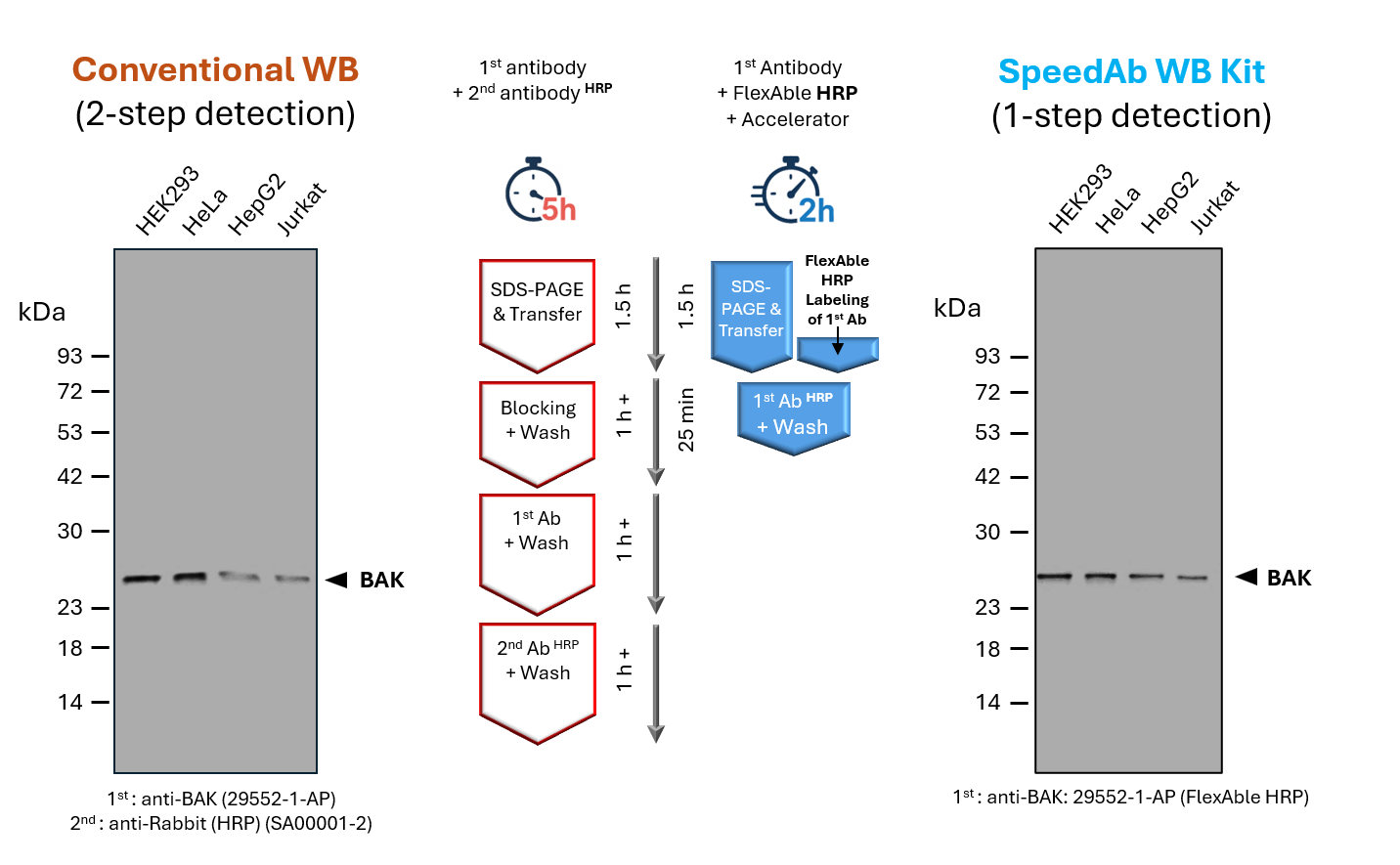
(left) Classical Western blot detection of BAK in lysates from different human cell lines. A primary anti-BAK antibody (rabbit) was used in combination with a secondary anti-rabbit antibody (HRP), after blocking and incubation with antibodies for 1 h, respectively. The totaltime of ~ 5 h includes SDS-PAGE, blotting, blocking, primary and secondary antibody incubation, and washing steps.
(right) Detection of BAK in lysates from different human cell lines via SpeedAb WB Kit. The primary anti-BAK antibody was labeled with FlexAble HRP (anti-rabbit: #KFA005) via a simple 15min protocol, enabling1-step detection. The blotting accelerator speeds up binding of the antibody to the target protein, cutting the antibody incubation time to 25minor less and the total time including SDS-PAGE, blotting, antibody incubation and a final washing step to ~2 h.
Available Reagents for fast Western Blot Workflows
| Product | Available Sizes | Specificity (towards primary WB detection antibody) |
|---|---|---|
| SpeedAb Western Blot Kit for Rabbit IgG | 20 rxns* | Rabbit IgG |
| SpeedAb Western Blot Kit for Mouse IgG1 | 20 rxns* | Mouse IgG1 |
| SpeedAb Western Blot Kit for Mouse IgG2a | 20 rxns* | Mouse IgG2a |
| SpeedAb Western Blot Kit for Mouse IgG2b | 20 rxns* | Mouse IgG2b |
| Blotting Accelerator | 100ml, 500ml | |
| FlexAble HRP | 10 rxns, 50 rxns, 200 rxns | Rabbit, Mouse IgG1, Mouse IgG2a, Mouse IgG2b, Human, Rat (Kappa Light Chain) |
* SpeedAb Kits provide sufficient reagent for usage of 20x 5 ml of Blotting Accelerator and labeling 20x 0.5 µg of primary antibody with FlexAble HRP
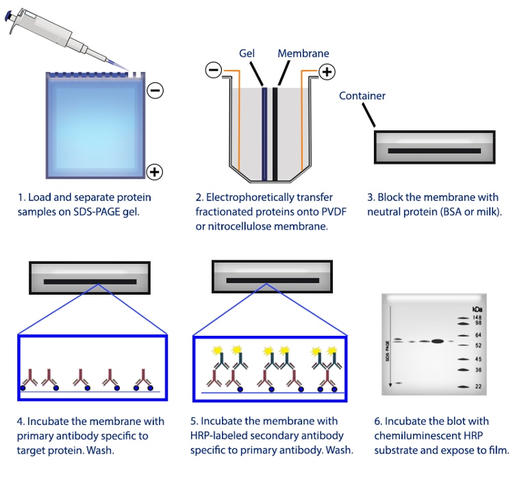
Proteintech Protocols: Conventional Western Blotting
Other Supporting reagents for Western Blot
Primary Antibodies
Polyclonal Antibodies
Recognize multiple epitopes on the protein of interest. Binds with very high affinity. Good for low abundant targets, rare species and mutated proteins.
Proteintech polyclonals are raised in rabbit.
Monoclonal Antibodies
Recognize a single epitope on the protein of interest. Low background, highly specific. Good for mid-high abundant targets.
Proteintech monoclonal antibodies are raised in mouse.
Recombinant Antibodies
Genetically defined monoclonals, recombinantly produced. Unrivalled reproducibility. Good for long-term, high-level projects.
Proteintech recombinant antibodies are raised in rabbit.
Tag/Control Antibodies
Our top cited antibodies for WB loading controls and popular expression tags.
Top Cited Antibodies
GAPDH Monoclonal Antibody
| Reactivity: | Human, Mouse, Rat, Yeast, Plant, Zebrafish, Bovine and more (9) |
| Applications: | WB, IP, IHC, IF, FC, CoIP, ChIP, ELISA |
TDP-43 Polyclonal Antibody
| Reactivity: | Human, Mouse, Rat, Zebrafish, Hamster, Pig and more (6) |
| Applications: | WB, RIP, IP, IHC, IF, IEM, FC, CoIP, chIP, ELISA |
GAPDH Polyclonal antibody
| Reactivity: | Human, Mouse, Rat, Pig, Arabidopsis, Corn, Cabbage, Rice and more (9) |
| Applications: | WB, RIP, IP, IHC, IF, FC, CoIP, ELISA |
Beta Actin Monoclonal antibody
| Reactivity: | Human, Mouse, Rat, Pig, Plant, Zebrafish And More (10) |
| Applications: | WB, IP, IHC, IF, FC, CoIP, ELISA |
BAX Polyclonal antibody
| Reactivity: | Human, Mouse, Rat, Chicken, Goat, Hamster, Pig, Rabbit and more (3) |
| Applications: | WB, IP, IHC, FC, CoIP, chIP, ELISA |
Caspase 3/p17/p19 Polyclonal antibody
| Reactivity: | Human, Mouse, Rat, Bovine, Chicken, Duck, Goat, Hamster and more (3) |
| Applications: | WB, RIP, IP, IHC, IF, FC, ELISA |
E-cadherin Polyclonal antibody
| Reactivity: | Human, Mouse, Rat, Bovine, Pig, Zebrafish |
| Applications: | WB. IF, WB, IP, IHC, IF, FC, CoIP, ELISA |
NF-κB p65 Polyclonal antibody
| Reactivity: | Human, Mouse, Rat, Bovine, Pig, Canine, Fish and more (4) |
| Applications: | WB, IP, IHC, IF, FC, chIP, ELISA |
Vimentin Polyclonal antibody
| Reactivity: | Human, Mouse, Rat, Bovine, Pig, Deer |
| Applications: | WB, IP, IHC, IF, FC, CoIP, ELISA |
P62,SQSTM1 Polyclonal antibody
| Reactivity: | Human, Bovine, Chicken, Goat, Hamster, Pig, Rabbit and more (3) |
| Applications: | WB, IP, IHC, IF, FC, CoIP, ELISA |
MMP9 (N-terminal) Polyclonal antibody
| Reactivity: | Human, Mouse, Rat, Pig, Rabbit, Hamster, Astragalus Membranaceus |
| Applications: | WB, IP, IHC, IF, FC, CoIP, ELISA |
Alpha Tubulin Polyclonal antibody
| Reactivity: | Human, Mouse, Rat, Bovine, Chicken, Goat, Hamster, Monkey, P. Lividus, Pig |
| Applications: | WB, IP, IHC, IF, FC, CoIP, ELISA |
ARL13B Polyclonal antibody
| Reactivity: | Human, Mouse, Rat, Canine, Chicken, Pig, Sheep, Xenopus, Zebrafish |
| Applications: | WB, IP, IHC, IF, ELISA |
PARK2/Parkin Polyclonal antibody
| Reactivity: | Human, Mouse, Rat, Pig, Canine, Chicken, Hamster |
| Applications: | WB, IP, IHC, IF, FC, ChIP, ELISA |
P53 Polyclonal antibody
| Reactivity: | Human, Rat, Mouse, Goat, Pig and more (3) |
| Applications: | WB, IP, IF, CoIP, chIP, ELISA |
Secondary Antibodies
When choosing a secondary antibody for WB, be mindful of:
- Host of your primary antibody, E.g. rabbit; and
- Your signal detection system
Popular and highly cited secondary antibodies for WB
Affinipure Goat Anti-Rabbit IgG (H+L)
| Conjugate: | HRP |
| Applications: | ELISA, WB |
Affinipure Goat Anti-Mouse IgG (H+L)
| Conjugate: | HRP |
| Applications: | ELISA, WB |
Affinipure Rabbit Anti-Goat IgG (H+L)
| Conjugate: | HRP |
| Applications: | ELISA, WB |
Nano-Secondaries
Nano-Secondaries have certain advantages over the conventional secondaries.
- Pre-incubation or parallel incubation of Nano-Secondaries with primary antibodies, i.e., one-step incubation, reduce incubation times and enhance multiplexing capabilities.
- The recombinant process of developing Nano-Secondaries ensures high lot to lot consistency.
- Smaller size of Nano-Secondaries has been proven to enhance image resolution in Super resolution Microscopy.
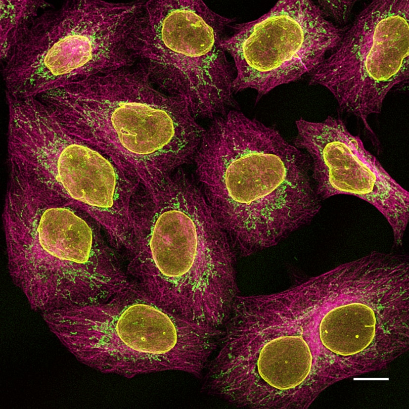
Multiplexed immunostaining of HeLa cells with two alpaca anti-mouse Nano-Secondaries and one anti-rabbit Nano-Secondary. Green: mouse IgG1 anti-COX4 + alpaca anti-mouse IgG1 VHH Alexa Fluor 488. Magenta: mouse IgG2b anti-Tubulin + alpaca anti-mouse IgG2b VHH Alexa Fluor 647. Yellow: rabbit anti-Lamin + alpaca anti-rabbit IgG VHH Alexa Fluor 568. Scale bar, 10 μm. Images were recorded at the Core Facility Bioimaging at the Biomedical Center, LMU Munich.
Chemiluminescent Substrates
SignalBright Enhanced Chemiluminescent substrates - luminol-based, chemiluminescent substrates for HRP (horseradish peroxidase) which provide a much higher sensitivity over traditional ECL reagents. We have two reagents available, enabling the detection of proteins at low to high femtogram levels.
| SignalBright Pro | SignalBright Max | |
|---|---|---|
| Sensitivity | Mid/high to high femtogram | Low to mid femtogram |
| Relative sensitivity to SignalBright Pro | 1x | 10x |
| Stable signal duration | >12 hours | >12 hours |
| Equivalent | SuperSignalTM West Dura | SuperSignalTM West Femto |
| Primary antibody concentration | 0.02 - 1 µg/ml | 0.01 - 0.2 µg/ml |
| Secondary antibody concentration | 4 - 20 ng/ml | 1 - 10 ng/ml |
Proteintech’s SignalBright Pro (PK10011) and SignalBright Max (PK10013) Chemiluminescent Substrates have different levels of sensitivity (see above table). In particular, our SignalBrights have comparable performance to top competitor products with highly sensitive detection, minimal optimization required, and long, stable signal duration.
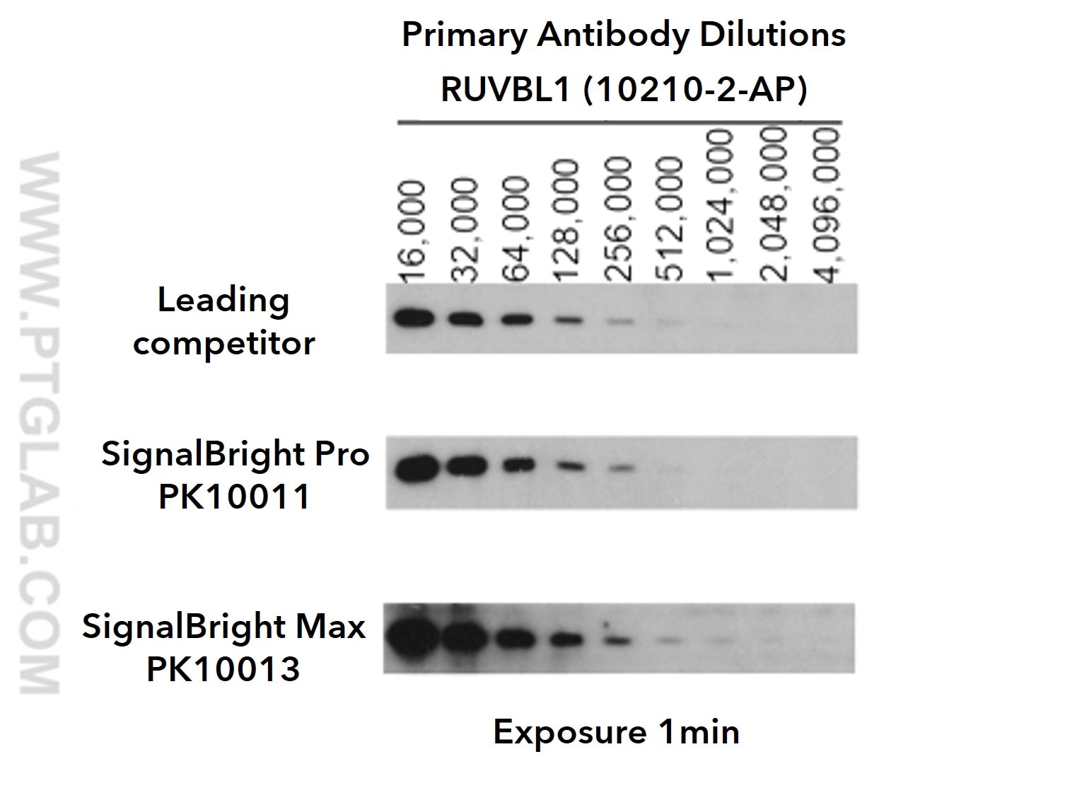 Figure 2a: Comparison of SignalBright Pro (PK10011) and SignalBright Max (PK10013) against leading competitor ECL. Serial dilutions of RUVBL primary antibody (10210-2-AP). Primary: Proteintech RUVBL1 (10210-2-AP) at various dilutions (see image). Secondary: Proteintech HRP-conjugated Affinipure Goat Anti-Rabbit IgG (SA00001-2); 1:6,000
Exposure time: 1 min
Figure 2a: Comparison of SignalBright Pro (PK10011) and SignalBright Max (PK10013) against leading competitor ECL. Serial dilutions of RUVBL primary antibody (10210-2-AP). Primary: Proteintech RUVBL1 (10210-2-AP) at various dilutions (see image). Secondary: Proteintech HRP-conjugated Affinipure Goat Anti-Rabbit IgG (SA00001-2); 1:6,000
Exposure time: 1 min
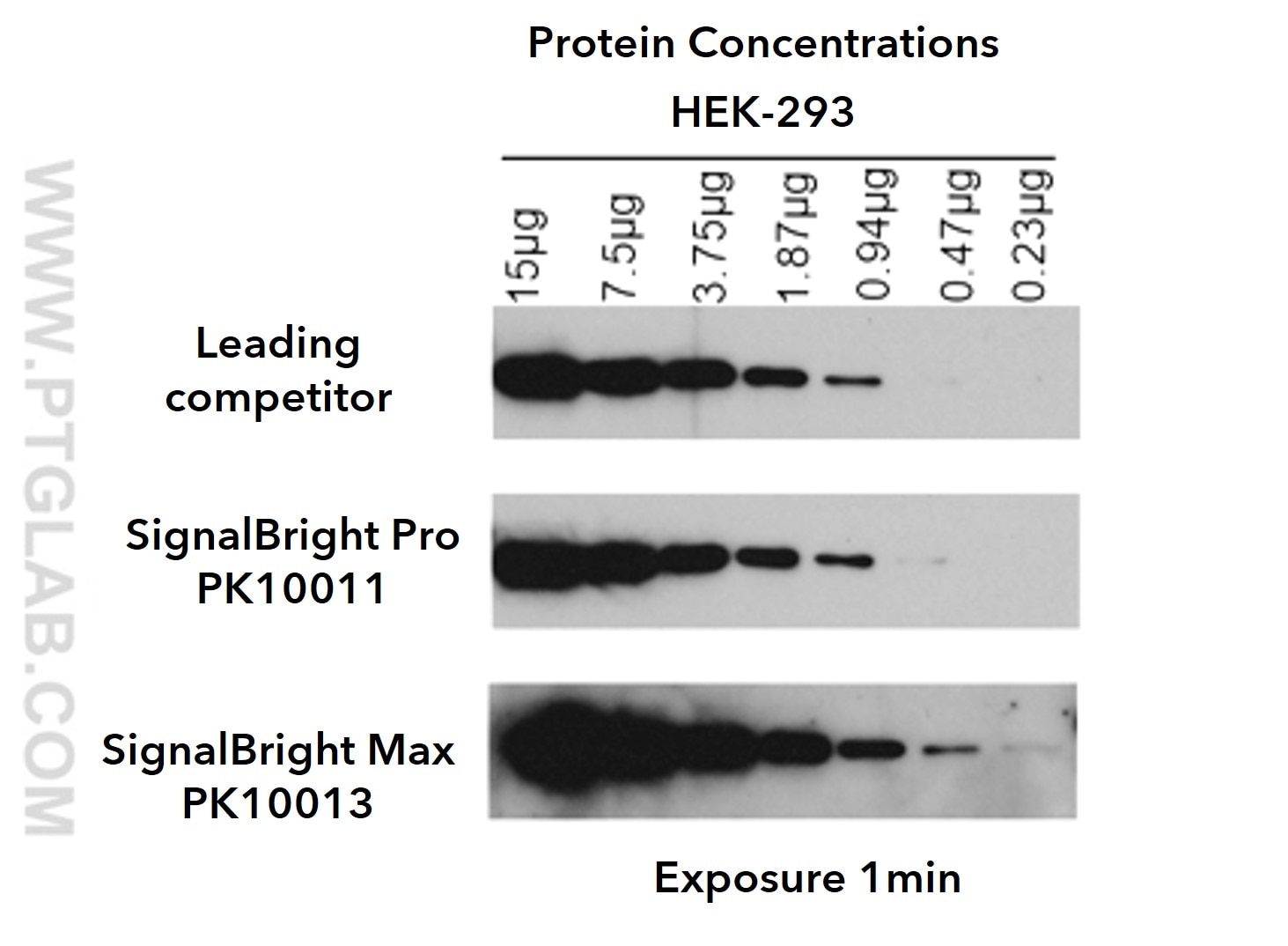 Figure 2b: Comparison of SignalBright Pro (PK10011) and SignalBright Max (PK10013) against leading competitor ECL. Serial dilutions of HEK-293 cell lysates. Primary: Proteintech RUVBL1 (10210-2-AP); 1:8,000
Secondary: Proteintech HRP-conjugated Affinipure Goat Anti-Rabbit IgG (SA00001-2); 1:6,000. Exposure time: 1 min
Figure 2b: Comparison of SignalBright Pro (PK10011) and SignalBright Max (PK10013) against leading competitor ECL. Serial dilutions of HEK-293 cell lysates. Primary: Proteintech RUVBL1 (10210-2-AP); 1:8,000
Secondary: Proteintech HRP-conjugated Affinipure Goat Anti-Rabbit IgG (SA00001-2); 1:6,000. Exposure time: 1 min
Buffers
RIPA Buffer
Non visible textGentle Cell Lysis Buffer
Non visible textProtein Ladders
Ready-to-use protein standards in three colours. Standard PL00001, broad range PL00002 and extra broad range PL00003 markers available. The perfect companion to every WB. Colorimetric detection. Also visible in red and far-red fluorescent wavelengths.
Prestained protein marker
| Molecular Weight: | 10-180 kDa |
| Number of markers: | 10 |
Broad range prestained protein marker
| Molecular Weight: | 3-245 kDa |
| Number of markers: | 13 |
Extra Range Prestained Protein Marker
| Molecular Weight: | 10-310 kDa |
| Number of markers: | 12 |
Technical Support for Western Blot
The Western blot is an incredibly common application. However, generating good WB data can be hard due to the many facets of a WB workflow that can interfere with results.
Our technical team have put together an excellent guide to Western blotting, describing the overall protocol and a comprehensive trouble shooting section. Download here.
Related
- Comparing BCA, Bradford, and UV quantification methods for scientists
- Proteases for Western Blotting: Choosing the right tools for the job
- How Do I Optimize My Extraction?
- Why is RIPA Buffer Best for Western Blot?
- Western Blot tips for low molecular weight proteins
- 7 Tips For Optimizing Your Western Blotting Experiments