
Apoptotic Cell Death
Introduction
Apoptosis is a form of programmed cell death characterized by a series of highly regulated biochemical events that lead to changes in cellular morphology and ultimately cell death. Apoptosis plays a central role in several physiological processes including development, cellular homeostasis, and aging. Moreover, it has been implicated in the pathogenesis of various human diseases including neurodegenerative disorders, autoimmune diseases, cancers, and viral infections. Not surprisingly, targeting the apoptotic pathway has emerged as a common therapeutic strategy in the treatment of such diseases.
Proteintech offers a wide range of products to help you with your apoptosis research.
Contents: |
Apoptotic pathways
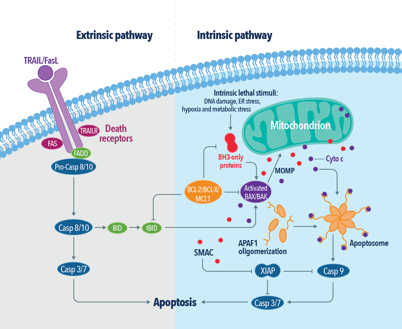
Apoptosis proceeds through one of two distinct signaling pathways known as the intrinsic and extrinsic pathways, both of which ultimately lead to the activation of proteolytic enzymes known as caspases. The intrinsic apoptotic pathway is activated in response to severe cellular damage or stress caused by various factors including DNA damage, hypoxia, and oncogenes resulting in mitochondrial outer membrane permeabilization (MOMP) and the consequent release of apoptogenic factors from the mitochondria. Initiation of the extrinsic apoptotic pathway relies on the activation of cell surface death receptors of the tumor necrosis factor (TNF) family of receptors by the binding of their corresponding ligands.
Related products
| DR5 | FLIP | TRADD |
| FADD | RIPK1 | TRAF2 |
| FAS | TNFR1 | TRAIL |
The Bcl-2 protein family
The Bcl-2 protein family consists of a group of structurally related proteins that are critical for the regulation of the mitochondria-mediated intrinsic apoptotic pathway. Members of the Bcl-2 protein family are characterized by the presence of one or more conserved regions of sequence homology (Bcl-2 homology domains) and can be classified into anti-apoptotic or pro-apoptotic depending on their ability to promote or inhibit apoptosis. The pro-apoptotic Bcl-2 members can be further classified into BH3-only proteins and pore-forming proteins. BH3-only proteins function by either binding anti-apoptotic Bcl-2 members to inactivate them or binding pore-forming proteins to activate them. Once activated, pore-forming proteins undergo a series of conformational changes leading to their homo-oligomerization followed by MOMP and the subsequent release of mitochondrial apoptogenic factors. Anti-apoptotic Bcl-2 members, on the other hand, inhibit apoptosis by binding and sequestering both the BH3-only proteins as well as the pore-forming proteins.
Related products
| BAD | Bcl-XL | MCL1 |
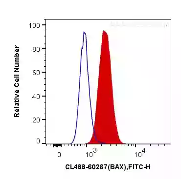
| BAX | BID | PUMA |
| BCL2 | BIM |
Caspases
Caspases are a group of serine proteases that play a central role in the initiation and execution of the proteolytic cascade during apoptosis. Depending on their role during apoptosis, caspases can be divided into initiator caspases including caspases 2, 8, 9, and 10, and effector caspases including caspases 3, 6, and 7. While initiator caspases cleave and activate the effector caspases, the effector caspases cleave other cellular protein targets to complete the apoptotic program. Some caspases are also known to regulate inflammation, including caspases 1, 4, 5, 11, and 12. Caspase-14 is not involved in apoptosis or inflammation but plays an important role in skin cell development.
Related products
| Caspase 1 | Caspase 6 | Caspase 9 |
| Caspase 2 | Caspase 7 | Caspase 10 |
| Caspase 3 | Caspase 8 | Caspase 12 |

Role of mitochondria in apoptosis
Mitochondria play a critical role in the regulation of apoptosis by initiating MOMP in response to the activation of the Bcl-2 family members, BAX and BAK. MOMP is followed by the release of soluble proteins such as cytochrome C from the mitochondrial intermembrane space into the cytoplasm leading to the activation of the apoptotic caspase cascade. During MOMP and cytochrome C release, mitochondria undergo massive fragmentation also known as mitochondrial fission. While healthy cells maintain a balance between mitochondrial fission and fusion, excessive mitochondrial fission is an integral part of apoptotic cell death. Therefore, not surprisingly, the mitochondrial fission machinery has been demonstrated to be actively involved in the induction of apoptosis.
Related products
| Cytochrome C | DRP1 | OPA1 |
| AIF | FIS1 | MFN1 |
| DIABLO | MFF | MFN2 |
| Endonuclease G | MID49 | COXIV |
| HTRA2 | MID51 | VDAC1 |
Measuring apoptosis
While detecting activated caspases is a common approach for studying apoptosis, several other assays can be used for quantifying apoptosis. The TUNEL method, for example, is a widely used apoptotic assay that detects fragmented DNA in apoptotic cells by incorporating fluorophore-labeled dUTP at the 3’-OH ends of broken DNA. The fluorophore-labeled DNA can then be quantified using a flow cytometer. The primary advantage of the TUNEL method is that it can selectively detect apoptotic cells, but not necrotic cells or cells with DNA strand breaks caused by irradiation and drug treatment.
Annexin V/propidium iodide double labeling is another widely used method for studying apoptosis that can also be used for differentiating apoptotic cells from necrotic cells. Annexin V detects early apoptotic cells by binding to phosphatidylserine (PS), which is flipped to the extracellular membrane surface during early apoptosis. PI on the other hand is a DNA-binding dye that can stain the nucleus of necrotic cells or cells that lose cell membrane integrity during the late stages of apoptosis.
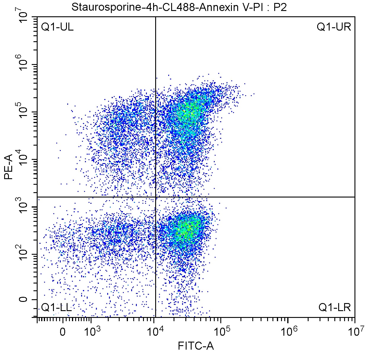
Flow cytometry analysis of Jurkat cells treated with staurosporine and stained using the CoraLite®488-Annexin V and PI Apoptosis Kit (PF00005). CL488-AnnexinV and PI are represented by the X and the Y axis of the scatterplot, respectively. Positively stained cells in the various regions of the scatterplot can be interpreted as follows:
- Q1-UL (CL488-AnnexinV)-/PI+, cells in this region are primarily necrotic.
- Q1-UR :(CL488-Annexinv)+/PI+, cells in this region are late apoptotic cells.
- Q1-LR :(CL488-Annexinv)+/PI-, cells in this region are early apoptotic cells.
- Q1-LL :(CL488-Annexinv)-/PI-, cells in this region are living cells.
Related products
CoraLite®488 TUNEL Assay Apoptosis Detection Kit (green)
CoraLite®594 TUNEL Assay Apoptosis Detection Kit
CoraLite®488-Annexin V and PI Apoptosis Kit
Viability/Cytotoxicity Assay Kit for Animal Live & Dead Cells (Calcein AM, EthD-1 Method)
Viability/Cytotoxicity Assay Kit for Animal Live & Dead Cells (Calcein AM, PI Method)
Related Content
What is the difference between necrosis and apoptosis?
Metabolic Regulation of Cell Death
Autophagy in the Pathogenesis of Disease

Support
Newsletter Signup
Stay up-to-date with our latest news and events. New to Proteintech? Get 10% off your first order when you sign up.
Able™