
Cardiovascular Research
Proteintech offers a wide range of markers and related antibodies for cardiovascular research
Introduction
The cardiovascular system is a general term for the heart and blood vessels. Coronary heart disease, cerebrovascular disease, peripheral arterial disease, rheumatic heart disease, congenital heart disease, or deep vein thrombosis and pulmonary embolism are only a few examples of cardiovascular diseases (CVDs). Since CVDs remain a consistently increasing cause of death worldwide, scientists are prioritizing the identification and understanding of the mechanisms of cellular and molecular basics related to CVDs. In 2012, WHO highlighted that more than 30% of our population die because of CVDs (with the majority due to coronary heart disease and stroke).
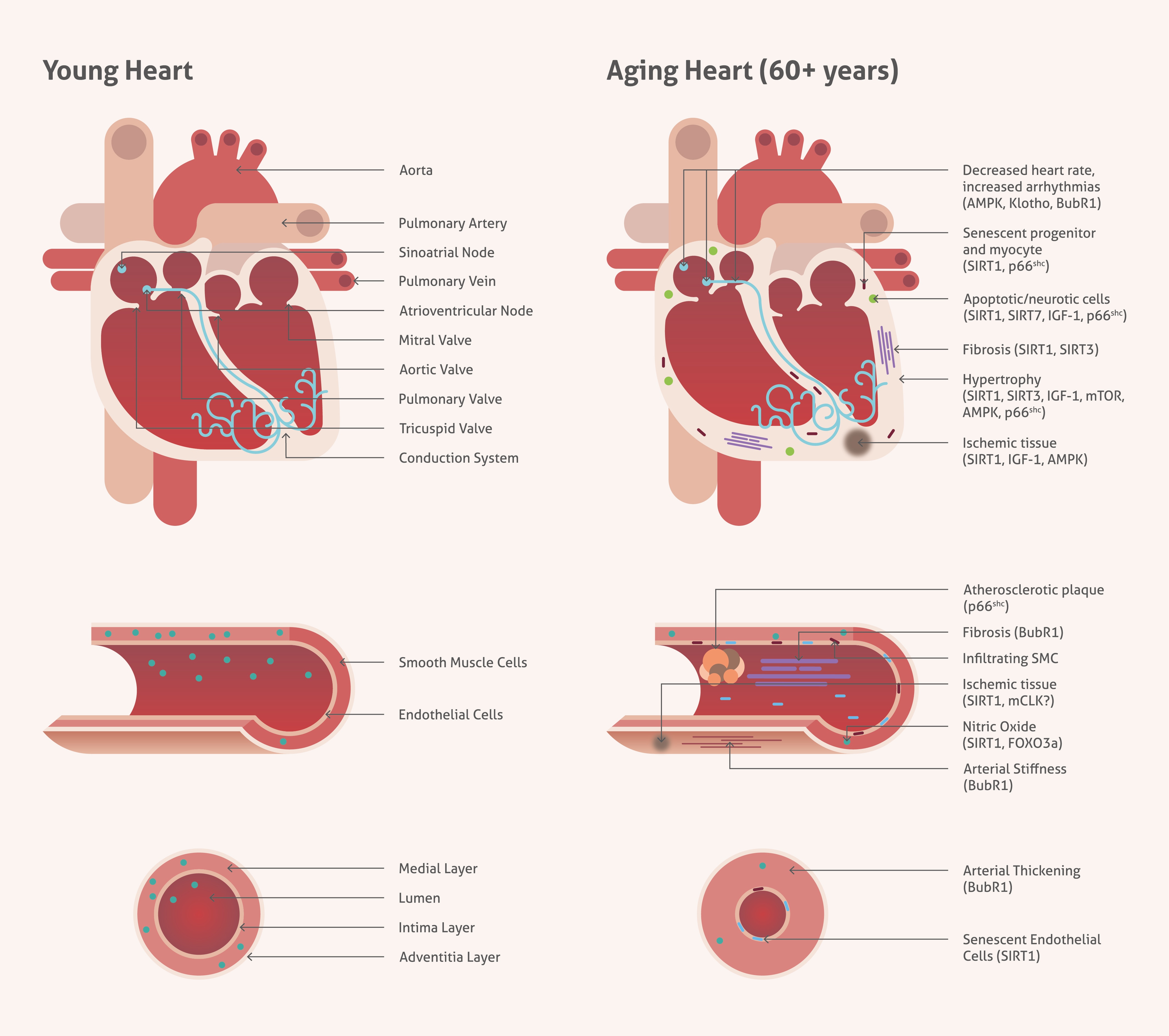
Age-Associated Changes In Cardiovascular Tissues
Age-associated changes in cardiovascular system are leading to deregulation of molecular pathways and pathological alterations (e.g., hypertrophy, altered left ventricular (LV) diastolic function, abnormalities in arterial stiffness or impaired endothelial function). That reflects in significant increase of CVD including atherosclerosis, hypertension or stroke. Inhibition of the mammalian target of rapamycin (mTOR) presents an evidence for being cardioprotective during aging and cardiac stress.
Blog post:Hidden Secrets Of The Heart |
Focus Antibodies
mTOR in Cardiac Physiology and Disease
| Catalog number: 20657-1-AP | Type: Rabbit Polyclonal |
| Publications: 15 | Applications: ELISA, IF, IHC, IP, WB |
mTOR (Mammalian or Mechanistic Target of Rapamycin) is an atypical serine/threonine kinase that exerts its main cellular functions interacting with specific protein adaptors to form two different complexes: mTORC1 and mTORC2.
The mTOR pathway is a key player in the cardiovascular regulation system. It appears crucial for the maintenance of cardiac structure and function in the postnatal period, adulthood, and also in the development of cardiac hypertrophy. mTOR is also deeply involved in the regulation of cardiac metabolism.
Moreover, the mTOR pathway that partners with Wnt and growth factor signaling are vital for endothelial and cardiomyocyte growth. From the other side, chronic mTOR activation appears to enhance the cardiac aging process.
 |
|
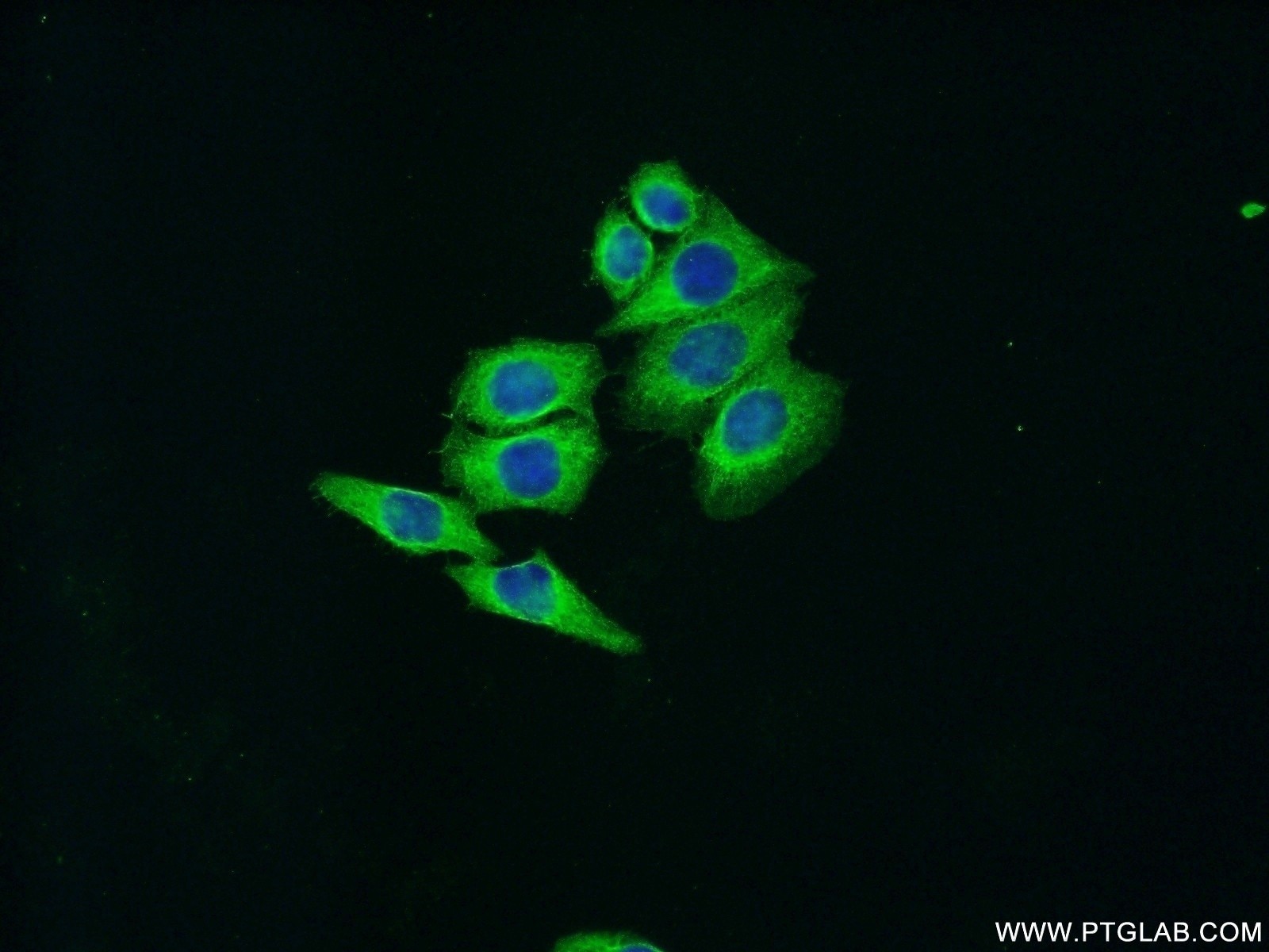
IF analysis of HeLa cells labeling mTOR with 20657-1-AP Proteintech antibody at a dilution of 1:50 and Alexa Fluor 488-conjugated AffiniPure Goat Anti-Rabbit IgG(H+L) |
 |
|
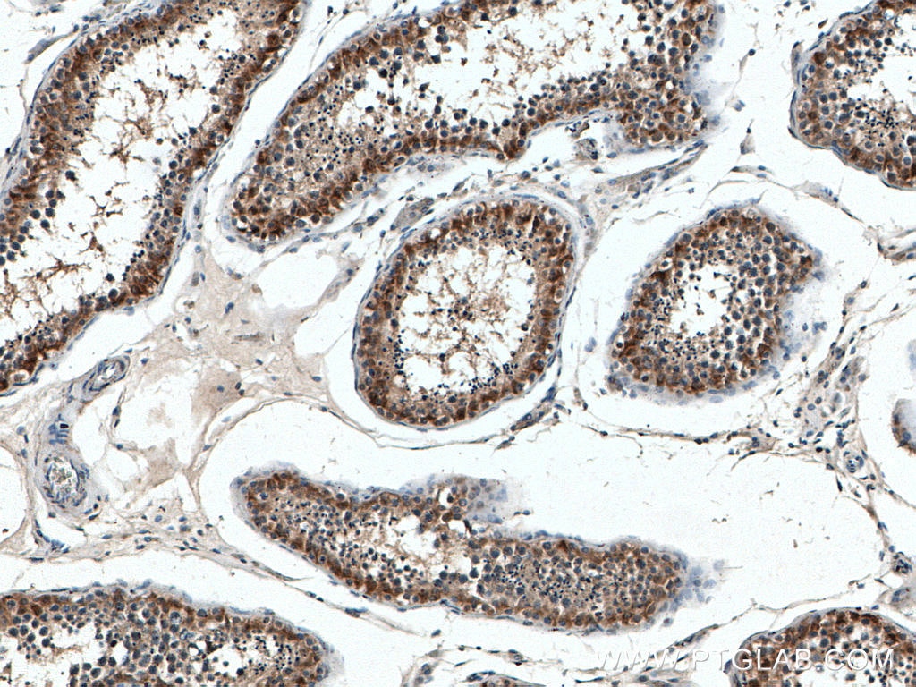
IHC analysis of paraffin-embedded human testis tissue slide using 20657-1-AP (mTOR antibody) at dilution of 1:200 (under 10x lens) heat mediated antigen retrieved with Tris-EDTA buffer(pH9). |
High-Sensitive Cardiac Troponin T
| Catalog number: 15513-1-AP | Type: Rabbit Polyclonal |
| Publications: 4 | Applications: ELISA, IHC, WB |
Defects in cTnT are the cause of cardiomyopathy familial hypertrophic type 2 (CMH2), cardiomyopathy dilated type 1D (CMD1D) and cardiomyopathy familial restrictive type 3 (RCM3). Statistically significant differences were also described in cTnT levels between newborns with heart defects and healthy individuals.
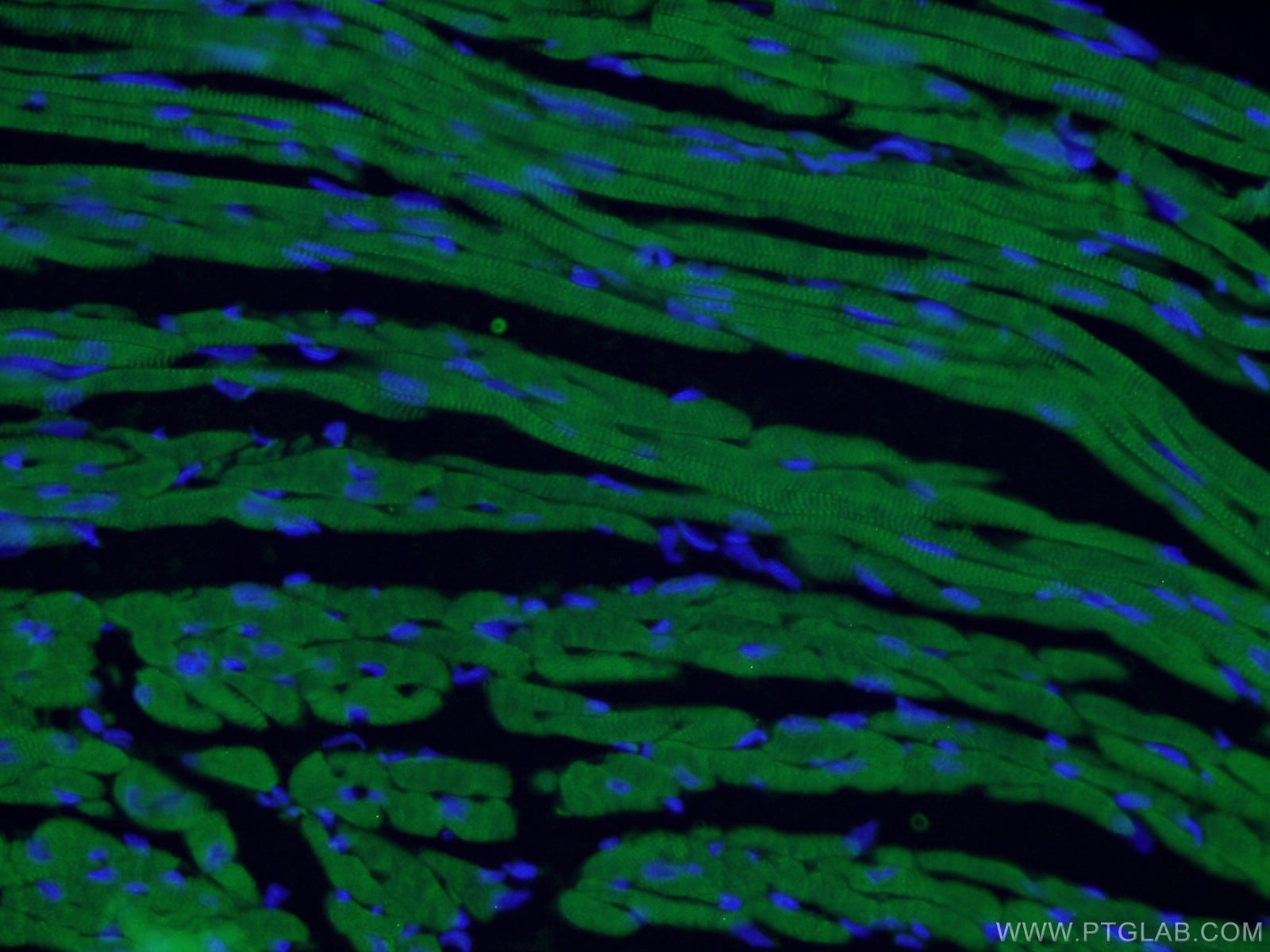 |
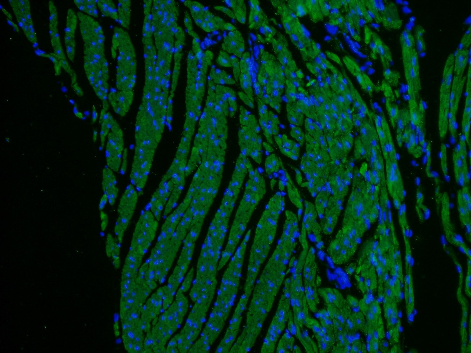
| IF analysis of (4% PFA) fixed mouse heart tissue using Cardiac Troponin T antibody (15513-1-AP; 1:50, 40x) and Alexa Fluor 488-conjugated AffiniPure Goat Anti-Rabbit IgG(H+L). |
 |
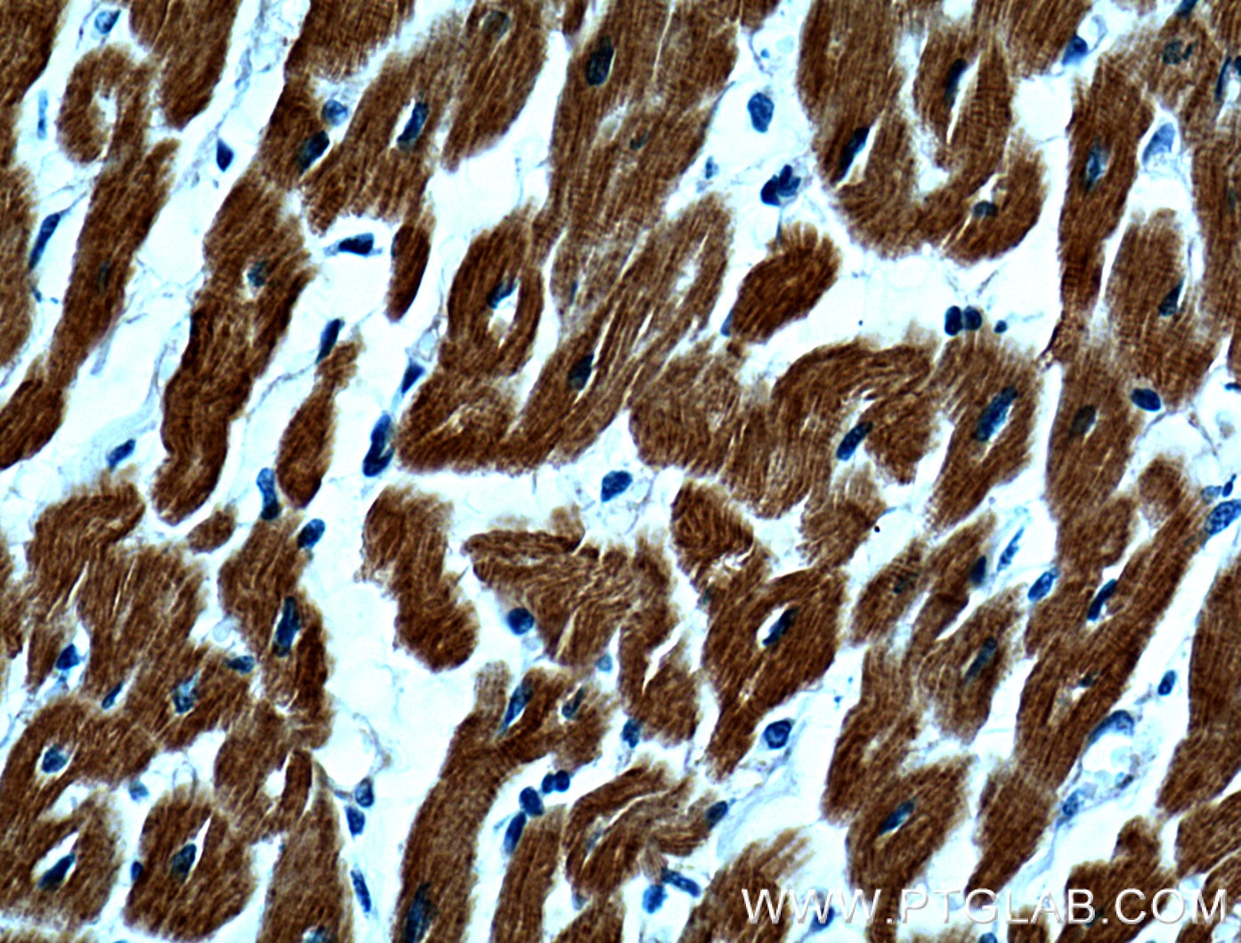
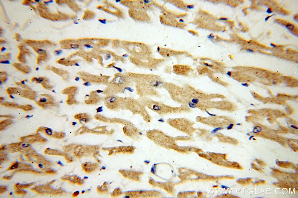
| Immunohistochemical analysis of paraffin-embedded mouse heart tissue slide using 15513-1-AP (Cardiac Troponin T antibody) at dilution of 1:200 (under 40x lens) heat mediated antigen retrieved with Tris-EDTA buffer(pH9). |
CD31 (PECAM-1): A Multi-Functional Molecule in Vascular Biology
| Catalog number: 11265-1-AP | Type: Rabbit Polyclonal |
| Publications: 15 | Applications: ELISA, FC, IF, IHC, IP, WB |
CD31 (PECAM-1 – platelet endothelial cell adhesion molecule-1) is an Ig gene superfamily member, composed of 6 extracellular Ig folds, with a molecular weight of 130 kDa. It is differentially glycosylated involving N-linked and O-linked glycosylation sites.
CD31 is expressed on all cells within the vascular compartment, on the surface of the endothelium. It plays a role in cell-cell adhesion, being an efficient signaling molecule. This reflects the diverse roles of CD31 in vascular biology including angiogenesis, platelet function, thrombosis, mechanosensing of endothelial cell response to fluid shear stress, and regulation of multiple stages of leukocyte migration through venular walls.
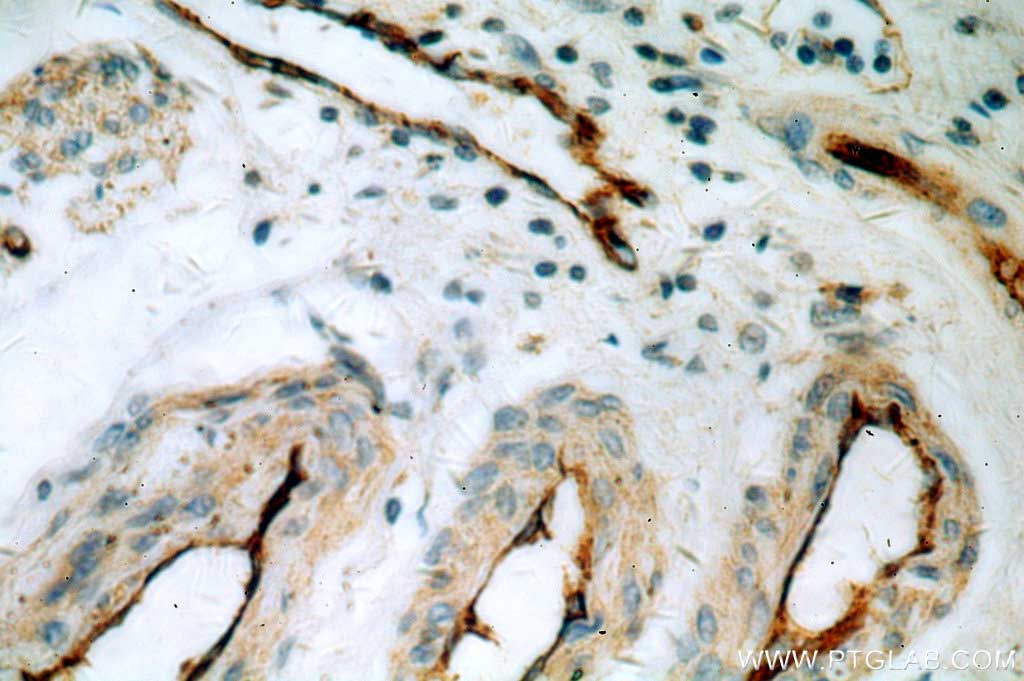 |
| IHC of paraffin-embedded human endometrial cancer sample using CD31 antibody (11265-1-AP) at a dilution of 1:50; under 40x. |
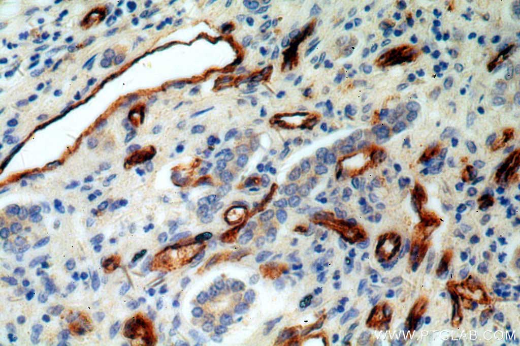 |
| IHC of paraffin-embedded human hepatocirrhosis sample using CD31 antibody (11265-1-AP) at a dilution of 1:50; under 40x. |
Myosin light chain 2 – Atrial (MYL7/MLC2a) & Ventricular (MYL2/MLC-2v) Cardiomyocytes
| Catalog number: 10906-1-AP | Type: Rabbit Polyclonal |
| Publications: 44 | Applications: ELISA, FC, IF, IHC, IP, WB |
MYL2/MLC-2v and MYL7/MLC2a are commonly known as two major isoforms of myosin light chain 2, which are essential for heart development.
MYL2/MLC-2v and MYL7/MLC2a are expressed in the heart in a defined manner. The ventricular myosin light chain-2 isoform (MYL2/MLC-2v) is restricted to the ventricular segment of the heart and is related to the ventricles throughout the developing and adult human heart. In contrast, the atrial myosin light chain-2 (MYL7/MLC-2a) is expressed in the presumptive ventricle prior to MYL2/MLC-2v. Its ventricular expression is subsequently down-regulated, instead, with abundant expression in the atrium postnatally.
The expression pattern of MYL2/MLC-2v and MYL7/MLC2a is considered as the specific marker for mature ventricle and atrial cardiomyocytes, commonly used for in vitro development of induced pluripotent stem cell-derived cardiomyocytes.
 |
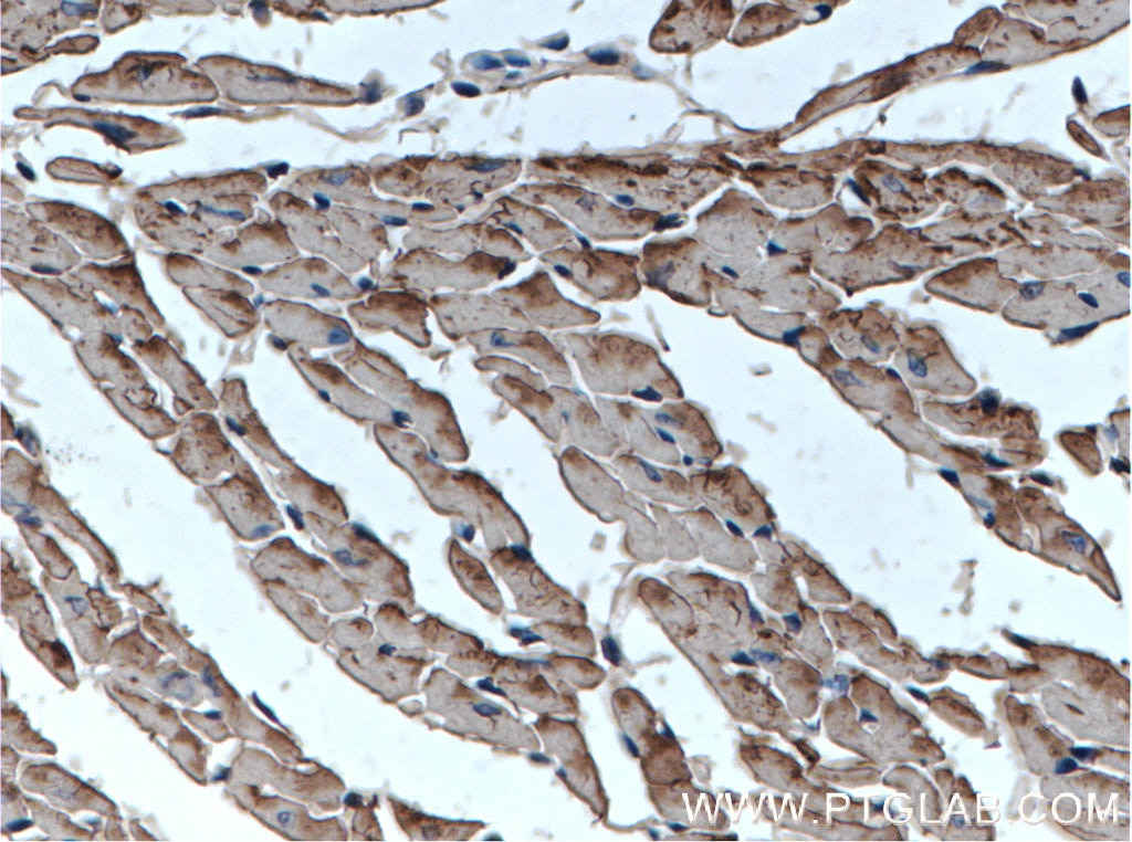
| IHC results of paraffin-embedded human heart tissue using MYL2 antibody (10906-1-AP; 1:200, 40x). |
 |
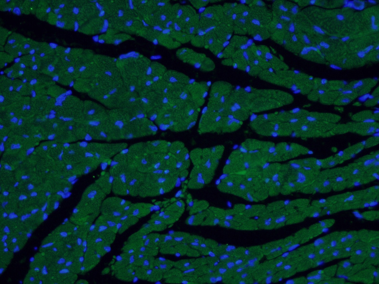
| IF analysis of (4% PFA) fixed mouse heart tissue using MYL2 antibody (10906-1-AP; 1:500, 40x) and Alexa Fluor 488-conjugated AffiniPure Goat Anti-Rabbit IgG(H+L). |
MYL7
| Catalog number: 17283-1-AP | Type: Rabbit Polyclonal |
| Publications: 1 | Applications: ELISA, IF, IHC, IP, WB |
 |
| IHC results of paraffin-embedded human heart tissue using MYL7 antibody (17283-1-AP; 1:100, 40x) |
 |
| IF analysis of (4% PFA) fixed mouse heart tissue using MYL7 antibody (17283-1-AP; 1:50, 40x) and Alexa Fluor 488-congugated AffiniPure Goat Anti-Rabbit IgG(H+L). |
More Related Antibodies
The full list can be found here.
| Antibody Name | Catalog number | Type | Applications | Publications |
| ACTA2/smooth muscle actin | 14395-1-AP | Rabbit Poly | ELISA, FC, IF, IHC, IP, WB | 57 |
| AKT1 | 10176-2-AP | Rabbit Poly | ELISA, FC, IF, IHC, IP, WB | 56 |
| ALDH2 | 15310-1-AP | Rabbit Poly | ELISA, IF, IHC, IP, WB | 7 |
| APEX1 | 10203-1-AP | Rabbit Poly | ELISA, IF, IHC, IP, WB | 5 |
| Catalase | 21260-1-AP | Rabbit Poly | ELISA, IF, IHC, WB | 10 |
| CD31 | 11265-1-AP | Rabbit Poly | ELISA, FC, IF, IHC, IP, WB | 14 |
| HEXIM1 | 15676-1-AP | Rabbit Poly | CoIP, ELISA, IF, IHC, IP, WB | 7 |
| Hexokinase 2 | 22029-1-AP | Rabbit Poly | ELISA, IHC, IP, WB | 7 |
| HEY2 | 10597-1-AP | Rabbit Poly | ELISA, IF, IHC, IP, WB | 15 |
| Icam1 | 10020-1-AP | Rabbit Poly | ELISA, FC, IF, IHC, WB | 15 |
| ICAM-1 | 10831-1-AP | Rabbit Poly | ELISA, FC, IHC, IP, WB | 13 |
| MEF2C | 10056-1-AP | Rabbit Poly | ELISA, IF, IHC, IP, WB | 15 |
| Mitofilin | 10179-1-AP | Rabbit Poly | ELISA, FC, IF, IHC, IP, WB | 14 |
| MTOR | 20657-1-AP | Rabbit Poly | ELISA, IF, IHC, IP, WB | 13 |
| Myosin Light Chain Antibody 2 | 10906-1-AP | Rabbit Poly | ELISA, FC, IF, IHC, IP, WB | 44 |
| NRF2, NFE2L2 | 16396-1-AP | Rabbit Poly | ELISA, FC, IF, IHC, IP, WB | 22 |
| PROX1 | 51043-1-AP | Rabbit Poly | ChIP, CoIP, ELISA, IF, IHC, WB | 8 |
| Sirt 1 | 13161-1-AP | Rabbit Poly | ELISA, IF, IHC, IP, WB | 25 |
| SNAI1 | 13099-1-AP | Rabbit Poly | ELISA, IHC, IP, WB | 17 |
| STAT3 | 10253-2-AP | Rabbit Poly | ChIP, ELISA, FC, IF, IHC, IP, WB | 13 |
| SUCLA2 | 12627-1-AP | Rabbit Poly | ELISA, IF, IHC, IP, WB | 8 |
| TFAM | 19998-1-AP | Rabbit Poly | ELISA, IHC, WB | 8 |
| transgelin/ SM22 | 10493-1-AP | Rabbit Poly | ELISA, FC, IF, IHC, WB | 22 |
| TWIST1 | 18125-1-AP | Rabbit Poly | ELISA, WB | 9 |
| VCAM1 | 11444-1-AP | Rabbit Poly | ELISA, FC, IF, IHC, WB | 10 |
Loading control antibodies
| GAPDH Antibody |  |
| Catalog no.: 60004-1-Ig | |
|
GAPDH is commonly used as a protein loading control in western blot due to its consistently high expression in most cell types. This enzyme participates in several cellular events such as glycolysis, DNA repair, and apoptosis. Proteintech monoclonal GAPDH antibodies are raised against a whole-protein antigen of human origin and have over 4,960 citations. |
| Beta Actin Antibody (KD/KO validated) | 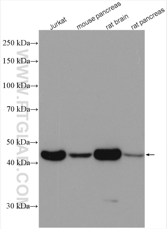 |
| Catalog no.: 66009-1-Ig | |
|
Beta-actin is usually used as a loading control due to its broad and consistent expression across all eukaryotic cell types and the fact that expression levels of this protein are not affected by most experimental treatments. 66009-1-Ig has been cited in over 2,460 publications and has wide species reactivity. |
Able™