![]()
Antibody Labeling Kits
FlexAble is a novel antibody labeling kit that uses an affinity linker to conjugate fluorochromes, enzymes, and molecules in any buffer condition - now with increased brightness to maximize your multiplex results.
FlexAble Antibody Labeling Kits revolutionized antibody labeling by allowing you to add dyes and molecules to any antibody, any time. We’ve improved our original FlexAble to allow for more dyes per antibody and a better, brighter signal with FlexAble 2.0 New.
-
 Compatible with primary antibodies from any supplier
-
Works with any antibody concentration and formulation
-
Fast and easy 2-step protocol – Ready to use in 10 minutes
-
Label as little as 0.5ug. No buffer exchange needed
-
Enable same species multiplexing
-
No additional equipment required
Research Area
Immunofluorescence Imaging of Live and Fixed Pancreatic Tissue Slices Using FlexAble 2.0 Antibody Labeling
Non visible textTestimonial
Scientist Stories: Léa Pechtimaldjian on Advancing 3D Vascular Imaging with FlexAble Technology
Non visible text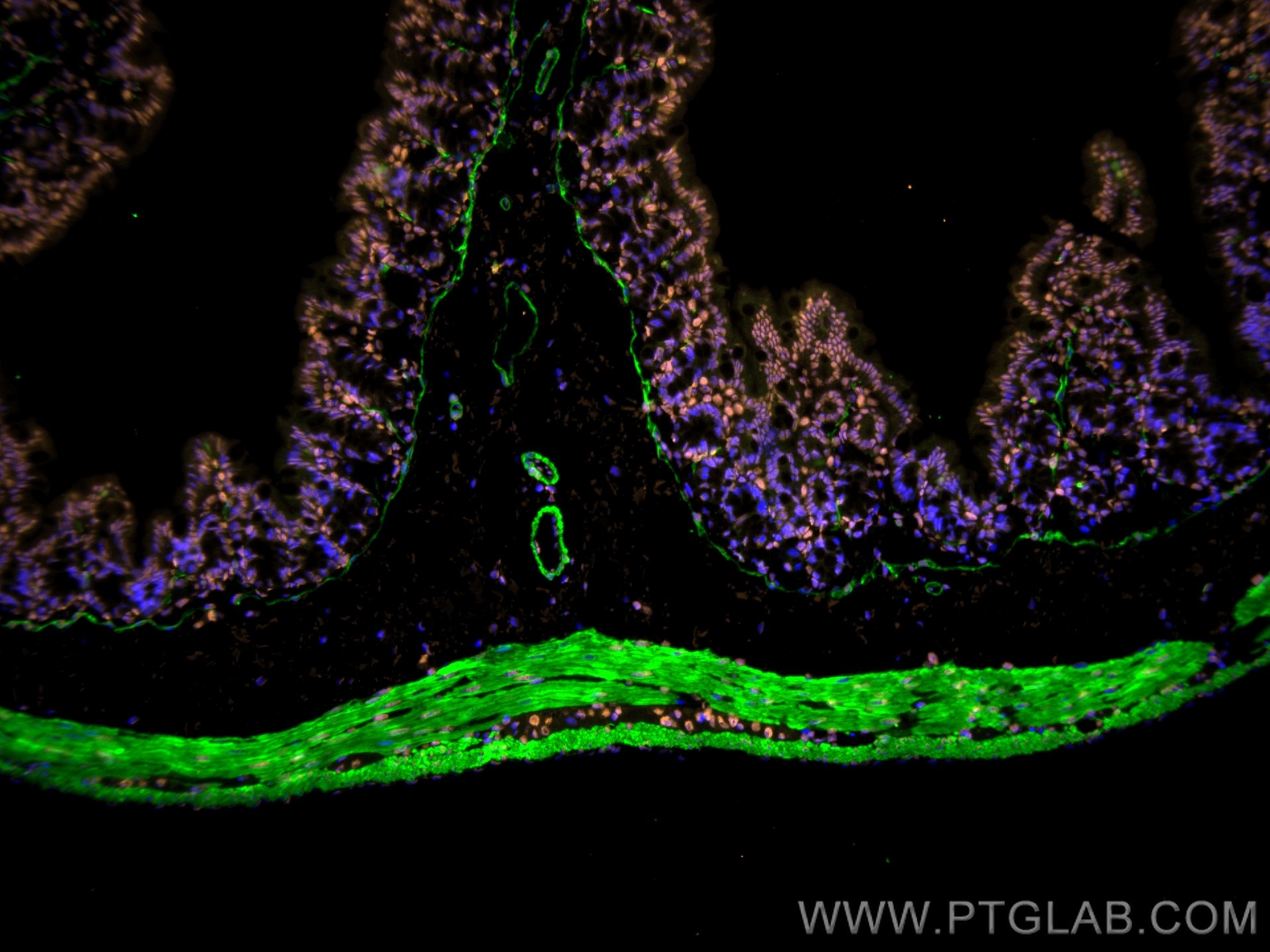
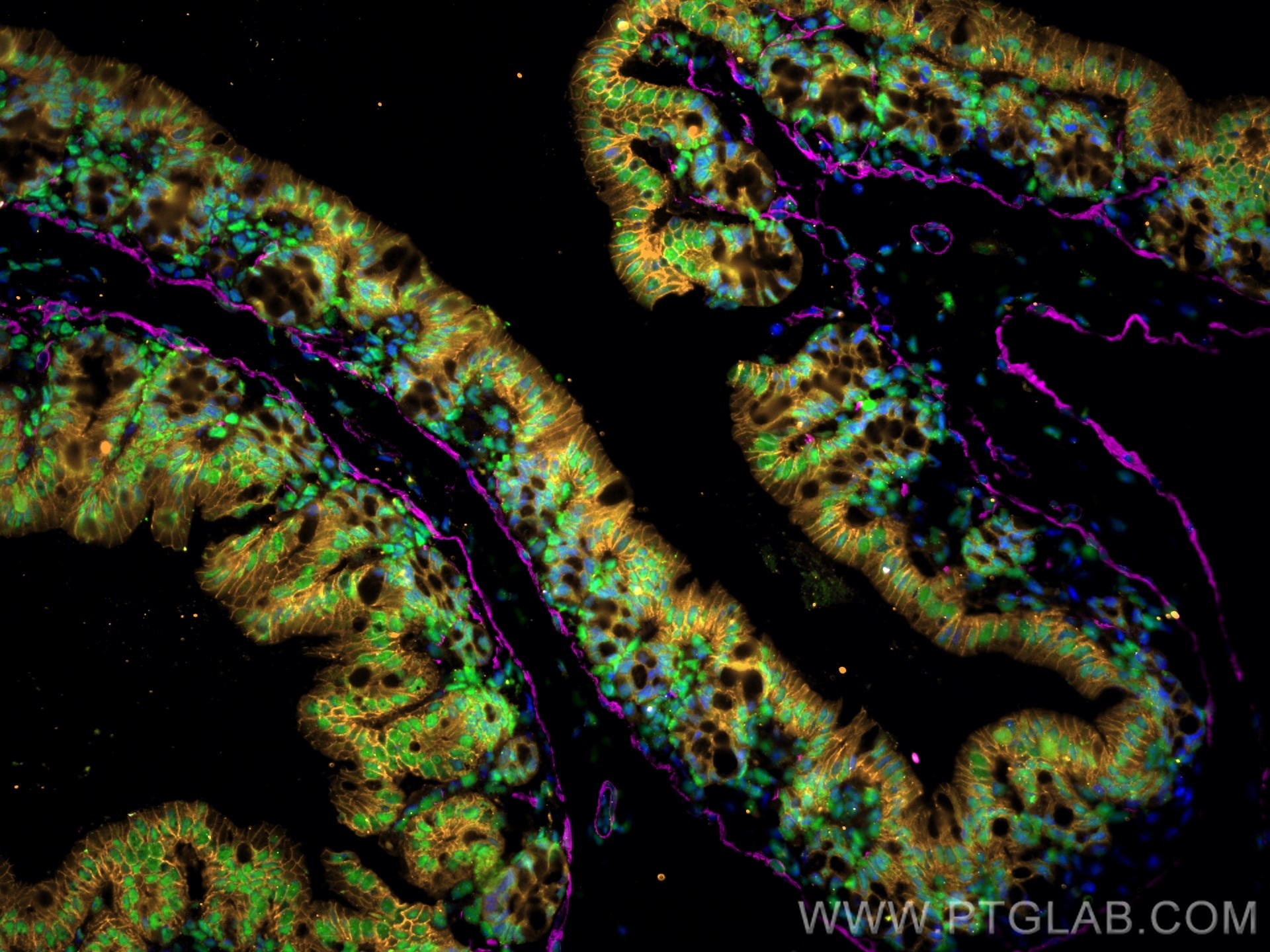
Immunofluorescence of Mouse colon: FFPE Mouse colon sections were stained with anti-FUS (11570-1-AP) labeled with FlexAble 2.0 CoraLite® Plus 555 Kit (KFA502, orange), anti-α-SMA (67735-1-Ig) labeled with FlexAble 2.0 CoraLite® Plus 488 Kit (KFA521, green) and DAPI (blue).
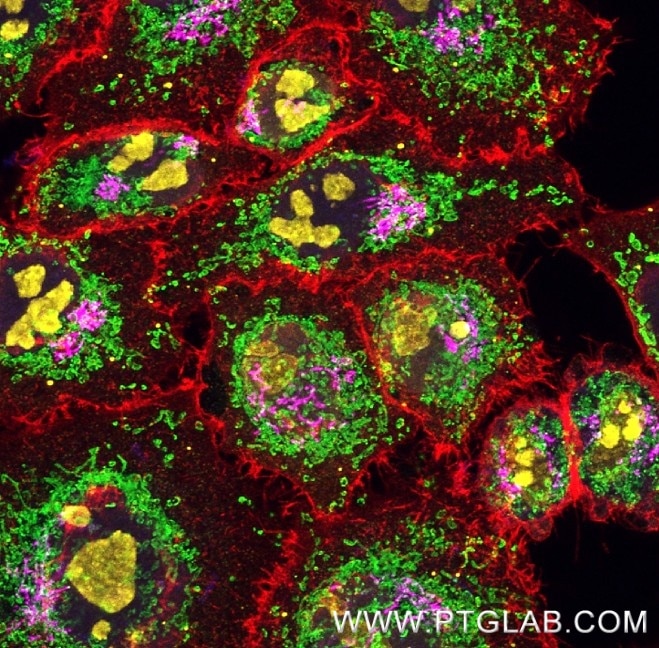
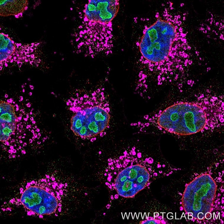
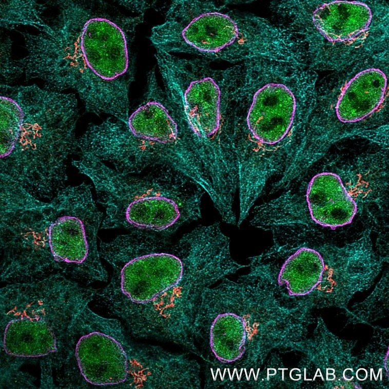
Immunofluorescence of HeLa cells with 5 rabbit primary antibodies: PFA-fixed cells were stained with anti-TDP-43 antibody (10782-2-AP, blue) labeled with FlexAble 2.0 CoraLite® Plus 405 Kit (KFA506), anti-TOM20 antibody (11802-1-AP, green) labeled with FlexAble 2.0 CoraLite® Plus 488 Kit (KFA501), anti-CD147 antibody (10186-R125, red) labeled with FlexAble 2.0 CoraLite® Plus 594 Kit (KFA509), anti-NCL antibody (10556-1-AP, yellow) labeled with FlexAble 2.0 CoraLite® Plus 647 Kit (KFA503), and anti-GOLGA2 antibody (11308-1-AP, magenta) labeled with FlexAble 2.0 CoraLite® Plus 750 Kit (KFA504). Confocal Images were acquired with a 60x oil objective and post-processed.
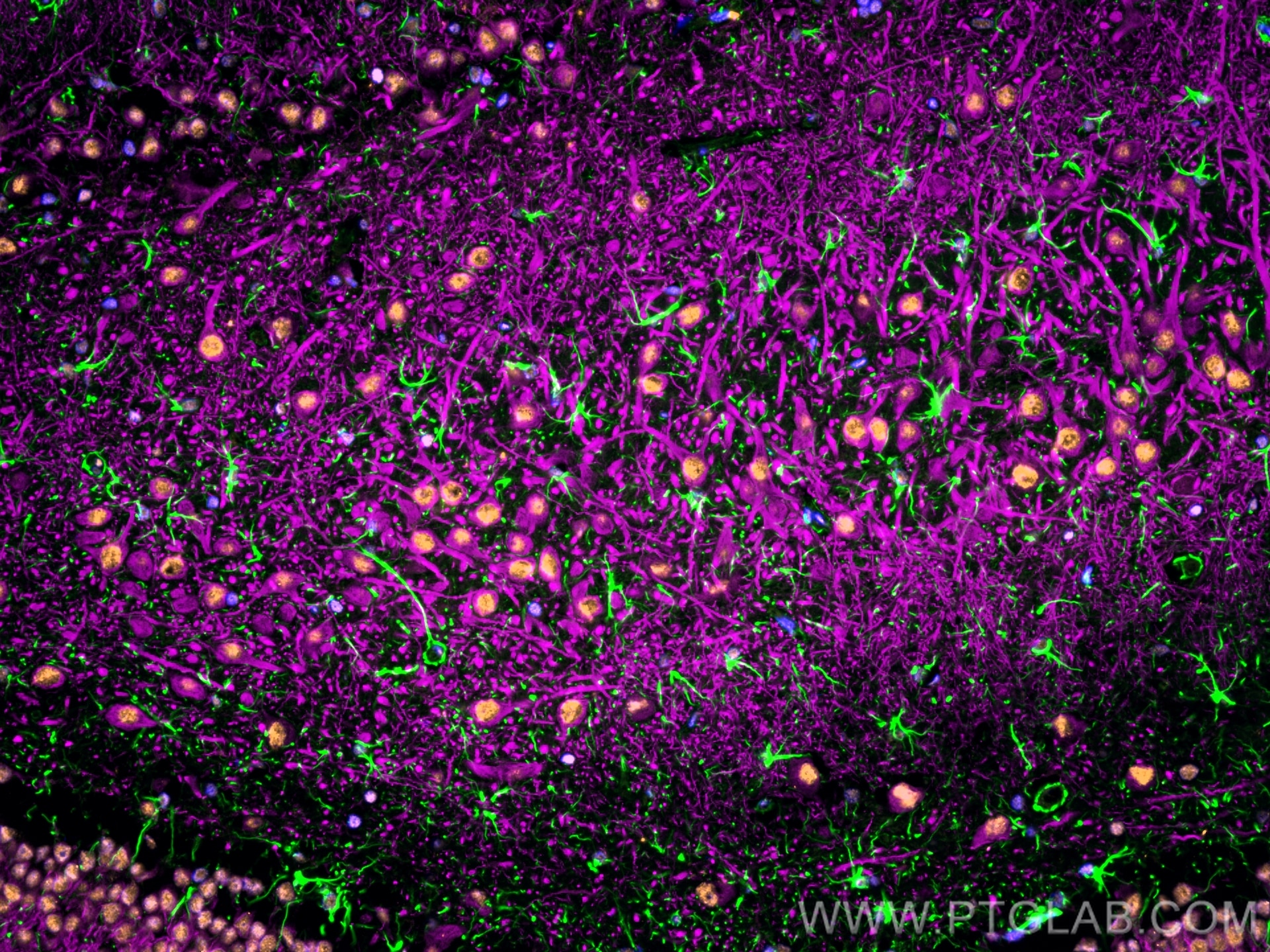
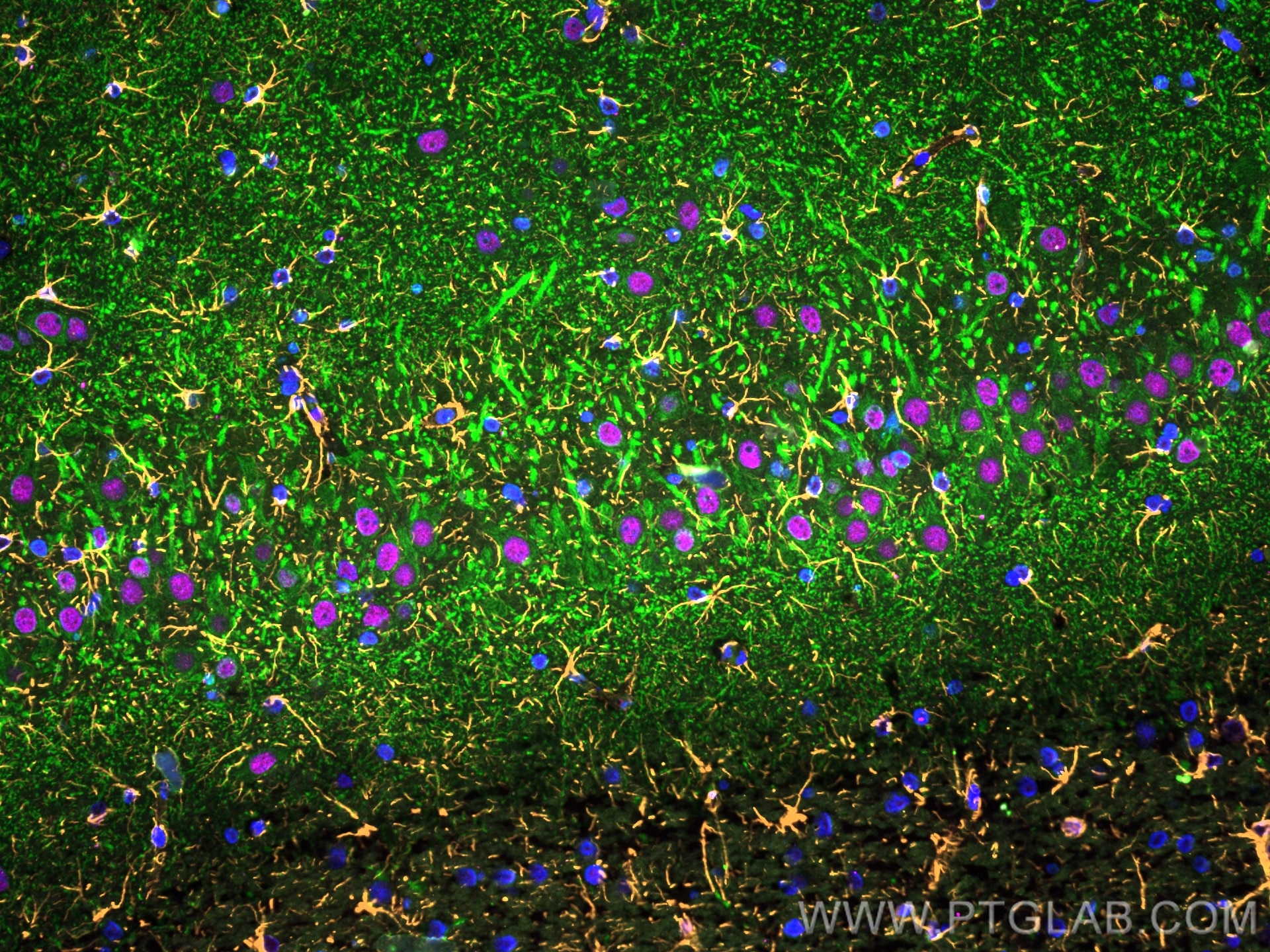
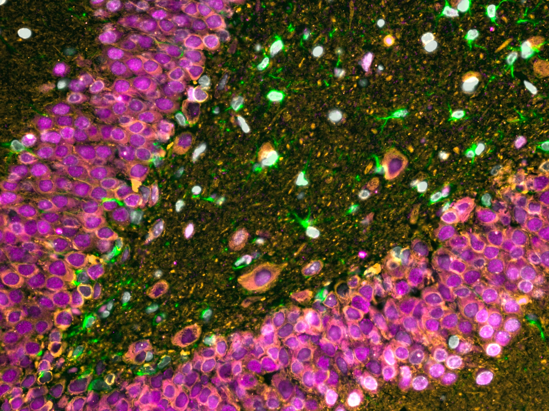
Immunofluorescence of Rat brain: FFPE Rat brain sections were stained with anti-MAP2 (17490-1-AP) labeled with FlexAble 2.0 CoraLite® Plus 647 Kit (KFA503, magenta), anti-GFAP (60190-1-Ig) labeled with FlexAble 2.0 CoraLite® Plus 488 Kit (KFA541, green), anti-TDP-43 (67345-1-Ig) labeled with FlexAble 2.0 CoraLite® Plus 555 Kit (KFA542,organge) and DAPI (blue).
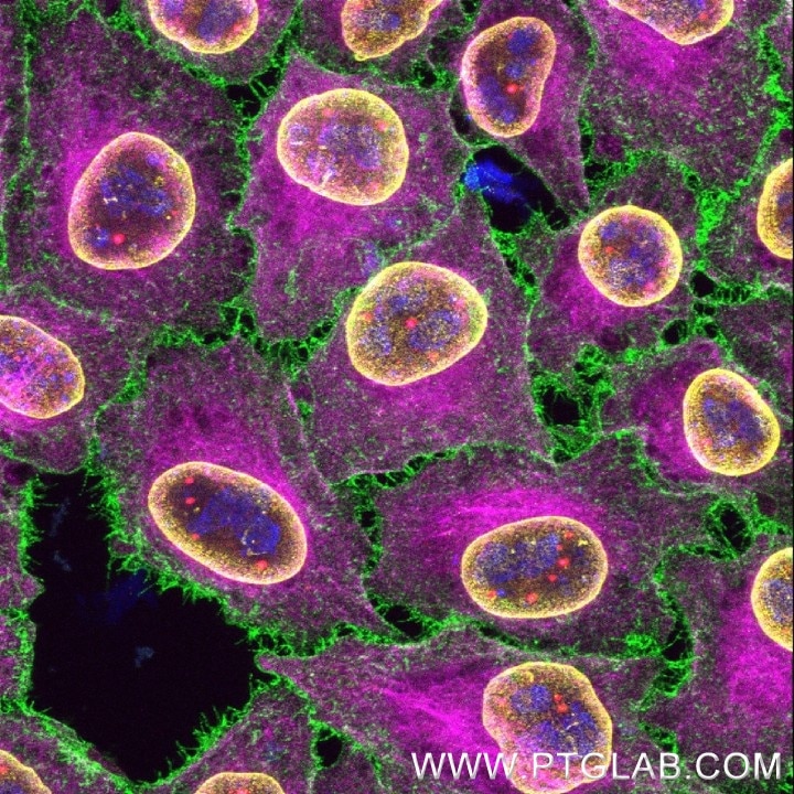
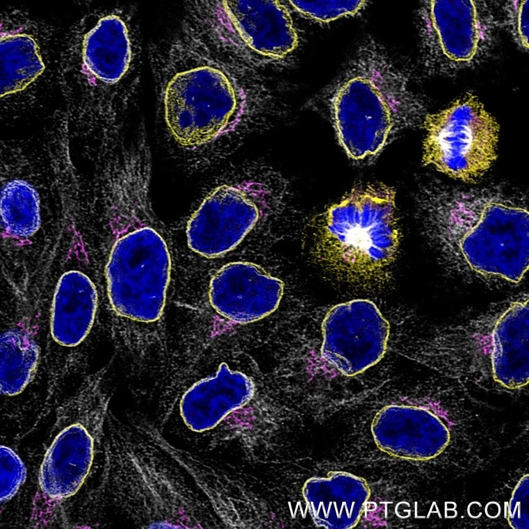
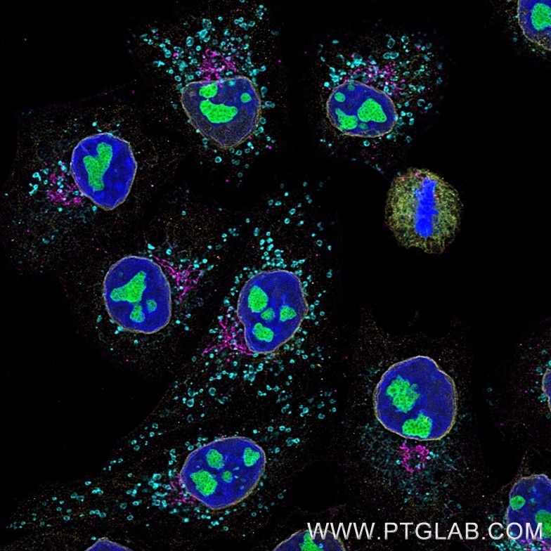
Immunofluorescence of HeLa cells with 5 rabbit primary antibodies: PFA-fixed cells were stained with anti-Ki-67 antibody (MA5-14520, blue) labeled with FlexAble 2.0 CoraLite® Plus 405 Kit (KFA506), anti-CD147 antibody (11989-1-AP, green) labeled with FlexAble 2.0 CoraLite® Plus 488 Kit (KFA501), anti-Lamin B1 antibody (12987-1-AP, yellow) labeled with FlexAble 2.0 CoraLite® Plus 555 Kit (KFA502), anti-Coilin antibody (10967-1-AP, red) labeled with FlexAble 2.0 CoraLite® Plus 647 Kit (KFA503), and anti-Tubulin antibody (80713-1-RR, magenta) labeled with FlexAble 2.0 CoraLite® Plus 800. Confocal Images were acquired with a 60x oil objective and post-processed.
Immunofluorescence of Mouse colon: FFPE Mouse colon sections were stained with anti-FUS (11570-1-AP) labeled with FlexAble 2.0 CoraLite® Plus 488 Kit (KFA501, green), anti-α-SMA (67735-1-Ig) labeled with FlexAble 2.0 CoraLite® Plus 647 Kit (KFA523, magenta), anti-E-cadherin (60335-1-Ig) labeled with FlexAble 2.0 CoraLite® Plus 555 Kit (KFA562, orange) and DAPI (blue).
Immunofluorescence of Hela cells: PFA-fixed cells were stained with rabbit IgG anti-Lamin antibody (12987-1-AP) labeled with FlexAble 2.0 CoraLite® Plus 555 Kit (KFA502, yellow), rabbit IgG anti-GM130 antibody (11308-1-AP) labeled with FlexAble 2.0 CoraLite® Plus 647 Kit (KFA503, magenta) and rabbit IgG anti-Tubulin antibody (80713-1-RR) labeled with FlexAble 2.0 CoraLite® Plus 750 Kit (KFA504, grey). Cell nuclei were stained with DAPI (blue).
Confocal images were acquired with a 63x oil objective and post-processed. Images were recorded at the Core Facility Bioimaging at the Biomedical Center, LMU Munich.
Immunofluorescence of Rat brain: FFPE Rat brain sections were stained with anti-MAP2 (17490-1-AP) labeled with FlexAble 2.0 CoraLite® Plus 488 Kit (KFA501, green), anti-GFAP (60190-1-Ig) labeled with FlexAble 2.0 CoraLite® Plus 555 Kit (KFA542, orange), anti-TDP-43 (67345-1-Ig) labeled with FlexAble 2.0 CoraLite® Plus 647 Kit (KFA543, magenta) and DAPI (blue).
Immunofluorescence of Hela cells: PFA-fixed cells were stained with mouse IgG1 anti-B23 antibody (60096-1-Ig) labeled with Multi-rAb CoraLite® Plus 488 Goat anti-Mouse Recombinant Secondary (RGAM002, green), mouse IgG1 anti-Lamin antibody (66095-1-Ig) labeled with FlexAble 2.0 CoraLite® Plus 555 Kit (KFA522, red) and mouse IgG1 anti-ATP5O antibody (66696-1-Ig) labeled with FlexAble 2.0 CoraLite® Plus 647 Kit (KFA523, magenta). Cell nuclei were stained with DAPI (blue).
Confocal images were acquired with a 63x oil objective and post-processed. Images were recorded at the Core Facility Bioimaging at the Biomedical Center, LMU Munich.
Immunofluorescence of Hela cells: PFA-fixed cells were stained with rabbit IgG anti-TDP43 antibody directly conjugated with CoraLite® Plus 488 (CL488-10782, green), rabbit IgG anti-GM130 antibody directly conjugated with CoraLite® Plus 555 (CL555-11308, red), rabbit IgG anti-Lamin antibody (12987-1-AP) labeled with FlexAble 2.0 CoraLite® Plus 647 Kit (KFA503, magenta) and rabbit IgG anti-Tubulin antibody (80713-1-RR) labeled with FlexAble 2.0 CoraLite® Plus 750 Kit (KFA504, cyan). Cell nuclei were stained with DAPI (blue).
Confocal images were acquired with a 63x oil objective and post-processed. Images were recorded at the Core Facility Bioimaging at the Biomedical Center, LMU Munich.
Immunofluorescence of Hela cells: PFA-fixed cells were stained with mouse IgG1 anti-B23 (60096-1-Ig) labeled with FlexAble 2.0 CoraLite® Plus 488 Kit (KFA521, green), mouse IgG1 anti-Lamin (66095-1-Ig) labeled with FlexAble 2.0 CoraLite® Plus 555 Kit (KFA522, yellow), mouse IgG1 anti-Gorasp2 (66627-1-Ig) labeled with FlexAble 2.0 CoraLite® Plus 647 Kit (KFA523, magenta) and mouse IgG1 anti-HSP60 (66041-1-Ig) labeled with FlexAble 2.0 CoraLite® Plus 750 Kit (KFA524, cyan). Cell nuclei were stained with DAPI (blue).
Confocal images were acquired with a 63x oil objective and post-processed. Images were recorded at the Core Facility Bioimaging at the Biomedical Center, LMU Munich.
Immunofluorescence of Hela cells: PFA-fixed cells were stained with mouse IgG1 anti-HSP60 antibody directly conjugated to CoraLite® Plus 488 (CL488-66041, green), mouse IgG1 anti-HDAC1 antibody directly conjugated to CoraLite® Plus 555 (CL555-66085, red), mouse IgG1 anti-Gorasp2 antibody (66627-1-Ig) labeled with FlexAble 2.0 CoraLite® Plus 647 Kit (KFA523, magenta) and mouse IgG1 anti-B23 antibody (60096-1-Ig) labeled with FlexAble 2.0 CoraLite® Plus 750 Kit (KFA524, cyan).
Confocal images were acquired with a 63x oil objective and post-processed. Images were recorded at the Core Facility Bioimaging at the Biomedical Center, LMU Munich.
Immunofluorescence of Hela cells: PFA-fixed cells were stained with mouse IgG2a anti-GNL3 antibody (67169-1-Ig) labeled with FlexAble 2.0 CoraLite® Plus 488 Kit (KFA541, green), mouse IgG2a anti-Tubulin antibody (66240-1-Ig) labeled with FlexAble 2.0 CoraLite® Plus 555 Kit (KFA542, red), mouse IgG2a anti-PAF49 antibody labeled with FlexAble 2.0 CoraLite® Plus 647 Kit (KFA543, magenta) and mouse IgG2a anti-Clec12a antibody labeled with FlexAble 2.0 CoraLite® Plus 750 Kit (KFA544, cyan). Cell nuclei were stained with DAPI (blue).
Confocal images were acquired with a 63x oil objective and post-processed. Images were recorded at the Core Facility Bioimaging at the Biomedical Center, LMU Munich.
FlexAble Kits have been validated in different multiplexing IF techniques – including cyclic IF, Iterative Bleaching Extends Multiplexity (IBEX) and tyramide signal amplification (TSA). They can also be used in expansion microscopy, and in tissue clearing/iDISCO+ clearing workflows.

Cyclic IF with HeLa cells: PFA-fixed HeLa cells were stained with mouse IgG1 anti-HSP60 antibody (66041-1-lg, Cycle 1) labeled with FlexAble 2.0 CoraLite® Plus 750 Kit (KFA504), imaged and bleached with 4.5 % H2O2 + 24 mM NaOH in PBS (1 h at RT, light). The next two cycle were performed accordingly using mouse IgG1 anti-B23 antibody (60096-1-lg, Cycle 2) labeled with FlexAble 2.0 CoraLite® Plus 750 (KFA504) and mouse IgG1 anti-Vimentin antibody (Sigma V6630, Cycle 3) labeled with FlexAble 2.0 CoraLite® Plus 750 (KFA504). Cell nuclei were stained with DAPI (blue).

Immunofluorescence of mouse cerebellum: FFPE mouse cerebellum sections were stained with anti-Calbindin antibody (14479-1-AP, green) labeled with FlexAble HRP Antibody Labeling Kit for Rabbit IgG (KFA005) and Tyramide-488, anti-Fus antibody (68262-1-Ig, orange) labeled with FlexAble HRP Antibody Labeling Kit for Mouse IgG1 (KFA025) and Tyramide-555, and anti-GFAP antibody (60190-1-Ig, magenta) labeled with FlexAble HRP Antibody Labeling Kit for Mouse IgG2a (KFA045) and Tyramide-650. Cell nuclei are in blue.
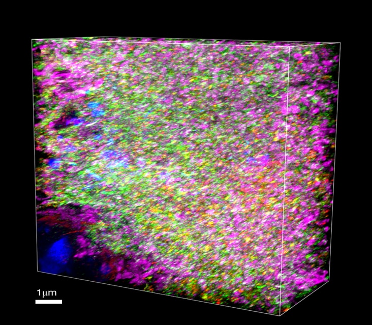
Volumetric image of a mouse brain specimen expanded using a Magnify™ Kit and stained with FlexAble 2.0 Antibody Labeling Kit. Expanded mouse brain tissue was stained with Rabbit anti-Syntaxin 1B (Proteintech, 83298-1-RR), Rabbit anti-Neuregulin (Proteintech, 83323-6-RR), and Rabbit anti-Ephrin A4 (Proteintech, 82951-1-RR). The antibodies were conjugated using the FlexAble 2.0 Antibody Labeling Kit (Syntaxin 1B conjugated with KFA501 CoraLite® Plus 488, Nuregulin conjugated with KFA502 CoraLite® Plus 555, and Ephrin A4 conjugated with KFA503 CoraLite® Plus 647). The sample was imaged using a Nikon Eclipse Ti2 epifluorescence microscope equipped with a CSU-W1 spinning disk confocal module using a CFI Plan Apochromat VC 60×C WI (1.2 NA) objective. Images were acquired by Magnify Biosciences.
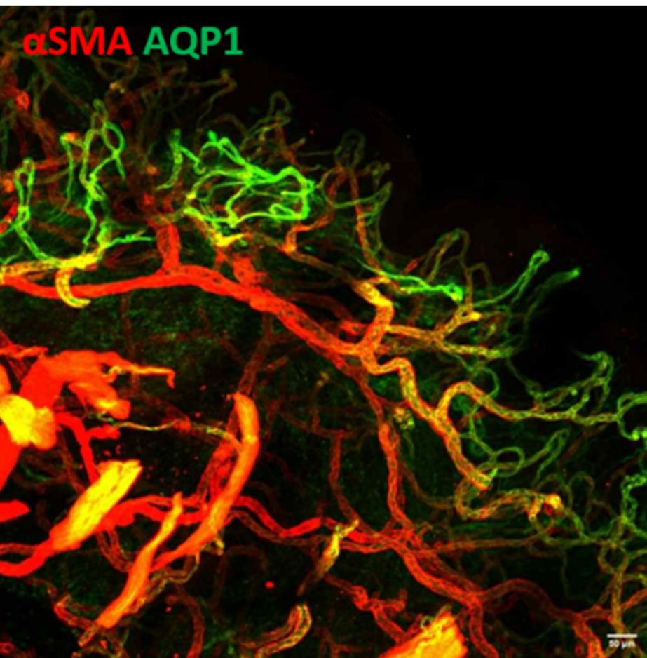
IF Imaging of 3D vascular structure in foreskin after Skin-iDISCO+ tissue clearing. Primary antibodies were labeled with FlexAble 2.0. Antibody Labeling Kits. Images were acquired by Léa Pechtimaldjian, PhD student at The Bordeaux Institute of oncology.
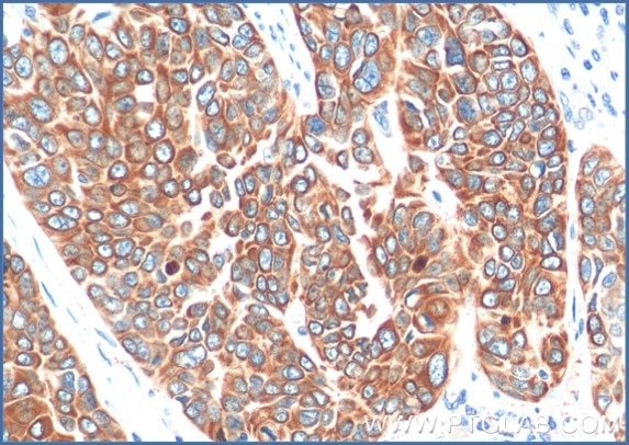
Immunohistochemical analysis of paraffin-embedded human oesophagus cancer tissue slide using anti-Cytokeratin 5 antibody (66727-1-Ig) labeled with FlexAble HRP Antibody Labeling Kit for Mouse IgG1 (KFA025). Antibody used at dilution of 1:400 under 10x & 40x lens. Heat mediated antigen retrieval performed with Tris-EDTA buffer (pH 9.0) and DAB substrate was used for detection.
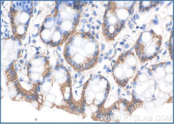
Immunohistochemical analysis of paraffin-embedded mouse colon tissue slide with anti-E-cadherin Antibody(20874-1-AP) labeled with FlexAble HRP Antibody Labeling Kit for Rabbit IgG (KFA005). Antibody used at a dilution of 1:1000 under 10x and 40x lens. Heat mediated antigen retrieval performed with Tris-EDTA buffer (pH 9.0) and DAB substrate was used for detection.
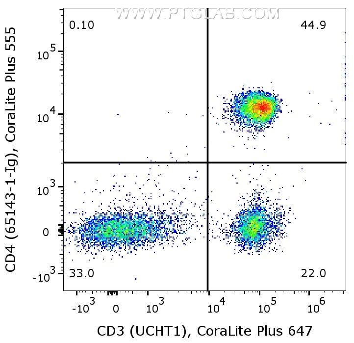
1X10^6 human PBMCs were surface co-stained with CoraLite® Plus 647 Anti-Human CD3 (CL647-65151, Clone:UCHT1), and Anti-Human CD4 (65143-1-Ig, Clone:RPA-T4) labeled with FlexAble 2.0 CoraLite® Plus 555 Kit (KFA522). Cells were not fixed. Lymphocytes were gated.
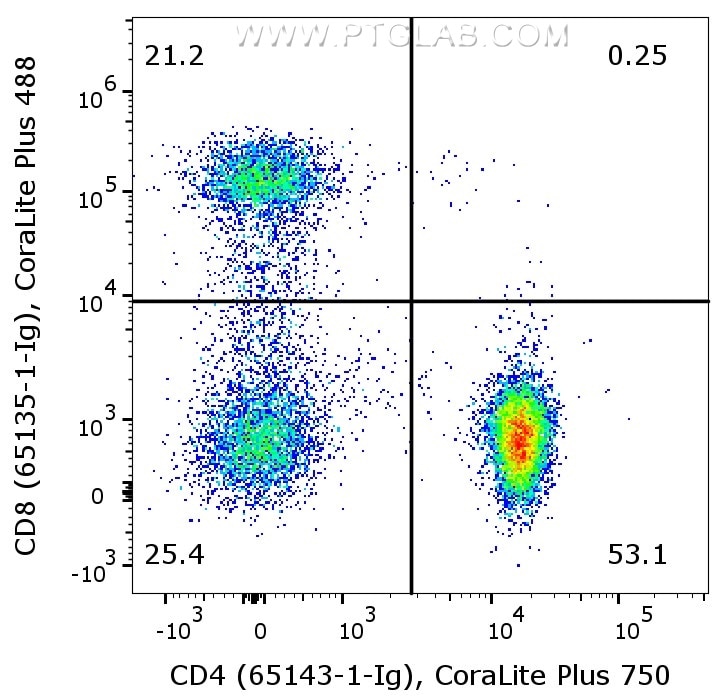
1X10^6 human PBMCs were surface co-stained with CoraLite® Plus 405 Anti-Human CD3 (CL405-65151, Clone: UCHT1), Anti-Human CD8 (65135-1-Ig, Clone:OKT8) labeled with FlexAble 2.0 CoraLite® Plus 488 Kit (KFA541), and Anti-Human CD4 (65143-1-Ig, Clone:RPA-T4) labeled with FlexAble 2.0 CoraLite® Plus 750 Kit (KFA524). Cells were not fixed. CD3+ lymphocytes were gated.
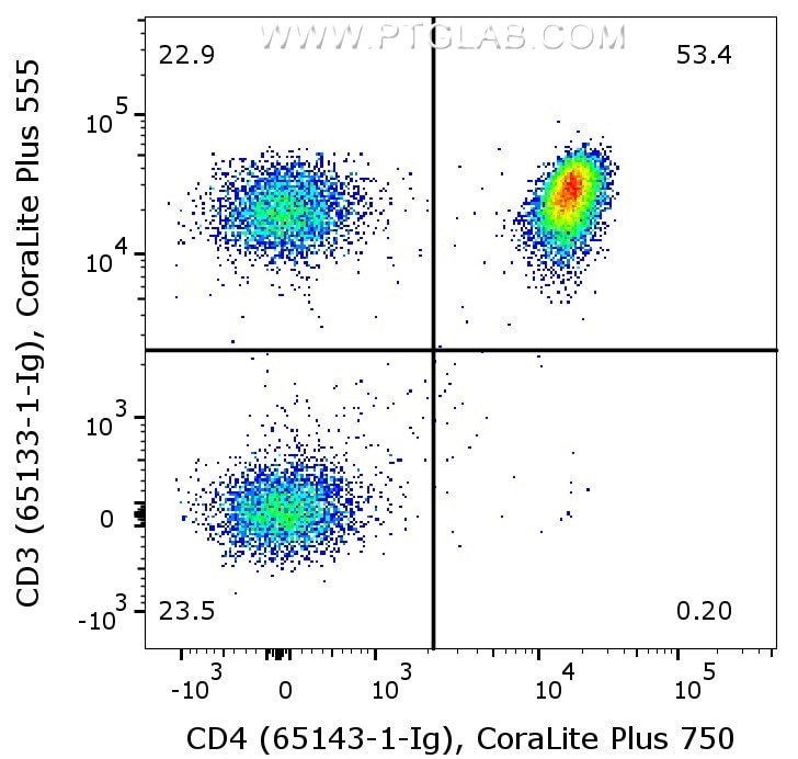
1X10^6 human PBMCs were surface co-stained with Anti-Human CD3 (65133-1-Ig, Clone:OKT3) labeled with FlexAble 2.0 CoraLite® Plus 555 Kit (KFA542), and Anti-Human CD4 (65143-1-Ig, Clone:RPA-T4) labeled with FlexAble 2.0 CoraLite® Plus 750 Kit (KFA524). Cells were not fixed. Lymphocytes were gated.
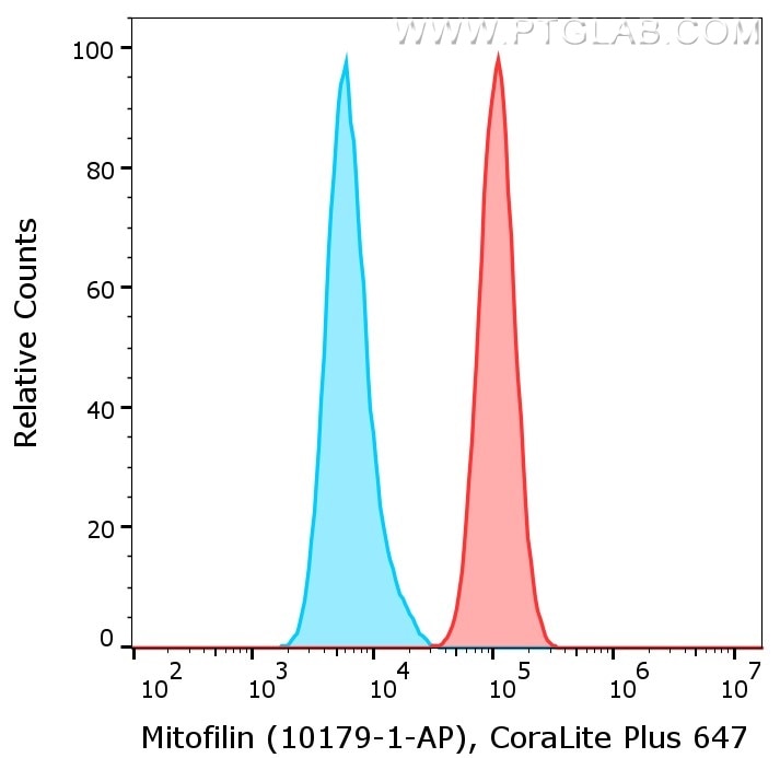
1X10^6 HeLa cells were intracellularly stained with anti-Mitofilin rabbit polyclonal antibody (10179-1-AP, red) or with isotype control antibody (30000-0-AP, blue) labeled with FlexAble 2.0 CoraLite® Plus 647 Kit (KFA503). Cells were fixed and permeabilized with Foxp3 /Transcription Factor Staining Buffer Kit (PF00011).
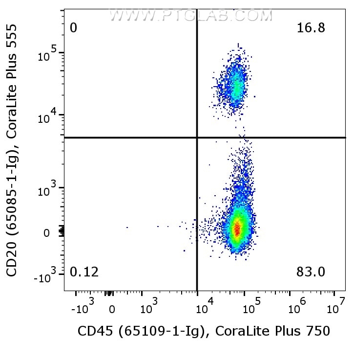
1X10^6 human PBMCs were surface co-stained with Anti-Human CD20 (65085-1-Ig, Clone:2H7) labeled with FlexAble 2.0 CoraLite® Plus 555 Kit (KFA562), and Anti-Human CD45 (65109-1-Ig, Clone:HI30) labeled with FlexAble 2.0 CoraLite® Plus 750 Kit (KFA524). Cells were not fixed. Lymphocytes were gated.
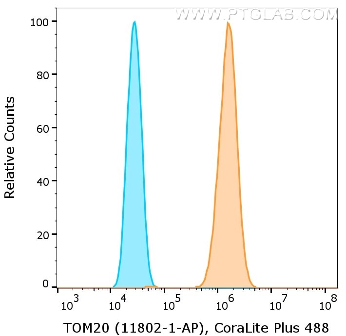
1X10^6 HeLa cells were intracellularly stained with anti-TOM20 rabbit polyclonal antibody (11802-1-AP, orange) or with isotype control antibody (30000-0-AP, blue) labeled with FlexAble 2.0 CoraLite® Plus 488 Kit (KFA501). Cells were fixed and permeabilized with Foxp3 /Transcription Factor Staining Buffer Kit (PF00011).
FlexAble Kits can be used to conjugate fluorophores or HRP to primary antibodies, enabling 1-step Western Blot detection. Accelerate your Western Blots even further with our SpeedAb WB Kits, which combine FlexAble HRP with Proteintech’s Blotting Accelerator cutting down your total experiment time to 2 h.
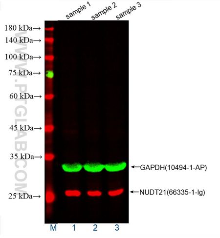
WB of HeLa cell lysates: HeLa cell lysates were detected with anti-GAPDH (10494-1-AP) labeled with FlexAble CoraLite® Plus 555 Kit (KFA002, green) and anti-NUDT21 (66335-1-Ig) labeled with FlexAble CoraLite® Plus 647 Kit (KFA023, red).
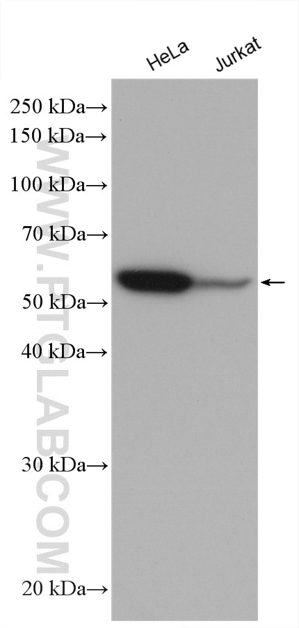
Various cell lysates were subjected to SDS PAGE followed by western blot with anti-GORASP2 antibody (66627-1-Ig) labeled with FlexAble HRP Antibody Labeling Kit for Mouse IgG1 (KFA025).
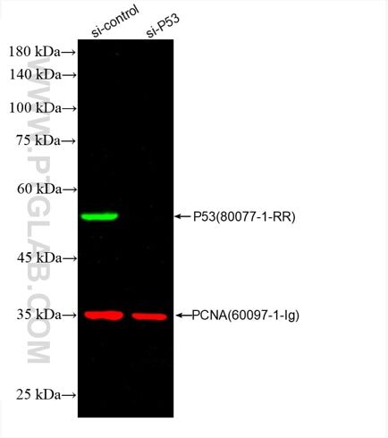
WB of A431 cell lysate: siRNA transfected A431 cell lysates were detected with anti-P53 (80077-1-RR) labeled with FlexAble CoraLite® Plus 555 Kit (KFA002, green) and anti-PCNA (60097-1-Ig) labeled with FlexAble CoraLite® Plus 647 Kit (KFA023, red).
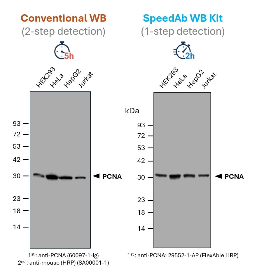
SpeedAb WB Kit for detection of mouse IgG1. (red) Classical Western blot detection of PCNA in lysates from different human cell lines. A primary anti-PCNA antibody (mouse IgG1) was used in combination with a secondary anti-mouse antibody (HRP), after blocking and incubation with antibodies for 1 h, respectively. The total time of ~ 5 h includes SDS-PAGE, blotting, blocking, primary and secondary antibody incubation, and washing steps of the membrane.(blue) Detection of PCNA in lysates from different human cell lines via SpeedAb WB Kit. The same primary anti-PCNA antibody was labeled with FlexAble HRP (anti-mouse IgG1) via a simple 10 min protocol, enabling a 1-step detection. The blotting accelerator speeds up binding of the antibody to the target protein, cutting the antibody incubation time to 15 min only and the total time including SDS-PAGE, blotting, antibody incubation and a final washing step to ~2 h.
FlexAble Kits can be used to label antibodies directly in culture supernatant (e.g., of hybridoma cells) without prior purification, concentration or buffer exchange simplifying the screening for positive clones. Read more here.
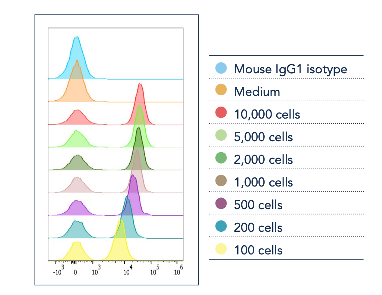
Readouts of labeled antibody harvested from the supernatant of CD3 (UCHT1) hybridomas. Cells were seeded in varying amounts into 96-well plates and FlexAble CoraLite Plus® 647 Kit for mouse IgG1 was used to label antibodies directly in the supernatant. The supernatants with labeled antibody were used to stain 1x10⁶ human PBMCs. Positively stained cells were detected by flow cytometry.
![]()
Oligo-Ready Antibody Labeling Kits
Combine the power of our FlexAble system wih the versatility of oligo labeling. Our kits use Fc-specific, high-affinity, conjugation-ready "FlexLinkers". Create your own set of up to three Oligo-FlexLinkers with one kit and use them for fast and easy antibody labeling.
-
Label 1 or up to 2 oligos per antibody
-
Same species multiplexing
-
Label 100 µg antibody per kit
-
60 – 80% recovery rate
-
Label as little as 0.5 µg antibody per reaction
-
Wide buffer compatibility range
Available FlexAble 2.0 Antibody Labeling Kits
Sizes: 10 rxn, 50 rxn, 200 rxn sizes.
Original FlexAble Kits are still available upon request here.
| Species | CoraLite® Plus | Other conjugates | |||||||
|---|---|---|---|---|---|---|---|---|---|
| Rabbit IgG | 405 | 488 | 555 | 594 | 647 | 750 | FITC Plus | HRP | Biotin |
| Mouse IgG1 | 405 | 488 | 555 | 594 | 647 | 750 | FITC Plus | HRP | Biotin |
| Mouse IgG2a | 405 | 488 | 555 | 594 | 647 | 750 | FITC Plus | HRP | Biotin |
| Mouse IgG2b | 405 | 488 | 555 | 594 | 647 | 750 | FITC Plus | HRP | Biotin |
| Human IgG | 405 | 488 | 555 | 594 | 647 | 750 | FITC Plus | HRP | Biotin |
| Rat Kappa Light Chain | 488 | 555 | 647 | FITC Plus | Biotin | ||||
CoraLite® and CoraLite® Plus dyes have equivalent brightness to the Alexa Fluor® dyes. Alexa Fluor® is a registered trademark of Molecular Probes, Inc, a Thermo Fisher Scientific Company.
Comparison of FlexAble labeled antibodies to secondary antibody detection
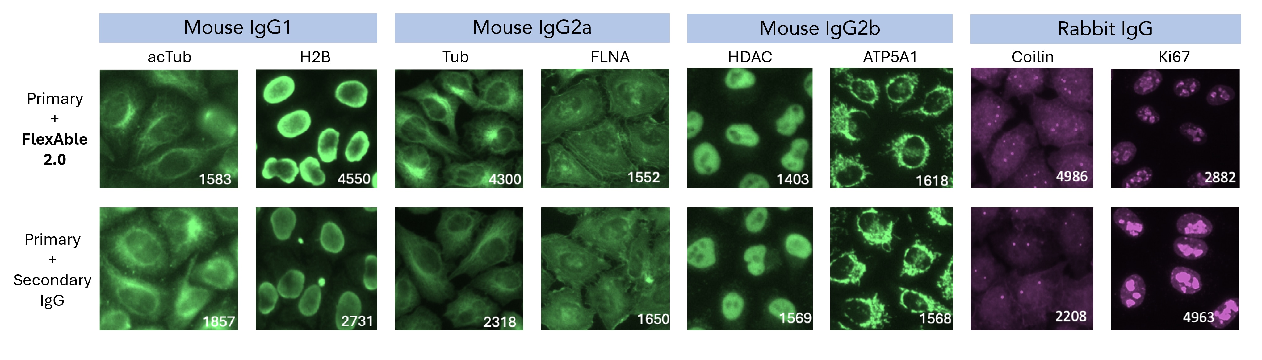
Eight different mouse and rabbit antibodies of different IgG subclasses were labeled with either Primary IgG + FlexAble 2.0, or with a primary and secondary IgG combination. Green: FlexAble 2.0 CoraLite Plus 488; Magenta: FlexAble 2.0 CoraLite Plus 647 or secondary antibodies labeled with the respective dyes. Mean fluorescent intensities are given to compare relative signals of each approach.
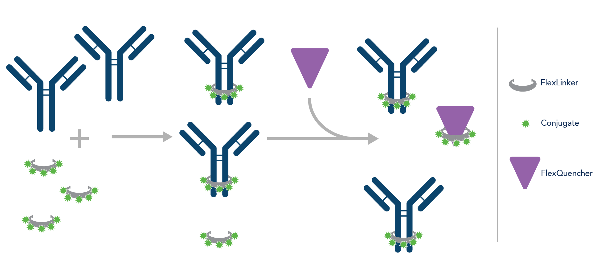
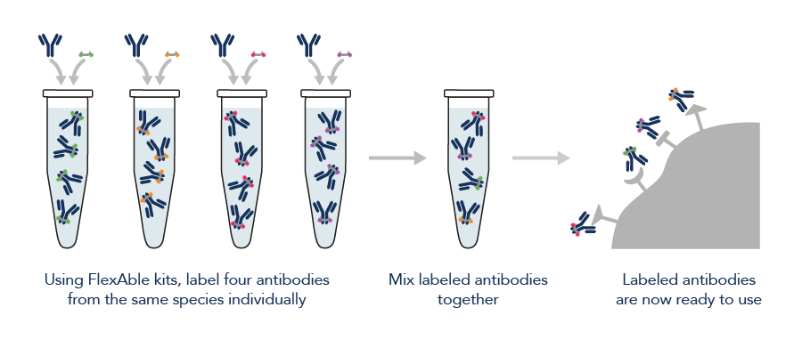
Antibody labeling with FlexAble is achieved by using a high-affinity, non-covalent FlexLinker. The FlexLinker binds tightly to the antibody within minutes and once bound does not release from the antibody. Unbound FlexLinker is neutralized by adding a FlexQuencher.
The labeling reaction using FlexAble yields a homogenous, well-defined antibody that is fully functional. Neither aggregation nor a loss of antibody is observed during the labeling.
FlexAble and FlexAble 2.0 Labeling kits can be used to label:
- Primary antibodies from any supplier
- Antibodies of any concentration
- Antibodies in any buffer formulation
FlexAble Antibody Labeling Kits have been thoroughly validated with primary antibodies from different suppliers. Primary antibodies with low concentration, antibodies stored in 50% glycerol, and antibodies supplemented with carriers like BSA are compatible for use with the FlexAble kits.
FlexAble and FlexAble 2.0 Antibody Labeling Kits work with any antibody formulation. Labeling with the high-affinity FlexLinker is not affected by stabilizers like BSA or glycerol, amine-containing buffers, or preservatives.
Antibody buffer formulation
FlexAble Antibody Labeling Kits work with any antibody formulation without the need to purify or buffer-exchange prior to labeling. This saves time and money and also avoids the loss of antibody that can happen during those processes. Since FlexAble utilizes a high-affinity FlexLinker molecule that binds site-specifically to an antibody, the binding reaction is not affected by carriers like BSA or glycerol, amine-containing buffers like Tris, or preservatives like sodium azide.
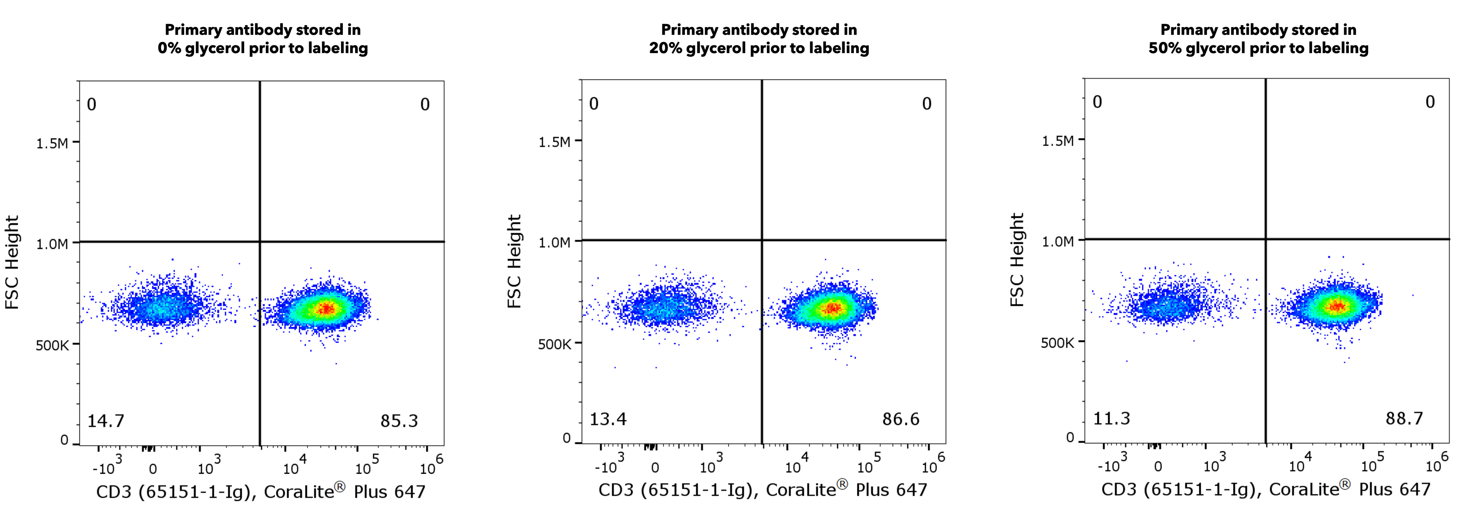
Anti-CD3 antibody, previously stored in the indicated concentration of glycerol, was efficiently labeled with FlexAble Antibody Labeling Kit regardless of the glycerol concentration during labeling. 1X10^6 human peripheral blood mononuclear cells (PBMCs) were stained with 0.5 µg anti-human CD3 antibody (clone UCHT1, 65151-1-Ig) labeled with FlexAble CoraLite® Plus 647 Kit (KFA023) . Cells were gated for live singlet lymphocytes.
Antibody Concentration
A highly concentrated antibody can be used without pre-dilution, or an antibody of low concentration can be used without further concentrating it. FlexAble is especially suitable for low-concentration antibodies due to the high affinity of the FlexLinker. Antibody concentrations as low as 0.06 mg/mL have been efficiently labeled with FlexAble.
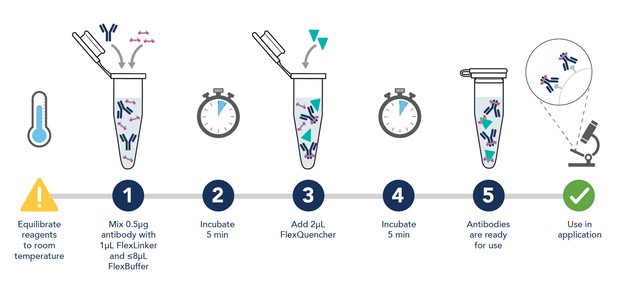
Labeling antibodies with FlexAble is fast and easy due to a simple 2-step protocol. The procedure only requires about 30 seconds of hands-on time and only a pipette is needed. Mix your primary antibody, FlexLinker, and FlexBuffer together and incubate for 5 minutes. Add FlexQuencher to neutralize unbound FlexLinker and incubate again for 5 minutes. The antibody is ready for use in the time it takes to label your tubes. The same protocol is suitable for both FlexAble and FlexAble 2.0 Antibody Labeling Kits.
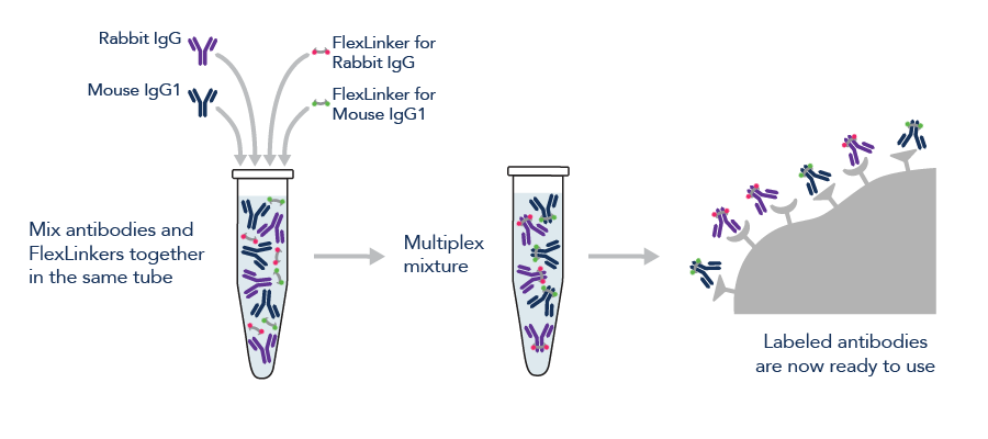
FlexAble Antibody Labeling Kits simplify multiplexing in immunofluorescence, western blotting, and flow cytometry. Save hands-on and incubation time by completing labeling of all your antibodies in 10 minutes before your experiment, followed by multiplex staining with all labeled antibodies at the same time. If primary antibodies of different species are used, they can even be labeled simultaneously in the same tube with FlexAble.
Antibodies labeled with FlexAble and FlexAble 2.0 Antibody Labeling Kits can be used alone or in multiplex experiments with:
- Other antibodies labeled with FlexAble Antibody Labeling Kits
- Commercially available pre-conjugated primary antibodies
- Other primary and secondary antibody combinations
Immunofluorescence of rat brain tissue: FFPE rat brain tissue sections were stained with anti-NeuN antibody (66836-1-Ig) labeled with FlexAble CoraLite® Plus 647 Kit (KFA023, magenta), anti-TUBB3 antibody 66375-1-Ig) labeled with FlexAble CoraLite® Plus 555 Kit (KFA022, orange), and CoraLite®488-conjugated GFAP antibody (CL488-60190, green).
FlexAble enables same species multiplexing
Same species multiplexing usually requires complex protocols that include primaries from different species/isotypes combined with highly specific and cross-adsorbed secondary antibodies. An alternative is pre-conjugated antibodies; however, not every antibody may be available in a pre-conjugated version.
FlexAble provides the easiest way to multiplex antibodies from the same species and isotype. FlexLinker binds to the primary antibody with high affinity and specificity and does not release from the antibody once bound. Therefore, antibodies from the same species or isotype can be incubated with the sample at the same time after being labeled with FlexAble. No leaking or cross-binding to other antibody isotypes has been observed.
![immunofluorescence staining of HeLa cells with TDP43 antibody,] and Lamin B1 antibody](/media/mqqpqn0g/1-immunofluorescence-of-hela-with-anti-tdp-43-antibody-anti-tom20-and-anti-lamin-b1-antibody.jpg)
Immunofluorescence of HeLa: PFA-fixed HeLa cells were stained with anti-TDP43 antibody (10782-2-AP) labeled with FlexAble CoraLite® 488 Kit (KFA001, green), anti-Tom20 antibody (11802-1-AP) labeled with FlexAble CoraLite® Plus 555 Kit (KFA002, red), and anti-Lamin B1 antibody (12987-1-AP) labeled with FlexAble CoraLite® Plus 647 Kit (KFA003, cyan). Confocal images were acquired with a 100x oil objective and post-processed. Images were recorded at the Core Facility Bioimaging at the Biomedical Center, LMU Munich.
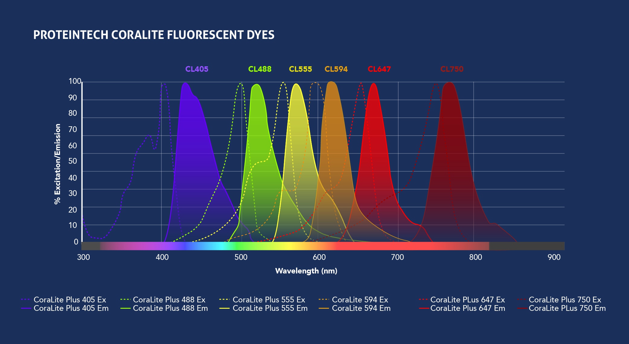
CoraLite Plus spectra
CoraLite® and CoraLite® Plus dyes are bright fluorescent dyes from Proteintech that deliver high photostability and minimal fluorescent bleed through, making it easy to combine two or more dyes in an experiment.
Our FlexAble and FlexAble 2.0 Kits are conjugated to multiple CoraLite® Plus dyes to offer more flexibility in multiple labeling experiments.