
Reverse Phase Protein Array – a high throughput proteomic tool
Discover the potential of RPPA and its many advantages over other proteomic techniques
Reverse Phase Protein Array (RPPA) is an antibody-based high throughput proteomic approach that enables the simultaneous quantification of large sample sizes (up to 1500 samples at once). Examples of RPPA use include biomarker quantification in clinical samples, mapping signaling interactions through phosphorylation events, and assessing molecular drug targets. This method has many advantages over other proteomic techniques such as western blotting, mass spectrometry, or ELISA.
The RPPA workflow:
Samples for RPPA are prepared in a similar way to western blot samples using SDS lysis and heat-mediated denaturation. These lysates are then plated into a 384- or 1536-well microtiter plate. Next, a microarrayer prints the samples onto a membrane-coated glass using a solid pin system that can handle the high viscosity of the concentrated samples, creating a “dot” on the membrane. Samples can be run in triplicates, and standard curves are often used. The slides are then blocked and probed with a primary antibody. Immunodetection is performed by an HRP-conjugated secondary antibody; however, a further signal amplification step is often required due to the low protein amount and small dots. The signal is then detected as either brightfield (DAB), a luminescent, or a fluorescent signal. Signal detection and quantification is automated, and the data can then be directly analyzed through software packages. See Figure 1 for an overview of the RPPA workflow.
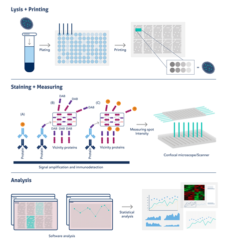
Fig 1: Simplified workflow of RPPA. Diagram adapted from Akbani et al, 2014 (PMID: 24777629).
Advantages of RPPA:
- The immunochemical signal amplification is greater (via DAB, luminescent, or fluorescent signal) and detection is more sensitive in RPPA, enabling a small amount of protein to be used.
- Signal readout and quantification is automated, increasing throughput and reducing human error.
- RPPA offers the possibility for multiplexing when using primary antibodies from different species (e.g., mouse and rabbit).
- RPPA offers signal uniformity as very large numbers of samples are read at once and can therefore be accurately compared for the same protein.
RPPA vs. other proteomic tools:
- Western blotting is limited in the number of samples that can be compared in a single experiment due to the multi-step protocol including SDS-PAGE and protein transfer to a membrane. Western blot requires 30-50ug of protein lysate, which can be challenging to extract from certain samples. In comparison, RPPA only requires 5ug extracted protein per sample.
- Mass spectrometry is another powerful proteomic tool and has the advantage of quantifying most of the sample’s proteome at once as well as detecting protein isoforms. It is best used during protein discovery rather than targeted research. However, mass spectrometry is limited to whole proteins and cannot detect post-translational modifications, therefore limiting its application in cell signaling research. RPPA is far more sensitive and cost-effective when investigating a known target. Mass-spec also requires extensive sample preparation, limiting the number of samples that can be processed at once.
- ELISAs are a sensitive and medium-throughput proteomic technique; however, they are limited to proteins in solution. RPPA can detect proteins from tissue/biopsies or even FFPE samples and therefore is better suited for high throughput clinical use than the other proteomic tools mentioned. ELISAs require pre-determined antibody pairs for detection, whereas RPPA is available to any antibody suited for western blot.
Summary Table:
|
Technique |
Advantages |
Disadvantages |
|
Western Blot |
Protein separation helps ensure the protein detected is the correct one. |
Labor intensive Low throughput High amount of protein |
|
ELISA |
Quantitative Sensitive |
Only for certain samples Specific antibody pairs required High amount of protein |
|
Mass spectrometry |
Unbiased for de novo protein discovery Protein isoforms are detected Thousands of proteins detected at once |
Complex sample preparation Low throughput Cannot detect PTMs |
|
IHC/IF |
Sub-cellular or tissue protein localization
|
Semi-quantitative Difficult to detect low-concentration targets |
|
RPPA |
Low amount of protein (5ug) High throughput High sensitivity PTMs detected Quantitative Possibility of multiplexing Signal uniformity across 1000s of samples. |
Special equipment required High-specificity antibody required for each slide |
The quality of the primary antibodies is at the center of high-quality data from RPPA.
As the protein lysate is not separated by molecular size before antibody probing, the antibody must be tried, tested, and trusted to detect the correct protein prior to large-scale arrays. It is recommended to carry out a western blot with positive and negative controls to confirm the specificity of the antibody for its target.
Proteintech antibodies can be used for RPPA!
A recent publication in The Journal of Immunology (Fan et al. 2022; PMID: 36150727) explores the role of metabolic rewiring in human macrophages and lung cancer tissue organoids. Macrophages can display opposing functions depending on their polarization status, and this translates into the cancer context. Tumor-associated macrophages are typically M2-like and tumor-promoting by creating an immunosuppressive environment through T-lymphocyte down-regulation. This is particularly true in Non-Small Cell Lung Carcinoma (NSCLC) where there are areas of T cell exclusion that lead to checkpoint therapy resistance.
The authors used radioactive tracers and Reverse Phase Protein Array (RPPA) to track immunomodulatory metabolic changes over time in healthy and tumor-associated macrophages in response to differential polarization and whole protein glucans (WPGs).
RPPA is a microarray antibody-based technique that allows for high throughput protein quantification. 80% of the primary antibodies used by the authors for RPPA were from Proteintech (42 Proteintech antibodies used in a single publication!). We are honored to help the researchers in the huge undertaking that is mapping macrophage metabolism over time. We are always thrilled to share our customers’ success.
Proteintech antibodies used by authors:
|
1 |
19677-1-AP |
|
|
2 |
60006-1-Ig |
|
|
3 |
16131-1-AP |
|
|
4 |
25949-1-AP |
|
|
5 |
17762-1-AP |
|
|
6 |
12842-1-AP |
|
|
7 |
11375-1-AP |
|
|
8 |
14299-1-AP |
|
|
9 |
22104-1-AP |
|
|
10 |
10566-1-AP |
|
|
11 |
20960-1-AP |
|
|
12 |
13333-1-AP |
|
|
13 |
66197-1-Ig |
|
|
14 |
15932-1-AP |
|
|
15 |
13268-1-AP |
|
|
16 |
15365-1-AP |
|
|
17 |
16806-1-AP |
|
|
18 |
66142-1-Ig |
|
|
19 |
66146-1-Ig |
|
|
20 |
60269-1-Ig |
|
|
21 |
66196-1-Ig |
|
|
22 |
12855-1-AP |
|
|
23 |
55013-1-AP |
|
|
24 |
15904-1-AP |
|
|
25 |
15212-1-AP |
|
|
26 |
16588-1-AP |
|
|
27 |
16754-1-AP |
|
|
28 |
14892-1-AP |
|
|
29 |
21829-1-AP |
|
|
30 |
13763-1-AP |
|
|
31 |
14718-1-AP |
|
|
32 |
60268-1-Ig |
|
|
33 |
55380-1-AP |
|
|
34 |
15851-1-AP |
|
|
35 |
25174-1-AP |
|
|
36 |
10620-1-AP |
|
|
37 |
21898-1-AP |
|
|
38 |
60291-1-Ig |
|
|
39 |
19003-1-AP |
|
|
40 |
25413-1-AP |
|
|
41 |
19987-1-AP |
|
|
42 |
18068-1-AP |
Blog written by Lucie Reboud, 4th year PhD Student at the University of Manchester and Science Marketing Intern for Proteintech.
Related Content
Detecting low abundance proteins via Western Blot
Go with the Flow - Make the most of this powerful technique
How to optimize your ELISA experiments
Loading control antibodies for Western Blotting

Support
Newsletter Signup
Stay up-to-date with our latest news and events. New to Proteintech? Get 10% off your first order when you sign up.
Able™