Neuroscience
Elevate your neuroscience research with purpose built reagents for discovery, validation, and beyond
Why choose Proteintech for your neuroscience research
-
 Comprehensive portfolio of extensively cited antibodies against key neural and neurodegenerative markers
-
Premium, bioactive cytokines and growth factors designed for organoid cultures and neuronal differentiation workflows
-
Specialized tissue multiplexing tools for spatial analysis and mapping neural complexity
-
Expert-level technical support and resources for workflow optimization and experimental troubleshooting
How to choose a neural cell-type marker
Choose the marker by cell type and the compartment you need to resolve, then confirm the antibody is validated in your species and application. The table below lists the most widely used CNS cell-type markers.
| Cell Type | Marker | Localization | Applications |
|---|---|---|---|
| Neuron | NeuN | Nucleus | WB, IHC, IF |
| MAP2 | Dendritic | WB, IHC, IF | |
| TUBB3 | Cytoplasm and neurites | WB, IHC, IF, IP | |
| Astrocyte | GFAP | Cytoplasm and cellular processes | WB, IHC, IF, IP |
| ALDH1L1 | Cytoplasm and cellular processes (pan-astrocytic) | WB, IHC, IF, IP | |
| AQP4 | End-feet | WB, IHC, IF, IP | |
| S100B | Cytoplasm and nucleus | WB, IHC, IF | |
| Microglia | IBA1 | Cytoplasm and processes (pan-microglial) | WB, IHC, IF |
| TMEM119 | Plasma membrane of resting microglia | WB, IHC, IF | |
| P2RY12 | Plasma membrane of resting microglia | WB, IF | |
| CD68 | Endosomal and lysosomal membranes | WB, IHC, IF, FC | |
| Oligodendrocyte | OLIG2 | Nucleus | WB, IHC, IF, IP |
| SOX10 | Nucleus | WB, IHC, IF, IP | |
| CNPase | Non-compacted myelin | WB, IHC, IF, FC (Intra) | |
| MBP | Compacted myelin | WB, IHC, IF | |
| MOG | Plasma membrane | WB, IHC, IF | |
| OPC | NG2 | Plasma membrane | WB, IHC, IF |
| PDGFRA | Plasma membrane | WB, IF, FC | |
| Neural Stem Cells | Nestin | Cytoplasm | WB, IHC, IF |
| SOX2 | Nucleus | WB, IHC, IF, FC (Intra) | |
| PAX6 | Nucleus | WB, IHC, IF, FC (Intra) | |
| FABP7 | Cytoplasm and nucleus | WB, IHC, IF |
Find the right marker
Filter Proteintech's neuroscience catalogue by what you study, the cell type or process you are interested in, species and application.
Antibodies for the targets that matter most
Browse through some of the most widely used targets in neuroscience research- TDP43, GFAP, NeuN, synaptophysin, and phospho Tau, with our broad of selection of high-cited antibodies, validated in multiple applications and available conjugated to multiple fluorescent dyes for easy multiplexing. Many of these antibodies have been 3D epitope mapped and feature whole slide IHC validation images.
TDP-43 — ALS and FTLD
TDP-43 (gene TARDBP) is the defining pathological protein of amyotrophic lateral sclerosis (ALS) and frontotemporal lobar degeneration (FTLD-TDP), and is increasingly implicated in Alzheimer's disease and LATE.
| Antibody (catalog no.) | Best for | Citations | Validated applications |
|---|---|---|---|
| TDP-43 Polyclonal Antibody (10782-2-AP) | Industry-standard detection reagent; played a seminal role in the original identification of TDP-43 pathology | 1,700+ | WB, IP, IHC, IF-Fro, IF/ICC, FC (Intra), ChIP |
| TDP-43 Monoclonal Antibody (60019-2-Ig) | High-affinity, human-specific detection; 3D epitope mapped | 120+ | WB, IP, IHC |
| TDP-43 Recombinant Antibody (80001-1-RR) | N-terminal-specific detection; 3D epitope mapped; whole-slide IHC plus advanced autostainer validation in clinical samples | — | WB, IHC, IF-P, IF, FC (Intra) |
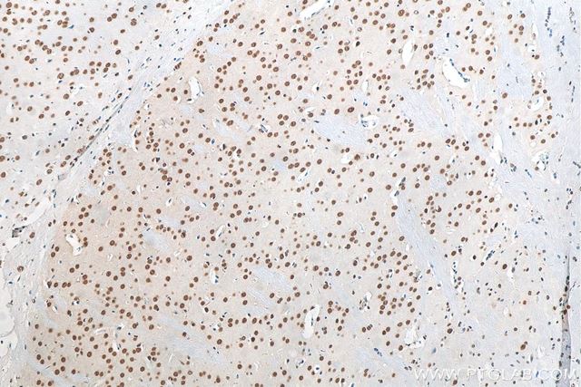
IHC staining of mouse brain tissue using TDP-43 (N-terminal) Polyclonal antibody.
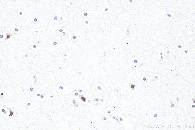
IHC staining of human brain tissue using TDP-43 (N-terminal) Polyclonal antibody.
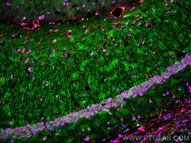
IF staining of mouse brain tissue using TDP-43 (N-terminal) Polyclonal antibody.
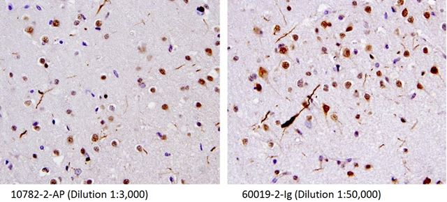
IHC staining of human brain (FTLD-U) tissue using TDP-43 (human-specific) Monoclonal antibody.
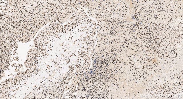
IHC staining of human glioma tissue using TDP-43 (human-specific) Monoclonal antibody.
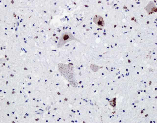
IHC staining of spinal cord from ALS patients using TDP-43 (N-terminal) Recombinant antibody.
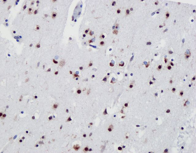
IHC staining of frontal cortex from FTLD-TDP type B using TDP-43 (N-terminal) Recombinant antibody.
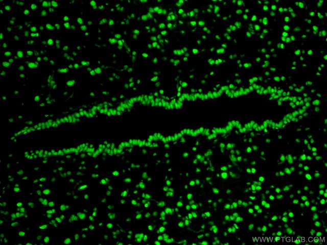
IF staining of rat brain tissue using TDP-43 (N-terminal) Recombinant antibody.
GFAP — Astrocytes
GFAP (glial fibrillary acidic protein) is the gold-standard marker for astrocytes. Increased expression is commonly associated with reactive astrocytes and astrogliosis. Circulating GFAP has also emerged as a promising blood-based biomarker of CNS injury and neurodegenerative disease.
| Antibody (catalog no.) | Best for | Citations | Validated applications |
|---|---|---|---|
| GFAP Polyclonal Antibody (16825-1-AP) | Top choice among researchers; multiple dye and biotin conjugate options available | 900+ | WB, IHC, IF-P, IF-Fro, IF/ICC |
| GFAP Monoclonal Antibody (60190-1-Ig) | High-affinity mouse monoclonal; 3D epitope mapped; whole-slide IHC data | 320+ | WB, IP, IHC, IF-P |
| GFAP Recombinant Antibody (81063-1-RR) | 3D epitope mapped; whole-slide IHC plus autostainer validation in healthy brain and glioma tissue | — | WB, IHC, IF-P, IF, IF/ICC |
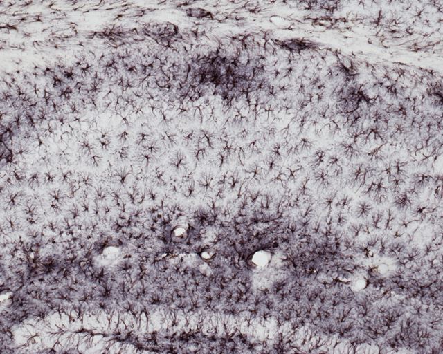
IHC staining of Alzheimer's disease mouse brain using GFAP Polyclonal antibody.
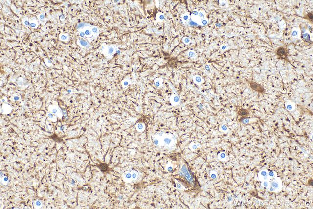
IHC staining of human brain tissue using GFAP Polyclonal antibody.
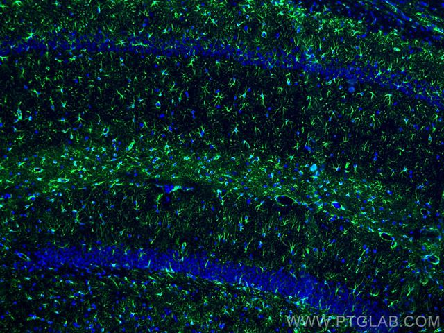
IF staining of rat brain tissue using GFAP Polyclonal antibody.
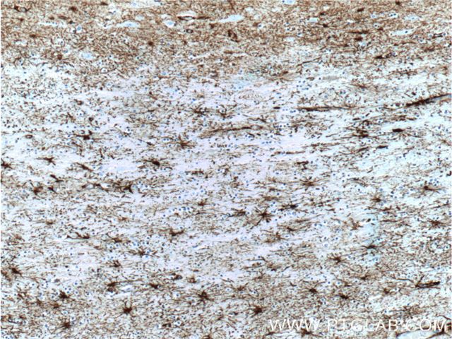
IHC staining of human brain tissue using GFAP Monoclonal antibody.
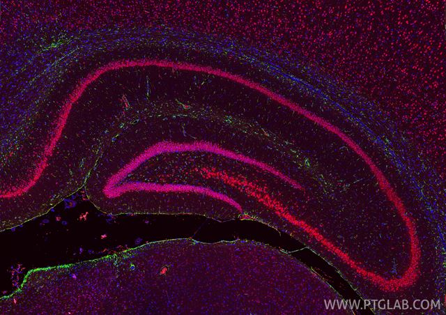
IF staining of rat brain tissue using GFAP Monoclonal antibody.
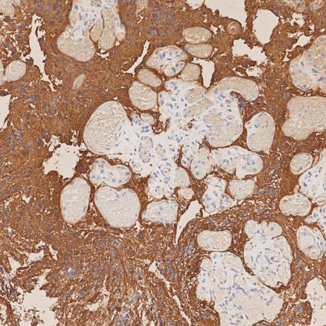
IHC staining of human glioma tissue using GFAP Recombinant antibody.
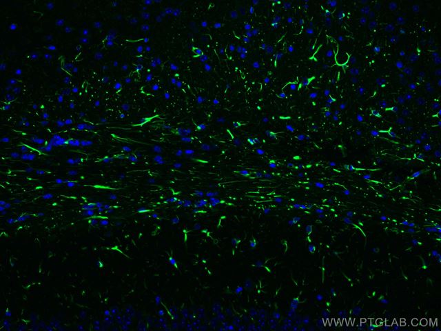
IF staining of mouse brain tissue using GFAP Recombinant antibody.
NeuN (RBFOX3) — Neuronal maturation
NeuN, encoded by RBFOX3, is the premier marker of mature, post-mitotic neurons and a mainstay of neuron counting, neuronal maturation studies.
| Antibody (catalog no.) | Best for | Citations | Validated applications |
|---|---|---|---|
| NeuN Polyclonal Antibody (26975-1-AP) | Highly versatile across tissue and cell applications | 500+ | WB, IHC, IF-P, IF-Fro, FC (Intra) |
| NeuN Monoclonal Antibody (66836-1-Ig) | High-affinity, optimised for IHC; 3D epitope mapped; whole-slide IHC plus autostainer validation in glioma and healthy brain tissue | 260+ | IHC, WB, IF |
| NeuN Recombinant Antibody (84401-4-RR) | 3D epitope mapped; high affinity and unmatched lot-to-lot consistency for long-term projects | — | WB, IHC, IF-P, IF-Fro |
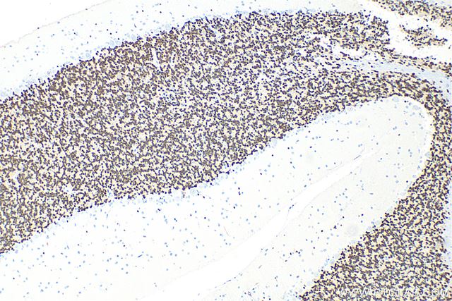
IHC staining of rat cerebellum tissue using NeuN Polyclonal antibody.
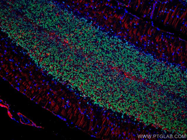
IF staining of rat cerebellum tissue using NeuN Polyclonal antibody.
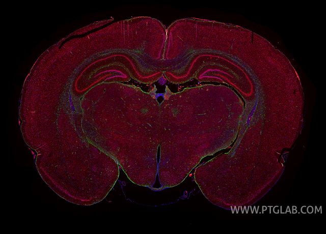
IF staining of rat brain tissue using NeuN Polyclonal antibody.
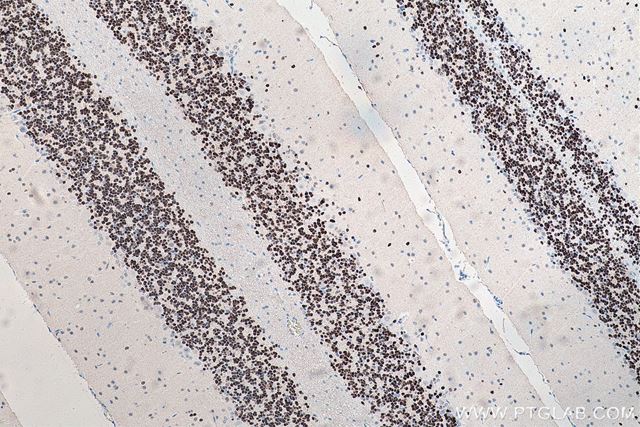
IHC staining of rat cerebellum tissue using NeuN Monoclonal antibody.
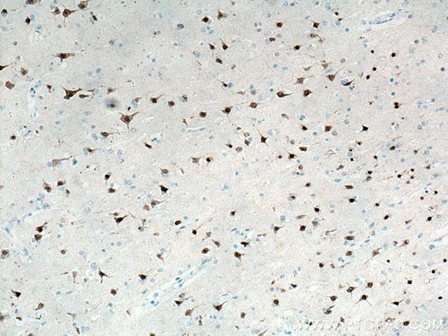
IHC staining of human brain tissue using NeuN Monoclonal antibody.
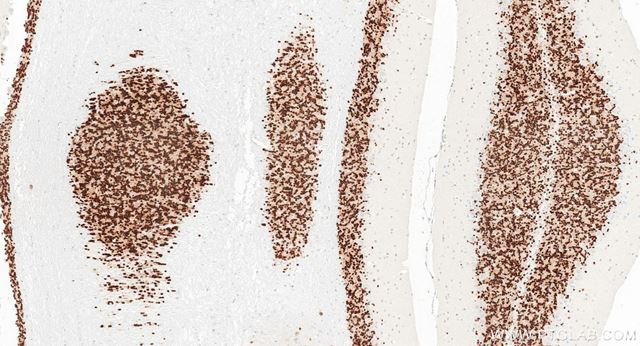
IHC staining of mouse cerebellum tissue using NeuN Recombinant antibody.
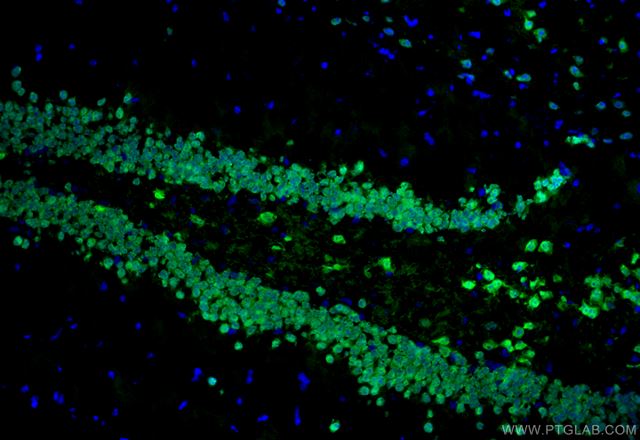
IF staining of mouse brain tissue using NeuN Recombinant antibody.
Synaptophysin — Presynaptic vesicles and synapse density
Synaptophysin (gene SYP) is an abundant synaptic vesicle glycoprotein used to quantify synaptic vesicle content and visualise synapse density in development, plasticity and neurodegeneration.
| Antibody (catalog no.) | Best for | Citations | Validated applications |
|---|---|---|---|
| Synaptophysin Polyclonal Antibody (17785-1-AP) | Top-cited synaptophysin antibody | 350+ | WB, IP, IHC, IF-P |
| Synaptophysin Monoclonal Antibody (67864-1-Ig) | Optimised for IHC; 3D epitope mapped; whole-slide IHC plus autostainer validation in multiple tissues | — | IHC, WB, IF |
| Synaptophysin Recombinant Antibody (82900-1-RR) | High-affinity antibody for use across multiple applications; 3D epitope mapped; unmatched lot-to-lot consistency for long-term projects | — | WB, IP, IHC, IF-P, IF/ICC, FC (Intra) |
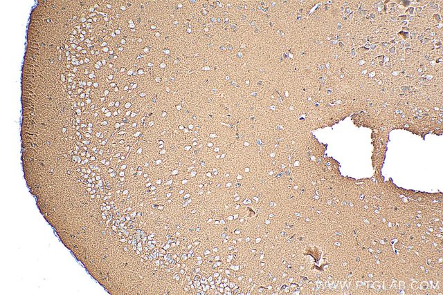
IHC staining of mouse brain tissue using Synaptophysin Polyclonal antibody.
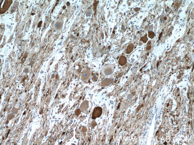
IHC staining of ganglioma tissue using Synaptophysin Polyclonal antibody.
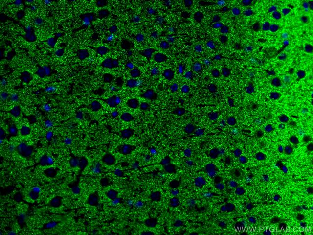
IF staining of mouse brain tissue using Synaptophysin Polyclonal antibody.
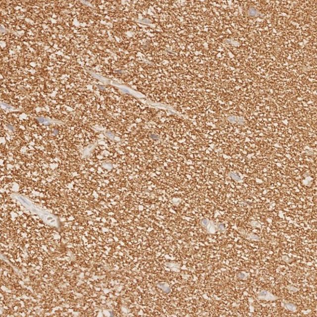
IHC staining of human brain tissue using Synaptophysin Monoclonal antibody.
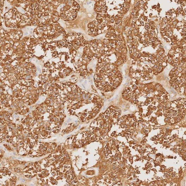
IHC staining of human pituitary adenoma tissue using Synaptophysin Monoclonal antibody.
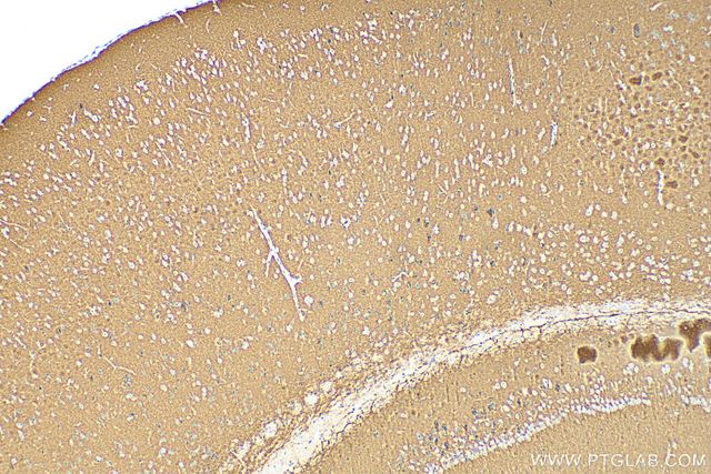
IHC staining of mouse brain tissue using Synaptophysin Recombinant antibody.
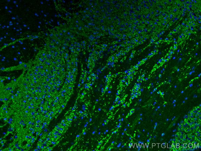
IF staining of mouse brain tissue using Synaptophysin Recombinant antibody.
View all synaptophysin antibodies →
PSD95 — Postsynaptic density
Postsynaptic density protein 95 (PSD95) is a scaffolding protein that serves as prominent marker for the postsynaptic end of excitatory synapses. It can be used to further study synaptic density and structural integrity
| Antibody (catalog no.) | Best for | Citations | Validated applications |
|---|---|---|---|
| PSD95-Specific Polyclonal Antibody (20665-1-AP) | Top-cited antibody; versatile across tissue and cell applications | 380+ | WB, IHC, IF-P, FC (Intra) |
| PSD95-Specific Recombinant Antibody (81106-1-RR) | 3D epitope mapped, high affinity and unmatched lot-to-lot consistency for long term projects | — | WB, IHC, IF-P |
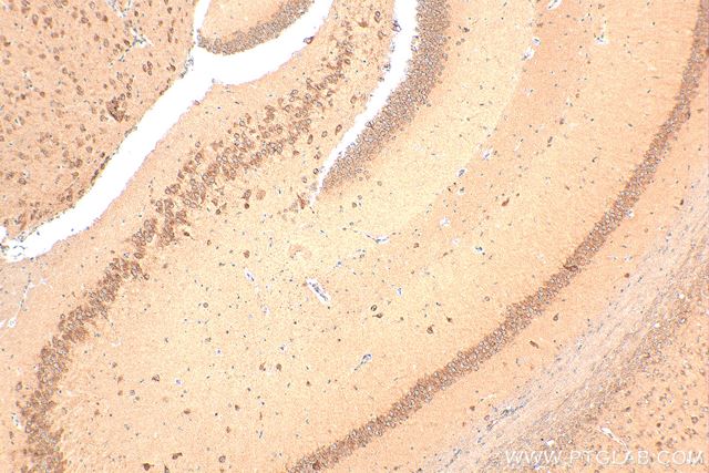
IHC staining of mouse brain using PSD95-Specific Polyclonal antibody.
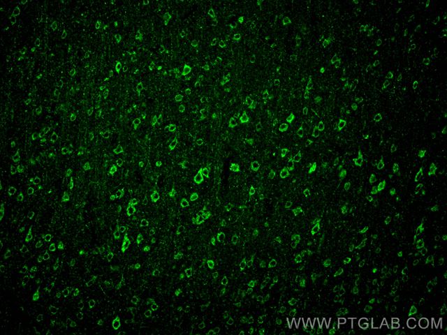
IF staining of mouse brain using PSD95-Specific Polyclonal antibody.
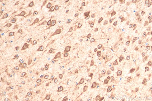
IHC staining of mouse brain using PSD95-Specific Recombinant monoclonal antibody.
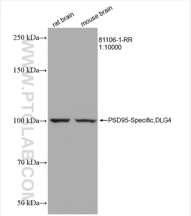
WB analysis of various lysates using PSD95-Specific Recombinant monoclonal antibody.
Phospho-Tau — 15 phosphorylation sites
Tau (gene MAPT) is abnormally hyperphosphorylated in Alzheimer's disease and other tauopathies, and different phospho-sites mark different disease stages. Proteintech's recombinant phospho-Tau portfolio covers 15 distinct sites; the three below span early tangles to late-stage neurofibrillary tangles (NFTs).
Proteintech’s phospho-Tau portfolio contains 20 antibodies against 15 distinct sites; the three antibodies below span sites that are markers for early to late-stage neurofibrillary tangles (NFTs)
| Antibody (catalog no.) | Phospho-site / stage | Validated applications |
|---|---|---|
| Phospho-Tau Thr231 Recombinant Antibody (80101-7-RR) | Thr231 — Critical early-stage Alzheimer's disease marker | WB, IHC, IF-P, FC (Intra); 3D epitope mapped |
| Phospho-Tau Ser202/Thr205 Recombinant Antibody (82568-1-RR) | Ser202/Thr205 — Gold-standard marker for paired helical filament (PHF) tau | WB, IF-P; 3D epitope mapped |
| Phospho-Tau Ser396/Ser404 Recombinant Antibody (87061-1-RR) | Ser396/Ser404 — Detects late-stage NFTs with high specificity; validated in phosphatase-inhibitor-treated and untreated brain tissue | WB, IHC, IF; 3D epitope mapped |
| Phospho-TAU (Thr217) Recombinant Antibody (87428-1-RR) | Thr217 — Gold standard fluid biomarker for Alzheimer’s | WB, IHC, Dot Blot |
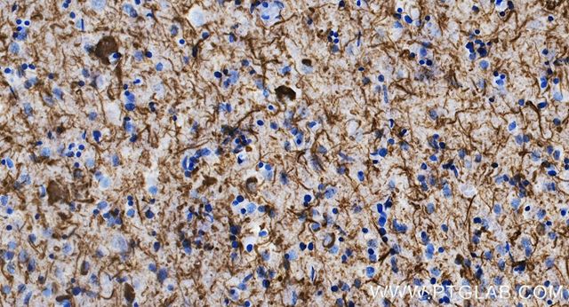
IHC staining of human brain (Alzheimer's disease) using Phospho-Tau (Thr231) Recombinant antibody.
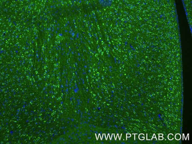
IF staining of mouse brain tissue using Phospho-Tau (Thr231) Recombinant antibody.
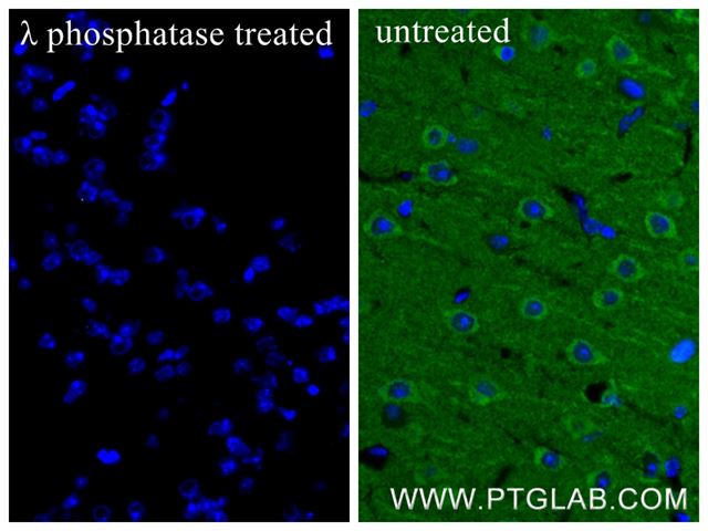
IF staining of mouse brain tissue using Phospho-Tau (Ser202/Thr205) Recombinant antibody.
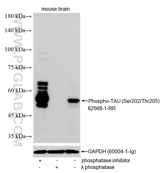
WB analysis of multiple lysates using Phospho-Tau (Ser202/Thr205) Recombinant antibody.
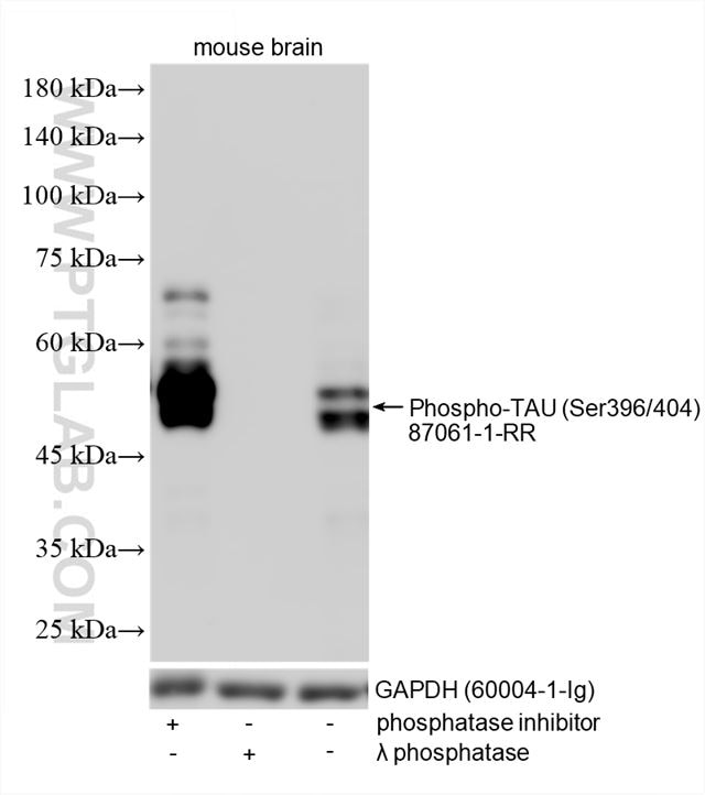
WB analysis of multiple lysates using Phospho-Tau (Ser396/Ser404) Recombinant antibody.
Growth factors for organoid and neuronal differentiation
HumanKine® growth factors are expressed in human cells (HEK293), so they carry native human conformation and post-translational modifications, giving high bioactivity and lot-to-lot stability. They are validated in-house across cerebral, cortical and midbrain organoid models, and every research-grade protein has a GMP-grade counterpart for a seamless transition to clinical manufacturing.
- Authentic human-cell-expressed proteins — native folding and glycosylation
- High specific bioactivity and stability
- In-house validation in multiple organoid differentiation models
- Research-grade to GMP-grade continuity for translational programmes
Essential growth factors for cerebral organoids
| Growth factor | Catalog No. |
|---|---|
| BDNF | HZ-1335 |
| FGF-basic (FGF2) | HZ-1285 |
| EGF | HZ-1326 |
| BMP-4 | HZ-1045 |
| Wnt3A | HZ-1296 |
| SHH (Sonic hedgehog) | HZ-1306 |
| Noggin | HZ-1118 |
| TGF-β1 | HZ-1011 |
| IGF-1 | HZ-1322 |
| GDNF | HZ-1311 |
| CNTF | HZ-1331 |
| NT-3 | HZ-1341 |
In-house validation includes midbrain organoid differentiation with HumanKine Noggin, SHH, FGF-8b, GDNF and BDNF: by day 33 the organoids express progenitor markers (SOX2, Nestin, FOXA2), proliferation and neuronal markers (Ki67, MAP2), midbrain identity markers (OTX2, NURR1, MASH1) and dopaminergic markers (TH, DAT).
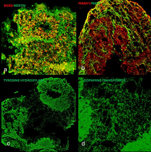
iPSC-derived midbrain organoids — SOX2/Nestin, MASH1/MAP2, TH and DAT — differentiated with HumanKine Noggin, SHH, FGF-8b, GDNF and BDNF.
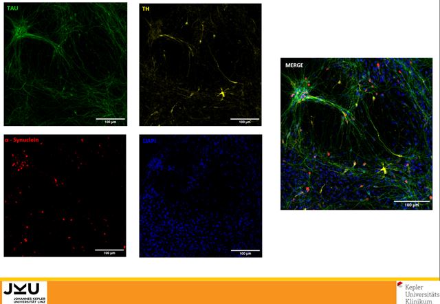
iPSC-derived dopaminergic neurons (tau, α-synuclein, tyrosine hydroxylase) — differentiated with HumanKine GDNF and FGF-8b.
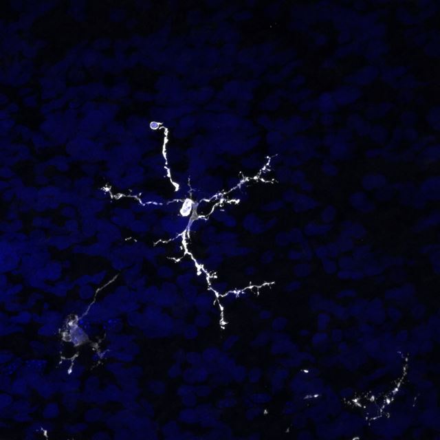
iPSC-derived microglia in a brain-like organoid — generated with HumanKine TGF-β1, BMP-4 and thrombopoietin.
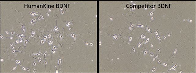
SH-SY5Y neuronal differentiation — more neurite outgrowth with HumanKine BDNF than a leading competitor.
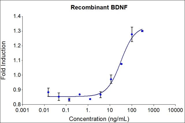
HumanKine BDNF drives dose-dependent proliferation of SH-SY5Y neuroblastoma cells (bioactivity assay).
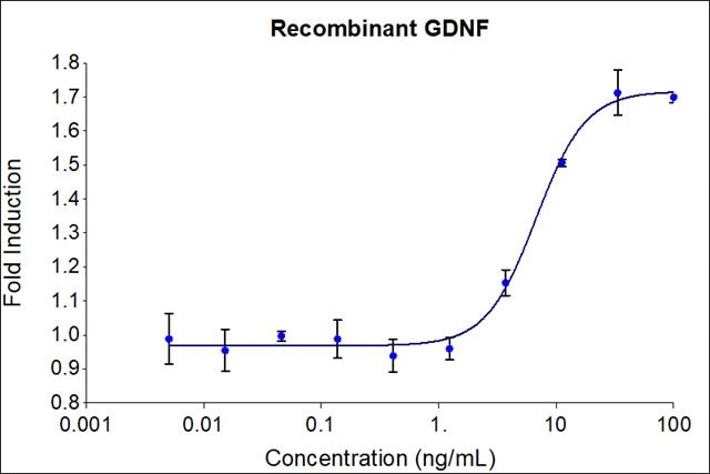
HumanKine GDNF drives dose-dependent proliferation of SH-SY5Y neuroblastoma cells (bioactivity assay).
Multiplexing tools for spatial neuroscience and mapping neural complexity
Tissue multiplexing lets you map many neural and glial markers in a single section. Proteintech supplies two complementary systems: FlexAble kits for rapid direct conjugation and same-species multiplexing, and TSA reagents for high-sensitivity, multiplexed detection of low-abundance targets.
FlexAble 2.0 antibody labeling kits
- Compatible with primary antibodies from any supplier, at any concentration or formulation
- Fast, easy 10-minute conjugation protocol
- Same-species multiplexing — combine several primaries raised in the same host
- Compatible with standard and advanced IF: cyclic IF, IBEX, TSA, expansion microscopy and tissue clearing
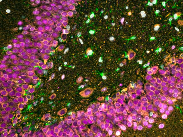
Rat brain — NeuN, β-III tubulin and GFAP, primaries conjugated with FlexAble kits.
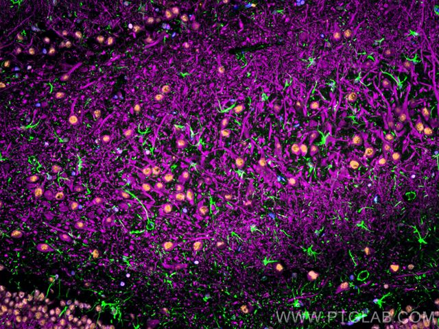
FFPE rat brain — MAP2, GFAP and TDP-43, conjugated with FlexAble 2.0 kits.
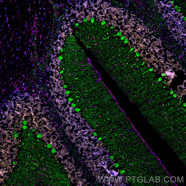
FFPE mouse cerebellum — Calbindin, FUS and GFAP, FlexAble HRP kits with tyramide.
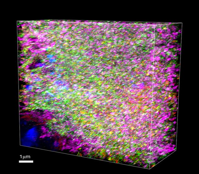
Expansion microscopy of mouse brain — Syntaxin 1B, Neuregulin and Ephrin A4, FlexAble 2.0 kits.
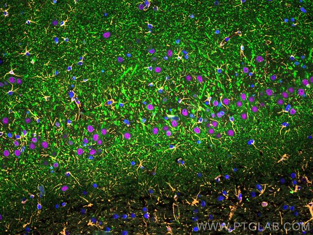
FFPE rat brain — MAP2, GFAP and TDP-43 (alternate kit combination), FlexAble 2.0 kits.
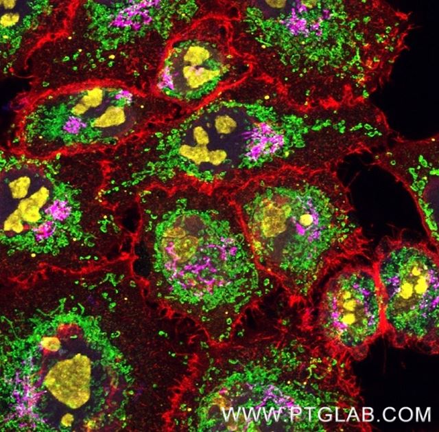
5-colour IF with five rabbit primaries incl. anti-TDP-43 (same-species multiplex) — FlexAble 2.0 kits.
Explore FlexAble antibody labeling kits →
TSA (tyramide signal amplification) reagents
- Up to 100× more sensitive than indirect staining methods
- Up to 5-plex imaging with no restriction on primary antibody host species
- Wide dynamic range for detecting low-abundance neural targets
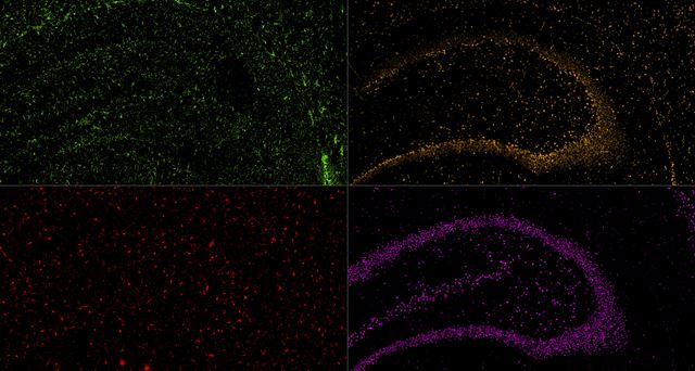
5-colour TSA, FFPE rat brain — Lamin B1, GFAP, IBA1 and TDP-43 with the PK10034 kit.
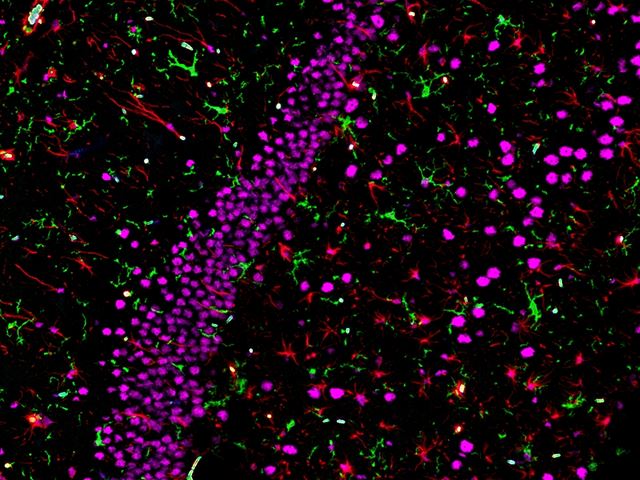
4-colour TSA, FFPE rat brain — IBA1, GFAP and TDP-43 with the PK10032 kit.
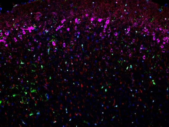
4-colour TSA, FFPE mouse brain — GFAP, MAP2 and WFS1 with the PK10032 kit.
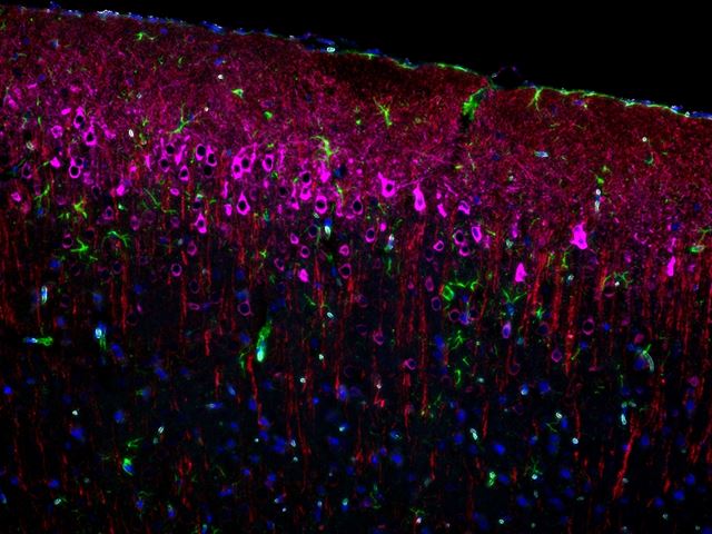
4-colour TSA (rabbit-only), FFPE mouse brain — GFAP, MAP2 and WFS1 with the PK10033 kit.
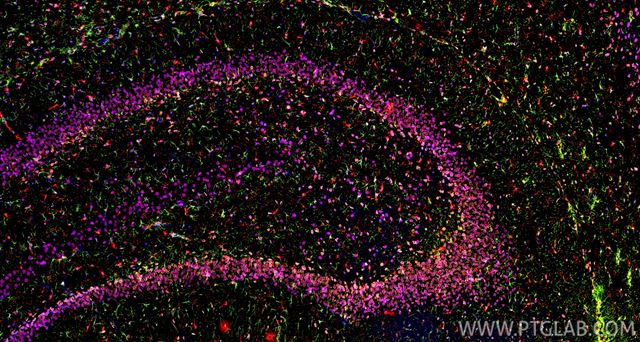
5-colour TSA, FFPE rat brain — PK10034 kit.
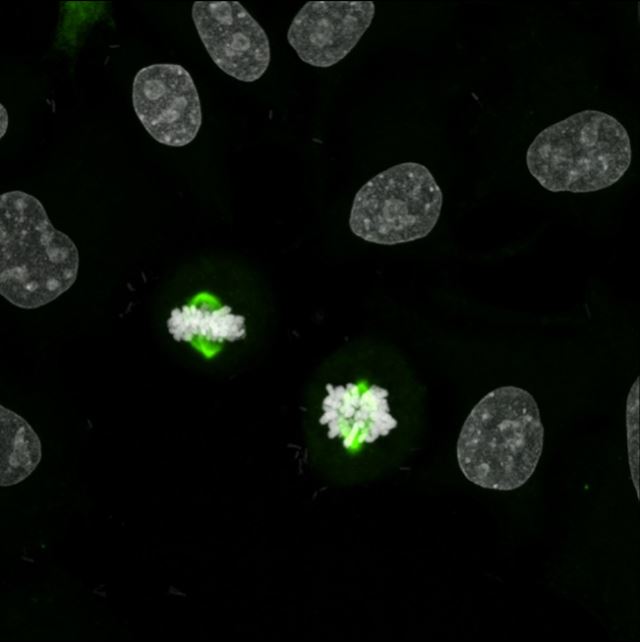
High-sensitivity TSA detection of a low-abundance target (AURKA) — Multi-rAb HRP secondary + CoraLite Plus 488 tyramide.
Applications for neuroscience research
Proteintech neuroscience antibodies are validated across the core techniques used to localise and quantify neural proteins in tissue, cells and biofluids.
| Application | Typical use in neuroscience |
|---|---|
| Immunohistochemistry (IHC) | Brain and spinal cord sections; free-floating and FFPE; cell-type mapping and pathology scoring |
| Immunofluorescence (IF / ICC) | Multiplex co-labelling of neurons and glia; subcellular localisation; primary neuron cultures |
| Western blot | Brain lysates, synaptosome and myelin fractions; phospho-protein quantification |
| Flow cytometry | Dissociated neural tissue, brain organoids and iPSC-derived neural cells |
| ELISA | Quantifying neural and neurodegeneration proteins in serum, plasma, CSF and lysates |
Neuroscience resources, posters and how-to guides
Guides and protocols
- How to identify activated microglia
- 7 tips to successfully culture primary rodent neurons
- Mastering the Transition: A Protocol for Differentiating SH-SY5Y Cells into Neurons
- Midbrain organoid generation: Practical tips from the bench
- A Guide to Markers for Oligodendrocyte Identification
- Markers for Astrocyte Identification
- A Guide to Immunostaining the Cerebellum
Product focus pages
- Antibodies for neural-related diseases: ALS and FTD
- Neurodegeneration antibodies
- Neuronal marker product focus
- Synaptic marker product focus
- Ion homeostasis product focus
- Cell polarity marker product focus
- Blood-brain barrier research
- Brain tumor markers
Pathway and marker posters
Frequently asked questions
What is the best marker for identifying neurons?
NeuN (RBFOX3) is the standard marker for mature, post-mitotic neurons and labels the nucleus and perinuclear cytoplasm. MAP2 marks dendrites and cell bodies, beta-III tubulin (TUBB3) marks early neuronal commitment, and neurofilament proteins (NEFL, NEFM, NEFH) label axons. Use NeuN to count neurons and MAP2 or beta-III tubulin for morphology.
Which markers distinguish resting from activated microglia?
IBA1 (AIF1) and TMEM119 label all microglia regardless of state. Homeostatic microglia express P2RY12 and CX3CR1, which fall on activation. Activated or phagocytic microglia upregulate CD68, CD11b (ITGAM) and MHC class II. Co-staining IBA1 with P2RY12 and CD68 lets you score the activation spectrum.
What is the difference between GFAP and S100B for astrocytes?
GFAP is an intermediate filament that labels astrocyte processes and is strongly upregulated in reactive astrogliosis, but it does not fill the whole cell and is low in some grey-matter astrocytes. S100B gives fuller cell-body labelling but is also present in some oligodendrocytes and NG2 cells. ALDH1L1 and AQP4 are more pan-astrocytic alternatives.
Which markers identify oligodendrocyte progenitors versus mature oligodendrocytes?
OLIG2, SOX10 and NKX2-2 label the whole oligodendrocyte lineage. Oligodendrocyte precursor cells (OPCs) express NG2 (CSPG4) and PDGFRA. Immature oligodendrocytes are marked by O4 and CNPase. Mature, myelinating oligodendrocytes express MBP, PLP1, MOG and GALC.
What antibodies and ELISA kits are used to study Alzheimer's and Parkinson's disease pathology?
Alzheimer's research uses antibodies and kits for amyloid-beta / APP, total and phospho-tau (MAPT), APOE, GFAP and UCH-L1. Parkinson's and other synucleinopathies use total and phospho-Ser129 alpha-synuclein (SNCA), LRRK2, PINK1 and parkin. TDP-43 (TARDBP) is central to ALS and frontotemporal dementia, and Proteintech offers TDP-43 ELISA kits for serum, plasma and CSF.
Are Proteintech neuroscience antibodies validated for knockout specificity?
Many are validated by knockout or siRNA knockdown. Each product page lists the validation data (Western blot, IHC, IF/ICC), tested species reactivity and published citations. Filter the catalogue by application and species and check the validation section on the product page before ordering.
Which TDP-43 antibody should I use for ALS and FTLD research?
For broad species reactivity and the widest application range, use the TDP-43 polyclonal antibody 10782-2-AP, the industry-standard reagent with over 1,700 citations, validated in WB, IP, IHC, IF/ICC, FC and ChIP. For human-specific detection use the monoclonal 60019-2-Ig (120+ citations; WB, IP, IHC). For N-terminal-specific, 3D epitope-mapped detection with whole-slide IHC and autostainer validation in clinical samples, use the recombinant antibody 80001-1-RR.
Which phospho-Tau antibody detects early versus late Alzheimer's disease pathology?
Phospho-Tau Thr231 (80101-7-RR) is a critical early-stage Alzheimer's marker. Phospho-Tau Ser202/Thr205 (82568-1-RR) is the gold-standard marker for paired helical filament (PHF) tau. Phospho-Tau Ser396/Ser404 (87061-1-RR) detects late-stage neurofibrillary tangles with high specificity. All three are recombinant, 3D epitope-mapped antibodies, part of a panel covering 11 distinct tau phosphorylation sites.
What is the best GFAP antibody for labelling astrocytes?
GFAP is the most widely used overall marker for astrocytes, and its expression level and cell morphology can also be quantified to study astrocyte reactivity. The GFAP polyclonal antibody 16825-1-AP is the most widely used option, with over 900 citations, validation in WB, IHC, IF-P, IF-Fro and IF/ICC, and multiple dye and biotin conjugates. For a mouse monoclonal use 60190-1-Ig (320+ citations, 3D epitope mapped); for maximum lot-to-lot consistency use the recombinant antibody 81063-1-RR, validated by whole-slide IHC and autostainer in healthy brain and glioma tissue.
Which NeuN antibody is best for neuronal maturation research?
NeuN (RBFOX3) is the premier marker of mature, post-mitotic neurons and is widely used to study neuronal maturation. The NeuN polyclonal antibody 26975-1-AP is the most versatile choice (500+ citations; WB, IHC, IF-P, IF-Fro, FC). The monoclonal 66836-1-Ig is optimised for IHC with whole-slide and autostainer validation, and the recombinant antibody 84401-4-RR offers high affinity and unmatched lot-to-lot consistency for long-term projects.
How do I quantify synaptic density by immunostaining?
Synaptophysin is a presynaptic vesicle glycoprotein used to quantify synaptic vesicle content and visualise synaptic density. The synaptophysin polyclonal antibody 17785-1-AP is top-cited (350+ citations; WB, IP, IHC, IF-P). Pair it with a postsynaptic marker such as PSD-95 for synapse colocalisation, and use the recombinant antibody 82900-1-RR when consistent signal across long imaging studies is required.
Which HumanKine growth factors are used for cerebral organoid differentiation?
Growth factors commonly used in cerebral organoid protocols include FGF-basic (HZ-1285), EGF (HZ-1326), BMP-4 (HZ-1045), Wnt3A (HZ-1296), SHH (HZ-1306), Noggin (HZ-1118), BDNF (HZ-1335), GDNF (HZ-1311), NT-3 (HZ-1341), IGF-1 (HZ-1322), TGF-β1 (HZ-1011) and CNTF (HZ-1331). The role of each factor depends on when it is added during differentiation, so concentrations and timing should follow your protocol. Midbrain dopaminergic organoids typically use Noggin, SHH, FGF-8b, GDNF and BDNF.
Can I multiplex two primary antibodies raised in the same host species?
Yes. FlexAble 2.0 antibody labelling kits conjugate a fluorophore directly to each primary antibody in a 10-minute protocol, so same-species multiplexing is possible without cross-reactivity. FlexAble works with primary antibodies from any supplier at any concentration or formulation and is compatible with cyclic IF, IBEX, TSA, expansion microscopy and tissue clearing. For low-abundance targets, TSA reagents add up to 100-fold sensitivity over indirect staining and support up to 5-plex imaging.